Review
. 2021 May 15:1159:338384.
doi: 10.1016/j.aca.2021.338384. Epub 2021 Mar 12.

Biosensing strategies for the electrochemical detection of viruses and viral diseases - A review

Affiliations
Review

Biosensing strategies for the electrochemical detection of viruses and viral diseases - A review

Laís Canniatti Brazaca et al. Anal Chim Acta. .

Abstract

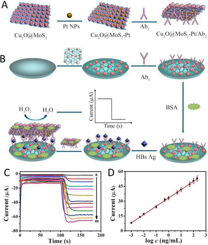
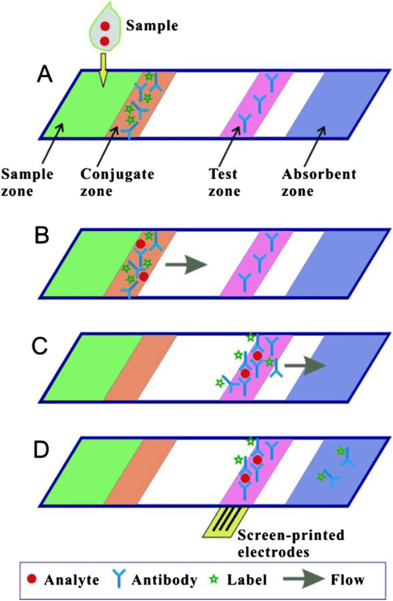
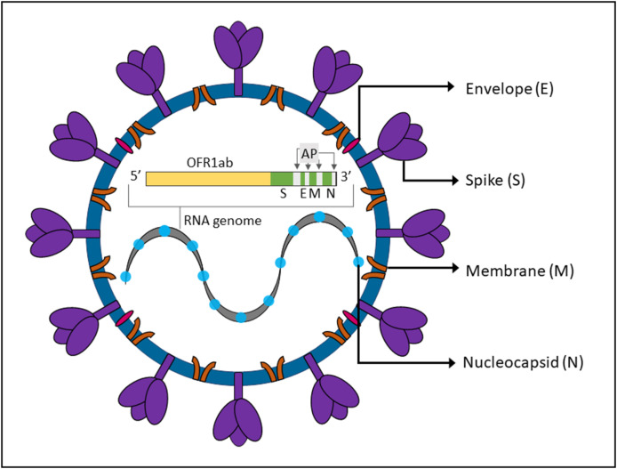
Viruses are the causing agents for many relevant diseases, including influenza, Ebola, HIV/AIDS, and COVID-19. Its rapid replication and high transmissibility can lead to serious consequences not only to the individual but also to collective health, causing deep economic impacts. In this scenario, diagnosis tools are of significant importance, allowing the rapid, precise, and low-cost testing of a substantial number of individuals. Currently, PCR-based techniques are the gold standard for the diagnosis of viral diseases. Although these allow the diagnosis of different illnesses with high precision, they still present significant drawbacks. Their main disadvantages include long periods for obtaining results and the need for specialized professionals and equipment, requiring the tests to be performed in research centers. In this scenario, biosensors have been presented as promising alternatives for the rapid, precise, low-cost, and on-site diagnosis of viral diseases. This critical review article describes the advancements achieved in the last five years regarding electrochemical biosensors for the diagnosis of viral infections. First, genosensors and aptasensors for the detection of virus and the diagnosis of viral diseases are presented in detail regarding probe immobilization approaches, detection methods (label-free and sandwich), and amplification strategies. Following, immunosensors are highlighted, including many different construction strategies such as label-free, sandwich, competitive, and lateral-flow assays. Then, biosensors for the detection of viral-diseases-related biomarkers are presented and discussed, as well as point of care systems and their advantages when compared to traditional techniques. Last, the difficulties of commercializing electrochemical devices are critically discussed in conjunction with future trends such as lab-on-a-chip and flexible sensors.

Keywords: Biomarkers; Biosensors; Electrochemistry; Genosensor; Immunosensor; Viral diseases.


Conflict of interest statement

Declaration of competing interest The authors declare that they have no known competing financial interests or personal relationships that could have appeared to influence the work reported in this paper.

Figures

Image 1
Graphical abstract
Fig. 1
Fig. 1
Basic device operation and results obtained by Carinelli et al. using RCA and C2CA combined with electrochemistry for the precise detection of Ebola virus cDNA. (A) Step-by-step schematic representation of the proposed genosensor. Step A1 - Coupling of the circularized biotinylated cDNA to streptavidin-magnetic particles; step A2 - amplification by RCA using ϕ29 DNA polymerase; step A3 - Hybridization step with a probe labeled with HRP and; step A4 - Electrochemical determination on SPCEs by SWV with an enzymatic reaction in the presence of hydroquinone as a mediator. (B) Square wave voltammograms for different concentrations of cDNA templates for C2CA (C) calibration curve obtained by one-site binding fitting and by (D) four-parameter logistic fitting. (Reprinted with permission from Carinelli et al. Yoctomole electrochemical genosensing of Ebola virus cDNA by rolling circle and circle to circle amplification. Biosens. and Bioelectron. 93, 65–71, © 2017 Elsevier BV [56]).
Fig. 2
Fig. 2
Strategy developed by Shariati et al. for the sensitive determination of hepatitis B virus. (A) Schematics showing the electrodes and Fet pattern; (B) Growth of ITO nanowires over Au patterns. C) Systems response upon the addition of different concentrations of complementary (red), mismatch (black) and non-complementary (blue) sequences. (Reprinted with permission from Shariati, M. et al. The field-effect transistor DNA biosensor based on ITO nanowires in label-free hepatitis B virus detecting compatible with CMOS technology. Biosens. and Bioelectron. 105, 58–64, © 2018 Elsevier BV [68]). (For interpretation of the references to color in this figure legend, the reader is referred to the Web version of this article.)
Fig. 3
Fig. 3
Schematic illustration of antibody covalent immobilization of (A) a carboxyl-functionalized surface activated with EDC/NHS, and (B) an amine-functionalized surface activated with glutaraldehyde.
Fig. 4
Fig. 4
Schematic representation of (A) label-free, (B) sandwich, and (C) competitive electrochemical immunosensors (Left) and schematic representation of the mechanism using enzymatic-labels (right): (D) ALP enzymatic-label for production of electrochemically active 1-naphthol, (E) ALP-catalyzed deposition of electroactive silver, and (F) HRP-catalyzed oxidation of the redox mediator.
Fig. 5
Fig. 5
Detailed construction of sandwich immunosensor using (A) MoS2@Cu2O–Pt/Ab2 and (B) GCE/GO-Au/Ab1. (C) Amperometric response obtained with different concentrations of hepatitis B surface antigen, and (D) its corresponding calibration curve. (Reprinted with permission from Li, F. et al. Facile synthesis of MoS2@Cu2O–Pt nanohybrid as enzyme-mimetic label for the detection of the Hepatitis B surface antigen. Biosens. and Bioelectron. 100, 512–518, © 2018 Elsevier BV [170]).
Fig. 6
Fig. 6
Detailed mechanism of a sandwich LFI (Reprinted with permission from Liu, B. et al. Paper-based electrochemical biosensors: from test strips to paper-based microfluidics. Electroanalysis. 26, 1214–1223, © 2014 WILEY-VCH [199]).
Fig. 7
Fig. 7
Structure and genomic organization of SARS-CoV-2. The open reading frame 1 ab gene (ORF1ab, in yellow) encodes non-structural proteins, including the RNA-dependent RNA polymerase (RdRp). The structural genes (in green) encode the structural proteins, spike (S), envelope (E), membrane (M), and nucleocapsid (N). The genes highlighted in gray encode the accessory proteins (AP). (Adapted from Alanagreh, L. et al. The human coronavirus disease COVID-19: its origin, characteristics, and insights into potential drugs and its mechanisms. Pathogens 9, 5, 331 © 2020 MDPI [236]). (For interpretation of the references to color in this figure legend, the reader is referred to the Web version of this article.)

Similar articles

Cited by

References

    1. Saylan Y., Erdem Ö., Ünal S., Denizli A. An alternative medical diagnosis method: biosensors for virus detection. Biosensors. 2019;9:65. doi: 10.3390/bios9020065. - DOI - PMC - PubMed
    1. Webster R.G., Bean W.J., Gorman O.T., Chambers T.M., Kawaoka Y. Evolution and ecology of influenza A viruses. Microbiol. Rev. 1992;56 http://mmbr.asm.org/content/56/1/152.abstract 152 LP – 179. - PMC - PubMed
    1. Feldmann H., Geisbert T.W. Ebola haemorrhagic fever. Lancet. 2011;377:849–862. doi: 10.1016/S0140-6736(10)60667-8. - DOI - PMC - PubMed
    1. Zumla A., Hui D.S., Perlman S. Middle East respiratory syndrome. Lancet. 2015;386:995–1007. doi: 10.1016/S0140-6736(15)60454-8. - DOI - PMC - PubMed
    1. Mellors J.W., Rinaldo C.R., Gupta P., White R.M., Todd J.A., Kingsley L.A. Prognosis in HIV-1 infection predicted by the quantity of virus in plasma. Science (80-. ) 1996;272 doi: 10.1126/science.272.5265.1167. 1167 LP – 1170. - DOI - PubMed