

Comment
doi: 10.1016/j.oooo.2021.03.010.
Epub 2021 Mar 28.
Painful palatal lesion in a patient with COVID-19
Affiliations
- PMID: 33867304
- PMCID: PMC8005255
- DOI: 10.1016/j.oooo.2021.03.010
Item in Clipboard
Comment
Painful palatal lesion in a patient with COVID-19
Oral Surg Oral Med Oral Pathol Oral Radiol.
2021 Jun.
No abstract available
Figures
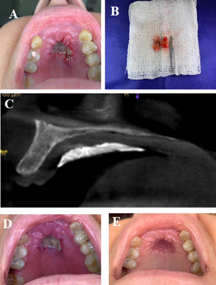
Clinical and imaging findings. (A) Clinical aspect of the lesion just after the incisional biopsy procedure, revealing a deep ulcerated lesion, with bone exposure in the hard palate. (B) Three fragments measuring about 0.5 × 0.3 × 0.1 cm to 1.0 × 1.0 × 0.5 cm removed during incisional biopsy. All of the material collected was sent for histopathological analysis. (C) Cone beam computed tomography scan taken immediately following incisional biopsy. No significant alteration was noted in the maxillary bone. Hyperdense image covering the soft tissue corresponds to surgical cement applied for homeostasis during the biopsy procedure. (D) Clinical presentation 7 days after incisional biopsy. (E) Ten days following discharge from hospitalization and approximately 60 days after initial evaluation. The lesion appears to be resolving and the patient is asymptomatic.
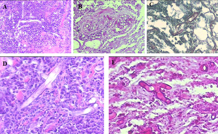
Histopathological findings. (A) and (D) Heavy mixed acute and chronic inflammatory infiltrate with necrosis and large nonseptate, thin-walled fungal hyphae that branch at a 90° angle. The microorganisms are aggregated around blood vessels. (A) Original magnification 20 ×, (D) original magnification 40 × . High-resolution versions of these images are available as eSlide VM06234 . (B) and (E) Hyphae stained with periodic acid–Schiff. (B) original magnification 20 ×, (E) original magnification 40 × . High-resolution versions of these images are available as eSlide VM06235 . (C) Grocott's methenamine. High-resolution versions of this image are available as eSlide VM06236 .
Comment on
-
Bacterial and Fungal Coinfection in Individuals With Coronavirus: A Rapid Review To Support COVID-19 Antimicrobial Prescribing.Clin Infect Dis. 2020 Dec 3;71(9):2459-2468. doi: 10.1093/cid/ciaa530. Clin Infect Dis. 2020. PMID: 32358954 Free PMC article. Review.
References
-
- Ledesma-Montes C, Garcés-Ortíz M, Salcido-García JF, Hernández-Flores F. Review of the literature on necrotizing sialometaplasia and case presentation. Quintessence Int. 2015;46:67–72. - PubMed
-
- de Camargo AR, Duarte BF, Lisboa ML, et al. Immunophenotyping for diagnosis of oral lesions: is it an important tool? Int J Med Pharm Case Reports. 2020;13:16–23.
-
- de Castro Costa M, de Carvalho MM, Sperandio FF, et al. Oral paracoccidioidomycosis affecting women: a systematic review. Mycoses. 2021;64:108–122. - PubMed
Publication types
MeSH terms
Substances
LinkOut - more resources
Full Text Sources
Other Literature Sources
Medical
