. 2021 Jul 10;83(6):990-993.
doi: 10.1292/jvms.20-0654. Epub 2021 Apr 19.

The formation process of button ulcers in pigs experimentally infected with a subgenotype 2.1 isolate of classical swine fever virus

Affiliations

The formation process of button ulcers in pigs experimentally infected with a subgenotype 2.1 isolate of classical swine fever virus

Kasumi Sudo et al. J Vet Med Sci. .

Abstract

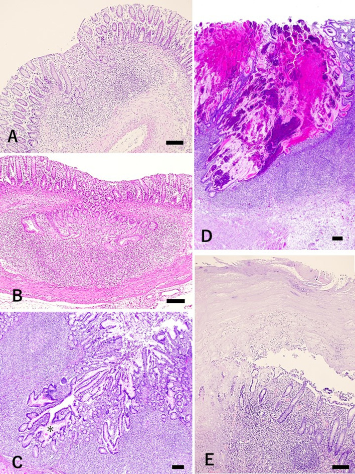
We evaluated the role of classical swine fever virus (CSFV) in the formation of button ulcers in the mucosa of the gastrointestinal tract. Histopathological and immunohistochemical analyses of pigs experimentally infected with a subgenotype 2.1 isolate of CSFV, which was isolated in Japan in 2019, revealed follicular necrosis in the submucosal mucosa-associated lymphoid tissue and herniation of crypts as factors that contribute to the development of button ulcers during CSFV infection. These findings indicate that CSFV induces follicular necrosis and is one of the causative agents of button ulcers in pigs.

Keywords: button ulcer; classical swine fever virus; herniation of crypt; histopathology.


Conflict of interest statement

The authors declare no conflicts of interest.

Figures

Fig. 1.
Fig. 1.
A button ulcer found in the ileocecal area.
Fig. 2.
Fig. 2.
Histopathology of the progression from herniation of crypts into the submucosal mucosa-associated lymphoid tissue to button ulcers. A & B: Follicular necrosis and herniation of crypts (A: mild, B: progressed). C: Herniated crypts are dilated by hypersecretion. Secretions are marked (*). D: Crypt abscesses fuse together and develop into massive necrotic lesions with accumulation of mucus, cell debris, inflammatory exudate, and bacterial colonies. E: Severe ulcers with pseudo-membranes mainly composed of exudative fibrin and mucus form a “button ulcer”. A, B and, E were obtained from a pig that developed the button ulcer, and C and D were obtained from the other pigs as representative images. Hematoxylin and eosin staining. Bar: 160 µm.
Fig. 3.
Fig. 3.
Detection of classical swine fever virus (CSFV) antigen by immunohistochemistry. A: CSFV antigen is consistent with the follicle but is absent in the herniated crypt epithelium. B: High magnification of A. Immunohistochemistry for CSFV antigen. Bar, A: 180 µm, B: 20 µm.

References

    1. Anonymous. Chapter 3.8.3. Classical swine fever (Infection with classical swine fever virus). Manual of diagnostic tests and vaccines for terrestrial animals 2019. https://www.oie.int/fileadmin/Home/eng/Health_standards/tahm/3.08.03_CSF... [accessed on November 14, 2020].
    1. Beer M., Goller K. V., Staubach C., Blome S.2015. Genetic variability and distribution of Classical swine fever virus. Anim. Health Res. Rev. 16: 33–39. doi: 10.1017/S1466252315000109 - DOI - PubMed
    1. Blome S., Staubach C., Henke J., Carlson J., Beer M.2017. Classical swine fever−an updated review. Viruses 9: 86. doi: 10.3390/v9040086 - DOI - PMC - PubMed
    1. Gelberg H. B.2017. Alimentary system and the peritoneum, omentum, mesentery, and peritoneal cavity. pp. 324–411. In: Pathologic Basis of Veterinary Disease, 6th ed. (Zachary, J. F. ed.), Elsevier, St. Louis.
    1. Kameyama K. I., Nishi T., Yamada M., Masujin K., Morioka K., Kokuho T., Fukai K.2019. Experimental infection of pigs with a classical swine fever virus isolated in Japan for the first time in 26 years. J. Vet. Med. Sci. 81: 1277–1284. doi: 10.1292/jvms.19-0133 - DOI - PMC - PubMed