Current Status of Familial LCAT Deficiency in Japan
- PMID: 33867422
- PMCID: PMC8265425
- DOI: 10.5551/jat.RV17051
Current Status of Familial LCAT Deficiency in Japan
Abstract
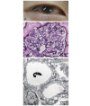
Lecithin cholesterol acyltransferase (LCAT) is a lipid-modification enzyme that catalyzes the transfer of the acyl chain from the second position of lecithin to the hydroxyl group of cholesterol (FC) on plasma lipoproteins to form cholesteryl acylester and lysolecithin. Familial LCAT deficiency is an intractable autosomal recessive disorder caused by inherited dysfunction of the LCAT enzyme. The disease appears in two different phenotypes depending on the position of the gene mutation: familial LCAT deficiency (FLD, OMIM 245900) that lacks esterification activity on both HDL and ApoB-containing lipoproteins, and fish-eye disease (FED, OMIM 136120) that lacks activity only on HDL. Impaired metabolism of cholesterol and phospholipids due to LCAT dysfunction results in abnormal concentrations, composition and morphology of plasma lipoproteins and further causes ectopic lipid accumulation and/or abnormal lipid composition in certain tissues/cells, and serious dysfunction and complications in certain organs. Marked reduction of plasma HDL-cholesterol (HDL-C) and corneal opacity are common clinical manifestations of FLD and FED. FLD is also accompanied by anemia, proteinuria and progressive renal failure that eventually requires hemodialysis. Replacement therapy with the LCAT enzyme should prevent progression of serious complications, particularly renal dysfunction and corneal opacity. A clinical research project aiming at gene/cell therapy is currently underway.
Keywords: Abnormal LDL; Corneal opacity; Enzyme replacement therapy; Lecithin cholesterol acyltransferase; Low HDL-cholesterol; Proteinuria.
Figures


Similar articles
-
Lipoprotein subfractions highly associated with renal damage in familial lecithin:cholesterol acyltransferase deficiency.Arterioscler Thromb Vasc Biol. 2014 Aug;34(8):1756-62. doi: 10.1161/ATVBAHA.114.303420. Epub 2014 May 29. Arterioscler Thromb Vasc Biol. 2014. PMID: 24876348
-
[LCAT deficiency: a nephrological diagnosis].G Ital Nefrol. 2011 Jul-Aug;28(4):369-82. G Ital Nefrol. 2011. PMID: 21809306 Review. Italian.
-
Identification and functional analysis of missense mutations in the lecithin cholesterol acyltransferase gene in a Chilean patient with hypoalphalipoproteinemia.Lipids Health Dis. 2019 Jun 5;18(1):132. doi: 10.1186/s12944-019-1045-0. Lipids Health Dis. 2019. PMID: 31164121 Free PMC article.
-
[Familial LCAT deficiency].Nihon Rinsho. 1994 Dec;52(12):3210-5. Nihon Rinsho. 1994. PMID: 7853712 Review. Japanese.
-
Compound heterozygosity (G71R/R140H) in the lecithin:cholesterol acyltransferase (LCAT) gene results in an intermediate phenotype between LCAT-deficiency and fish-eye disease.Atherosclerosis. 2006 Jul;187(1):101-9. doi: 10.1016/j.atherosclerosis.2005.08.038. Epub 2005 Oct 10. Atherosclerosis. 2006. PMID: 16216249
Cited by
-
Familial LCAT Deficiency and Low HDL-C Levels: In silico Characterization of Two Rare LCAT Missense Mutations.Appl Clin Genet. 2024 Feb 20;17:23-32. doi: 10.2147/TACG.S438135. eCollection 2024. Appl Clin Genet. 2024. PMID: 38404612 Free PMC article.
-
Two Cases of Acquired High-Density Lipoprotein Deficiency with Immunoglobulin G4-Related Lecithin-Cholesterol Acyltransferase Autoantibody.J Atheroscler Thromb. 2023 Aug 1;30(8):1070-1082. doi: 10.5551/jat.63616. Epub 2022 Nov 16. J Atheroscler Thromb. 2023. PMID: 36384970 Free PMC article.
-
Uncommon Factors Leading to Nephrotic Syndrome.Biomedicines. 2025 Aug 5;13(8):1907. doi: 10.3390/biomedicines13081907. Biomedicines. 2025. PMID: 40868160 Free PMC article. Review.
-
LCAT-trial-24 weeks: Protocol for a clinical study to evaluate the safety of regenerative medicine and gene therapy by the autologous transplantation of human lecithin:cholesterol acyltransferase gene-transduced human pre-adipocytes.Contemp Clin Trials Commun. 2022 Jun 9;28:100946. doi: 10.1016/j.conctc.2022.100946. eCollection 2022 Aug. Contemp Clin Trials Commun. 2022. PMID: 35734220 Free PMC article.
-
A Rare Case of Autoimmune-Mediated Lecithin:Cholesterol Acyltransferase Insufficiency Manifesting as the Acute Onset of Extremely Hypo-High-Density Lipoprotein-Cholesterolemia and Spontaneous Improvement: A Case Report with a Review of the Literature.J Atheroscler Thromb. 2025 May 1;32(5):649-659. doi: 10.5551/jat.65298. Epub 2024 Dec 10. J Atheroscler Thromb. 2025. PMID: 39662947 Free PMC article. Review.
References
-
- Glomset JA, Parker F, Tjaden M, Williams RH. The esterification in vitro of free cholesterol in human and rat plasma. Biochim Biophys Acta, 1962; 58:398-406 - PubMed
-
- Glomset JA, Janssen ET, Kennedy R, Dobbins J. Role of plasma lecithin: cholesterol acyltransferase in the metabolism of high density lipoproteins. J Lipid Res, 1966; 7: 638-648 - PubMed
-
- Carlson LA., Philipson B. Fish-eye disease: a new familial condition with massive corneal opacities and dyslipoproteinem. Lancet, 1979; 314: 921-924 - PubMed
-
- Glomset JA, Assmann G, Gjone E, Norum KR. Lecithin cholesterol acyltransferase deficiency and fish eye disease. In: The Metabolic and Molecular Bases of Inherited Disease, 7th ed. by Scriver CR, Beaudet AL, Sly WS, Valle D, Stanbury JB, Wyngaarden JB, Fredrickson DS., McGraw-Hill Inc., New York, 1995, 1933-1951
-
- Santamarina-Fojo S, Hoeg JM, Assman G, Brewer Jr HB. Lecithin cholesterol acyltransferase deficiency and fish eye disease. In: The metabolic and molecular bases of inherited disease, 8th ed. by Scriver CR, Beaudet AL, Sly WS, Valle D, Childs B, Kinzler KW, Vogelstein B., McGraw-Hill Inc., New York, 2001, 2817-2833
Publication types
MeSH terms
Substances
LinkOut - more resources
Full Text Sources
Other Literature Sources
Miscellaneous

