. 2018 Mar 21;28(12):1700239.
doi: 10.1002/adfm.201700239. Epub 2017 Jun 7.

Neuro-Nano Interfaces: Utilizing Nano-Coatings and Nanoparticles to Enable Next-Generation Electrophysiological Recording, Neural Stimulation, and Biochemical Modulation

Affiliations

Neuro-Nano Interfaces: Utilizing Nano-Coatings and Nanoparticles to Enable Next-Generation Electrophysiological Recording, Neural Stimulation, and Biochemical Modulation

Ashlyn T Young et al. Adv Funct Mater. .

Abstract

Neural interfaces provide a window into the workings of the nervous system-enabling both biosignal recording and modulation. Traditionally, neural interfaces have been restricted to implanted electrodes to record or modulate electrical activity of the nervous system. Although these electrode systems are both mechanically and operationally robust, they have limited utility due to the resultant macroscale damage from invasive implantation. For this reason, novel nanomaterials are being investigated to enable new strategies to chronically interact with the nervous system at both the cellular and network level. In this feature article, the use of nanomaterials to improve current electrophysiological interfaces, as well as enable new nano-interfaces to modulate neural activity via alternative mechanisms, such as remote transduction of electromagnetic fields are explored. Specifically, this article will review the current use of nanoparticle coatings to enhance electrode function, then an analysis of the cutting-edge, targeted nanoparticle technologies being utilized to interface with both the electrophysiological and biochemical behavior of the nervous system will be provided. Furthermore, an emerging, specialized-use case for neural interfaces will be presented: the modulation of the blood-brain barrier.

Keywords: blood-brain barrier; brain; nanoparticle; neural interface; neurostimulation.


Conflict of interest statement

Conflict of Interest The authors declare no conflict of interest.

Figures

Figure 1.
Figure 1.
Neural interfaces provide a window into the workings of the nervous system—enabling both biosignal recording and modulation. Nano-particles are used to improve current electrophysiological interfaces, as well as enable new nano-interfaces to modulate neural activity via alternative mechanisms. (i) Nanoparticle coatings of implanted electrodes reduce interface impedance with dramatically improved recording and stimulation fidelity. Alternatively, nanoparticles can be introduced intravenously and engineered to cross the blood brain barrier and excite neurons. (ii) Targeted-nanoparticles can remotely transduce external fields, e.g., infrared light or alternating magnetic fields, to modulate and stimulate neuronal function. (iii) An emerging, specialized use case for neural interfaces: Using nanoparticles and external fields to control the permeability of the blood-brain barrier for delivery of therapeutic and diagnostic materials into the central nervous system.
Figure 2.
Figure 2.
Micrographs illustrate the myriad nanomaterial functionalization of neural interface electrodes. a-b) CNTs are grown on the tip of a micro-electrode via chemical vapor deposition and a gold-CNT composite is coated on an electrode via a metal plating technique, respectively Reproduced with permission.[49] Copyright 2011, American Chemical Society. CNTs have been deposited on microelectrodes to improve performance in neural recording by increasing the surface-area of the electrodes, which decreases impedance. The CVD electrode displays smaller surface structures and less lamellar structures when compared to the gold-CNT composite, resulting in a greater surface area and mechanical stability, c) Microstructure of PEDOT/MWCNT film deposited in galvanostatic mode on microelectrode (inset) for neuronal recording. Reproduced with permission.[65] Copyright 2013, Elsevier. This deposition technique resulted in longitudinal film growth with lessened electrical cross-talk, resulting in increased spatial resolution of the MEA. d-e) Gold particle and PEDOT:PSS-CNT coated microelectrodes and corresponding microstructures (below), respectively. Reproduced with permission.[51] Copyright 2016, Frontiers Publishing. PEDOT:PSS-CNT coated microelectrodes show a fine nano-structure as compared to the microstructure present on the gold particle coated electrodes. f-g) Morphology of PEDOT and PEDOT-CNT coated electrodes and their microstructures, respectively. Reproduced with permission.[66] Copyright 2012, Frontiers Publishing. The porous structure of the PEDOT-CNT coated electrodes results in a larger surface area, therefore a lower impedance, when compared to the electrodes coated in PEDOT alone.
Figure 3.
Figure 3.
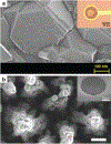
Electrodes fabricated with boron-doped diamond have improved stability and biocompatibility, a) SEM image of boron-doped diamond microstructure and microelectrode (inset), Reproduced with permission,[74] Copyright 2013, Institute of Electrical and Electronics Engineers. b) 3D-nanostructured boron-doped diamond electrode before addition of insulation layer and microelectrode (inset). Reproduced with permission.[73] Copyright 2015, Elsevier.
Figure 4.
Figure 4.
Metallic nanomaterials are incorporated onto neural interface electrodes to improve functionality and stability. a-c) Gold nanopillars are fabricated on MEAs. A cross-section of an HL-1 cell grown on the gold nanopillar array illustrates contact between electrodes and cell membrane, with gaps between the cell membrane and electrode less than 100 nm. Reproduced with permission.[81] Copyright 2011, IOP Publishing. d,e) Schematic for the nanowire geometry and structure for gallium-phosphide nanowire array on MEA. Reproduced with permission.[235] Copyright 2013, Public Library of Science. f) AuNPs are deposited on a Pt electrode and functionalized with a self-assembled monolayer. Reproduced with permission.[79] Copyright 2012, Royal Society of Chemistry. h,k) Silver nanowire electrodes are fabricated using photolithography. Reproduced with permission.[86] Copyright 2014, American Chemical Society. i) Micrographs of nanoporous Au-Pt electrodes, which exhibited high neuron coverage (inset, red) and reduced astrocyte coverage (inset, green). Reproduced with permission.[85] Copyright 2015, American Chemical Society. j) Microelectrodes are coated in AuNP-CNT composite. Reproduced with permission.[84] Copyright 2012, American Chemical Society.
Figure 5.
Figure 5.
Polymer-derived nanoparticles can be used to interface with the brain without implanted electrodes. a-b) Zebrafish microinjected with PFPBA nanoparticles were used to optically detect dopamine by fluorescence. Reprinted with permission.[125] Copyright 2015, American Chemical Society, c-d) Neurons were thermally activated when nanoparticles were excited by IR radiation, resulting in remotely controlled depolarization of neurons. Reproduced with permission.[123] Copyright 2016, American Chemical Society. e-g) Polymer nanoparticles functionalized with neuro-active molecules directed the growth of axons, and neural activity was regulated by remote NIR irradiation. Reproduced with permission.[122] Copyright 2015, Elsevier.
Figure 6.
Figure 6.
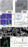
Semiconductor nanoparticles can be excited by external light sources to stimulate growth and activity of neurons. a-d) Polymer-coated AuNRs excited by NIR resulted in neurite outgrowth. Current-clamp recording of a neuron showed action potentials fired in response to a single laser pulse, which is attributed to hyperthermal response of the AuNRs. Increased laser pulse produced an increased mean peak temperature change. Representative temperature profiles for various pulse lengths (inset). Reproduced with permission.[131] Copyright 2013, John Wiley & Sons. e-g) Fluorescent micrographs of Ca2+ concentration in neurons (green) and AuNPs (red). Representative images of varying Ca2+ concentration (green) in response to NIR excitation and corresponding spectral intensity response, illustrating the activation of neurons with NIR excitation at time points indicated by dashed lines. Reproduced with permission.[130] Copyright 2016, Nature Publishing Group. h-j) Functionalized AuNPs are robustly localized to neurons, and continue to stimulate neuronal activity after multiple washings. Increasing irradiance induces greater cell depolarization, triggering action potentials. The NIR-activated depolarization is attributed to the recorded, localized hyperthermia induced by excitation of the functionalized AuNPs. Reproduced with permission.[133] Copyright 2015, Cell Press.
Figure 7.
Figure 7.
Upconversion nanoparticles can be used to excite genetically modified neurons. a,b) A schematic of the energy transfer mechanism of YbEr UCNPs, and results from their application in the NIR activation of channelrhodopsin illustrate NIR activated neuronal firing and neuronal depolarization. Reproduced with permission.[164] Copyright 2016, American Chemical Society. c,d) Representative electron micrograph of polymer-coated UNCPs which were utilized to induce neurite outgrowth when incubated with cells and excited with NIR. Reproduced with permission.[163] Copyright 2014, John Wiley & Sons.
Figure 8.
Figure 8.
External fields can be applied to remotely stimulated neurons via nanoparticle transducers. a-d) Magnetic nanoparticles generate localized hyperthermia when exposed to an alternating magnetic field. This effect was employed to stimulate the heat-sensitive receptor TRPV1, and heat maps show the activation of TRPV1 when magnetic nanoparticles and an external field are introduced into the neuronal culture. Upon application of the magnetic field, both TRPV1 was activated and there was significant temperature increase, illustrating the operation of the magnetic nanoparticles to stimulate the TRPV1 activation via remote controlled hyperthermia. Reproduced with permission.[168] Copyright 2015, American Association for the Advancement of Science. e-g) Piezoelectric barium titanate nanoparticles associate with neuronal cell membranes and calcium flux can be modulated by applied ultrasound. Arrow indicated application of ultrasound pulse. Reproduced with permission.[172] Copyright 2015, American Chemical Society.
Figure 9.
Figure 9.
Physiochemical properties of nanoparticles determine how they cross the BBB. a) Silica nanoparticles traverse a BBB model based on particle size. Reproduced with permission.[182] Copyright 2014, MDPI. b) More polymer-coated nanoparticles accumulated in brain endothelial cells when compared to dermal endothelial cells, showing potential preference for the BBB. Reproduced with permission.[187] Copyright 2014, Royal Society of Chemistry. c) Albumin-coated magnetite nanoparticles were localized in the brain after a transient breakdown of the BBB, illustrating the potential for delivery of large nanoparticles into neural tissue by temporal modulation of BBB properties. Reproduced with permission.[238] Copyright 2016, Nature Publishing Group.
Figure 10.
Figure 10.
Receptor-mediated transport across the BBB can be facilitated by protein, antibody, peptide, or hormone conjugation to the surface of nanoparticles. a,b) Lactoferrin-functionalized nanoparticles are localized in the brain; whereas, bare nanoparticles do not cross the BBB. TEER values are reduced during the initial exposure to lactoferrin-functionalized nanoparticles but quickly recover and are attributed to measurement fluctuation, suggesting the receptor-mediated crossing of the BBB does not permanently disrupt the barrier function. Reproduced with permission.[166] Copyright 2012, American Chemical Society. c-e) RVG-targeted nanoparticles accumulate in the hemisphere of the brain with a disrupted BBB. Quantification of nanoparticles in the contralateral (C) and injured hemisphere (I) illustrate significant accumulation when the BBB is disrupted. Astrocytes (GFAP+), microglia (Iba1), and neurons (NeuN) localized at the injury site, where there were reduced astrocytes and microglia after application of the neuron-targeted nanoparticles. Reproduced with permission.[236] Copyright 2016, American Chemical Society. f-i) PBCA nanoparticles functionalized with APOE across the BBB, but increase DTPA signal intensity is not measured in the brain, suggesting the nanoparticles do not disrupt the BBB. This is further illustrated by an intact BBB, and APOE knockout mice did not display significant uptake of the nanoparticles without the APOE functionalization. Reproduced with permission.[237] Copyright 2011, National Academy of Science.
Figure 11.
Figure 11.
Nanoparticle delivery across the BBB can be facilitated by temporarily disrupting the BBB using external stimuli. a) Applying a magnetic force externally allows the transport of magnetic nanoparticles into the brain. Reproduced with permission.[223] Copyright 2012, Elsevier. b) MRI-assisted ultrasound facilitates the delivery of gadolinium nanoparticles to the brain, and more gadolinium entered the brain at higher pressures. Reproduced with permission.[204] Copyright 2014, Elsevier. (c) Temporary disruption in the BBB due to injected magnetic nanoparticles and an applied external field which induces localized hyperthermia and is illustrated by the transport of Evans blue dye into the treated hemisphere of the brain. Reproduced with permission.[165] Copyright 2015, Elsevier.
Figure 12.
Figure 12.
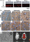
Neural interface stability can be compromised due to BBB gap formation and release of macromolecules causing an exacerbated immune response. a) Chronic neural implant failure cause chronic BBB failure, which induces a cascade of deleterious immune responses including glial scarring and neurodegeneration which results in recording failures. In general, the SNR increases over the course of the implantation, and soon after implantation the presence of reactive astrocytes is high (GFAP+, red). After 16 weeks of implantation, reactive astrocytes are still localized around the implanted electrode interface. Reproduced with permission.[173] Copyright 2013, Elsevier. b) By employing mechanically compliant nanocomposites, the neuroinflammatory response is mitigated and recruitment of reactive astrocytes and subsequent glial scarring is reduced. Reproduced with permission.[100] Copyright 2014, Institute of Physics.

Similar articles

Cited by

  • Noble metal sensitized invasive porous bioelectrodes: advanced medical device for enhanced neuronal activity and chronic alcohol treatment.
    Kim HS, Choi H, Flores MC, Razzaq A, Gwak YS, Ahn D, Kim MS, Gurel O, Lee BH, In SI. Kim HS, et al. RSC Adv. 2020 Dec 9;10(71):43514-43522. doi: 10.1039/d0ra07922g. eCollection 2020 Nov 27. RSC Adv. 2020. PMID: 35519706 Free PMC article.
  • Development of Neuronal Guidance Fibers for Stimulating Electrodes: Basic Construction and Delivery of a Growth Factor.
    Wille I, Harre J, Oehmichen S, Lindemann M, Menzel H, Ehlert N, Lenarz T, Warnecke A, Behrens P. Wille I, et al. Front Bioeng Biotechnol. 2022 Jan 24;10:776890. doi: 10.3389/fbioe.2022.776890. eCollection 2022. Front Bioeng Biotechnol. 2022. PMID: 35141211 Free PMC article.
  • Tumor diagnosis using carbon-based quantum dots: Detection based on the hallmarks of cancer.
    Wang H, Yang S, Chen L, Li Y, He P, Wang G, Dong H, Ma P, Ding G. Wang H, et al. Bioact Mater. 2023 Nov 15;33:174-222. doi: 10.1016/j.bioactmat.2023.10.004. eCollection 2024 Mar. Bioact Mater. 2023. PMID: 38034499 Free PMC article. Review.
  • Improving the Biocompatibility and Functionality of Neural Interface Devices with Silica Nanoparticles.
    Shi D, Narayanan S, Woeppel K, Cui XT. Shi D, et al. Acc Chem Res. 2024 Jun 18;57(12):1684-1695. doi: 10.1021/acs.accounts.4c00160. Epub 2024 May 30. Acc Chem Res. 2024. PMID: 38814586 Free PMC article.
  • Interfacing with the Brain: How Nanotechnology Can Contribute.
    Ahmed AAA, Alegret N, Almeida B, Alvarez-Puebla R, Andrews AM, Ballerini L, Barrios-Capuchino JJ, Becker C, Blick RH, Bonakdar S, Chakraborty I, Chen X, Cheon J, Chilla G, Coelho Conceicao AL, Delehanty J, Dulle M, Efros AL, Epple M, Fedyk M, Feliu N, Feng M, Fernández-Chacón R, Fernandez-Cuesta I, Fertig N, Förster S, Garrido JA, George M, Guse AH, Hampp N, Harberts J, Han J, Heekeren HR, Hofmann UG, Holzapfel M, Hosseinkazemi H, Huang Y, Huber P, Hyeon T, Ingebrandt S, Ienca M, Iske A, Kang Y, Kasieczka G, Kim DH, Kostarelos K, Lee JH, Lin KW, Liu S, Liu X, Liu Y, Lohr C, Mailänder V, Maffongelli L, Megahed S, Mews A, Mutas M, Nack L, Nakatsuka N, Oertner TG, Offenhäusser A, Oheim M, Otange B, Otto F, Patrono E, Peng B, Picchiotti A, Pierini F, Pötter-Nerger M, Pozzi M, Pralle A, Prato M, Qi B, Ramos-Cabrer P, Genger UR, Ritter N, Rittner M, Roy S, Santoro F, Schuck NW, Schulz F, Şeker E, Skiba M, Sosniok M, Stephan H, Wang R, Wang T, Wegner KD, Weiss PS, Xu M, Yang C, Zargarian SS, Zeng Y, Zhou Y, Zhu D, Zierold R, Parak WJ. Ahmed AAA, et al. ACS Nano. 2025 Mar 25;19(11):10630-10717. doi: 10.1021/acsnano.4c10525. Epub 2025 Mar 10. ACS Nano. 2025. PMID: 40063703 Free PMC article. Review.

References

    1. Kovacs GTA, Science 1990, 248, 1280. - PubMed
    1. Hodgkin AL, Huxley AF, Nature 1939, 744, 710.
    1. Vidal JJ, Annu. Rev. Biophys. Bioeng 1973, 2, 157. - PubMed
    1. Vidal JJ, Proc. IEEE 1977, 65, 633.
    1. Bartholow R, Am. J. Med. Sci 1874, 66, 305.

LinkOut - more resources