Review
. 2021 Apr 1:15:662453.
doi: 10.3389/fncel.2021.662453. eCollection 2021.

Transduction and Adaptation Mechanisms in the Cilium or Microvilli of Photoreceptors and Olfactory Receptors From Insects to Humans

Affiliations
Review

Transduction and Adaptation Mechanisms in the Cilium or Microvilli of Photoreceptors and Olfactory Receptors From Insects to Humans

Fatima Abbas et al. Front Cell Neurosci. .

Abstract

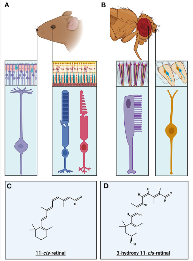
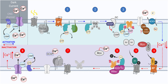
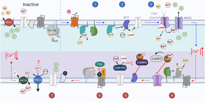
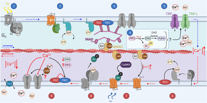
Sensing changes in the environment is crucial for survival. Animals from invertebrates to vertebrates use both visual and olfactory stimuli to direct survival behaviors including identification of food sources, finding mates, and predator avoidance. In primary sensory neurons there are signal transduction mechanisms that convert chemical or light signals into an electrical response through ligand binding or photoactivation of a receptor, that can be propagated to the olfactory and visual centers of the brain to create a perception of the odor and visual landscapes surrounding us. The fundamental principles of olfactory and phototransduction pathways within vertebrates are somewhat analogous. Signal transduction in both systems takes place in the ciliary sub-compartments of the sensory cells and relies upon the activation of G protein-coupled receptors (GPCRs) to close cyclic nucleotide-gated (CNG) cation channels in photoreceptors to produce a hyperpolarization of the cell, or in olfactory sensory neurons open CNG channels to produce a depolarization. However, while invertebrate phototransduction also involves GPCRs, invertebrate photoreceptors can be either ciliary and/or microvillar with hyperpolarizing and depolarizing responses to light, respectively. Moreover, olfactory transduction in invertebrates may be a mixture of metabotropic G protein and ionotropic signaling pathways. This review will highlight differences of the visual and olfactory transduction mechanisms between vertebrates and invertebrates, focusing on the implications to the gain of the transduction processes, and how they are modulated to allow detection of small changes in odor concentration and light intensity over a wide range of background stimulus levels.

Keywords: activation; adaptation; inactivation; olfaction; phototransduction cascade.


Conflict of interest statement

The authors declare that the research was conducted in the absence of any commercial or financial relationships that could be construed as a potential conflict of interest.

Figures

Figure 1
Figure 1
Primary Sensory Neurons of the invertebrate and vertebrate visual and olfactory sensory systems: (A) The primary sensory neurons in the vertebrate olfactory and visual systems and their respective circuitry. Rods (blue) and cones (magenta) mediate vision at different light intensities. Rods mediate low light vision and having specializations enabling this, detailed within this review. Cones mediate daylight color vision and have specific specializations that mediate this. The olfactory sensory neurons (purple) have multiple cilia protruding from the end of the neuron into the olfactory lumen, in which the olfactory transduction machinery, including olfactory receptors, are localized. (B) The primary sensory neurons in the invertebrate (Drosophila) olfactory and visual systems, within their respective circuitry. The invertebrate ommatidium is the structure in which invertebrate photoreceptors (rhabdomeres) are coupled with the support and pigment cells. The rhabdomere (purple) is primary sensory neuron in which the visual transduction machinery is localized within the villi. The olfactory sensory neurons (OSNs) are located within sensory hairs, sensilla. OSNs (orange) project dendrites into the sensillum, with the olfactory receptors expressed on the surface. (C) The chemical structure of the chromophore responsible for vertebrate visual transduction, 11-cis-retinal. (D) The chemical structure of the chromophore present in Microvillar photoreceptors, 3-hydroxy-11-cis-retinal.
Figure 2
Figure 2
The vertebrate phototransduction cascade, inactivation, and adaptation mechanisms. The key steps within the vertebrate phototransduction cascade (blue numbering from top left to right) and its subsequent inactivation (red numbering from bottom right to left). In the dark, the CNG channel maintains a dark current, in which Ca2+ ions influx through open CNG channels as the Guanyl Cyclase enzyme is constituently active. Upon absorption of a photon, the chromophore 11-cis-retinal is isomerized to all-trans-retinal, causing the receptor to become activated (R*). In turn, the associated G protein, transducin, is activated with the exchange of GDP for GTP, GTα* (step 1). GTα* then displaces PDEβγ subunits (step 2). The displaced PDEγ subunit allows cGMP to be reduced to GMP, reducing the concentration of cGMP, closing CNG channels, causing a hyperpolarization of the photoreceptor (step 3). The activated receptor, R*, is inactivated by GRK mediated phosphorylation (step 4). Followed by Arrestin binding (step 5). The GAP protein complex formed of RGS9, R9AP, and Gβ5 bound to both PDEγ and activated GTα* causes hydrolysis of GTP on the GTα to GDP (step 6). GTα then dissociates from PDE, reassociating to the βγ subunits, and PDEγ also inhibits the PDEαβ to prevent cGMP hydrolysis (step 7). Intracellular Ca2+ concentration is raised as CNG channels reopen in response to the increasing cGMP concentration, reestablishing the dark current (step 8).
Figure 3
Figure 3
The vertebrate olfactory transduction cascade, inactivation, and adaptation mechanisms: OSNs maintain a high intracellular Cl ion concentration through the constitutive activity of the NKCC1 transporter, which transports Cl ions into the cell using the energy from the export of Na+ and K+ ions. An odorant binds to the G-protein coupled olfactory receptor (OR) on the cilium of the sensory neuron. The activated receptor in turn activates Golf (step 1). The Gαolf subunits then activate adenylyl cyclase III (AC3) which catalyzes the conversion of ATP to cAMP (step 2). The increasing concentration of cAMP causes the opening of CNG channels on the membrane. This causes an influx of Ca2+ ions. The increasing Ca2+ concentration also causes the opening of the calcium activated chloride ion channel, ANO2, which further depolarizes the cell as the increased intracellular chloride gradient causes an efflux of Cl. The increased Ca2+ concentration leads to several inactivation mechanisms (step 3). GTP bound to the active Gαolf subunit is hydrolyzed to GDP, inactivating it, and causing it to re-associate with the βγ subunits. Ca2+ binds to Calmodulin (CaM) which in turn activates CaMKII. Calmodulin potentially reduces CNG channel affinity for cAMP through interaction with the A-subunit of the channel (step 4). CaMKII inhibits the activity of AC3 by phosphorylating it, preventing the generation of cAMP. PDE1C is also activated by Ca2+ bound CaM and accelerates the hydrolysis of cAMP to AMP (step 5). GRK mediated phosphorylation of the OR (step 6). Intracellular Ca2+ concentration is re-established through the activity of ion exchangers (NCKX4 and possibly PMCAs), and the high intracellular Cl concentration is reestablished through NKCC1 activity and the closure of ANO2 channels (step 7).
Figure 4
Figure 4
The invertebrate phototransduction cascade, inactivation, and adaptation mechanisms: A photon causes isomerization of the chromophore 3-hydroxy 11-cis-retinal to 3-hydroxy all-trans-retinal (step 1). This activates the opsin (step 2), which also activates the Gqα subunit through GDP-GTP exchange. The Gqα subunit then activates membrane bound PLC (step 3), which hydrolyzes PIP2 to produce IP3, DAG and H+ (step 4), and potentially DAG is further catalyzed to produce PUFAs. Some or all the products from step 4 activate the membrane bound TRP and TRP-L channels in step 5, causing an influx of Ca2+ and Na+ ions, depolarizing the microvillus. PLC can act as a GTPase, hydrolyzing the GTP bound to Gα to GDP (step 6). Invertebrate chromophore is bistable and is converted back from 3-hydroxy 11-cis retinal to 3-hydroxy-all-trans retinal at via the absorption of a second photon (step 7). CaMKII phosphorylates Arrestin 2 allowing it to dissociate from R* after the chromophore returns to its inactive state (step 8).
Figure 5
Figure 5
The invertebrate olfactory transduction cascade: In invertebrates, the ORs are 7 transmembrane domain receptors, which associate with a co-receptor, Orco. Upon binding of an odorant (step 1), the OR/Orco co-receptor channel pore opens, allowing the influx of cations, Ca2+ and Na+ (step 2). It is possible that a G protein mediated pathway regulates the sensitivity of the channels, odorant binding activates Gs (step 3). The Gsα subunit then in turn activates adenylyl cyclase to create cAMP from ATP, which may mediate sensitization of the odorant response (step 4). Phosphorylation by PKC of the Orco receptor may mediate the olfactory neuron sensitization (step 3).
Figure 6
Figure 6
The visual pigment cycle reactions in vertebrate and invertebrate photoreceptors: (A) Pathways for visual pigment regeneration in the vertebrate retina, including both the classical and non-classical pathways. A cone photoreceptor (dark blue) and its' interactions with the muller glial cells (magenta) and Retinal pigment epithelium (RPE, light blue). Red arrows with red text annotation indicate enzyme mediated reactions and their respective enzymes. The classical pathway occurs via the RPE, all-trans-retinol is taken up by IRBP into the RPE, where LRAT, RPE65, and 11-cis-RDH mediates its conversion to 11-cis-retinal which is then taken up by the photoreceptor via IRBP again, which reassociated with the apo-opsin. The intrinsic light mediated pathway combining all-trans-RAL with phosphatidylethanolamine (PE) to form all-trans-N-retinyl-PE (N-retPE), which is then converted to 11-cis-retinal via a blue photon. Light mediated pigment regeneration can occur both in Müller cells and RPE, whereby all-trans retinal is converted to 11-cis retinal mediated by RGR opsin and the absorption of a photon. Stores of 11-cis-retinoids as well as all-trans-RE are present in both the RPE and Müller cells. (B) Summary of the visual pigment cycle in invertebrates. The invertebrate visual pigment, 3-OH 11-cis retinal is a bistable pigment, and remains associated to the opsin, upon absorption of a second photon, it can convert back from 3-OH all-trans retinal to 3-OH 11-cis retinal. An external pathway is only required in conditions in which the animal is calorie restricted. This pathway is mediated by the retinal pigment cells that surround the rhabdomeres and involves the Photoreceptor retinol dehydrogenase (PDH) and retinol dehydrogenase B (RDHB) enzymes.

Similar articles

Cited by

References

    1. Acharya J. K., Jalink K., Hardy R. W., Hartenstein V., Zuker C. S. (1997). InsP3 receptor is essential for growth and differentiation but not for vision in Drosophila. Neuron 18, 881–887. 10.1016/S0896-6273(00)80328-1 - DOI - PubMed
    1. Alloway P. G., Dolph P. J. (1999). A role for the light-dependent phosphorylation of visual arrestin. Proc. Natl. Acad. Sci. U.S.A. 96, 6072–6077. 10.1073/pnas.96.11.6072 - DOI - PMC - PubMed
    1. Andrea Yao C., Carlson J. R. (2010). Role of G-proteins in odor-sensing and CO2-sensing neurons in Drosophila. J. Neurosci. 30, 4562–4572. 10.1523/JNEUROSCI.6357-09.2010 - DOI - PMC - PubMed
    1. Antolin S., Reisert J., Matthews H. R. (2010). Olfactory response termination involves Ca2+-ATPase in vertebrate olfactory receptor neuron cilia. J. Gen. Physiol. 135, 367–378. 10.1085/jgp.200910337 - DOI - PMC - PubMed
    1. Antunes G., Simoes De Souza F. M. (2016). Olfactory receptor signaling. Methods Cell Biol 132, 127–145. 10.1016/bs.mcb.2015.11.003 - DOI - PubMed

LinkOut - more resources