. 2021 Mar 31:12:613449.
doi: 10.3389/fphar.2021.613449. eCollection 2021.

Abacavir Increases Purinergic P2X7 Receptor Activation by ATP: Does a Pro-inflammatory Synergism Underlie Its Cardiovascular Toxicity?

Affiliations

Abacavir Increases Purinergic P2X7 Receptor Activation by ATP: Does a Pro-inflammatory Synergism Underlie Its Cardiovascular Toxicity?

Víctor Collado-Díaz et al. Front Pharmacol. .

Abstract

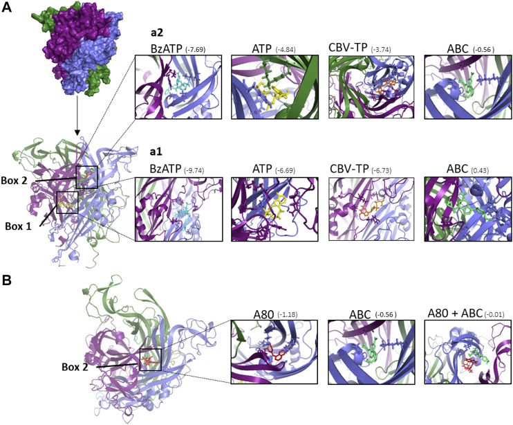
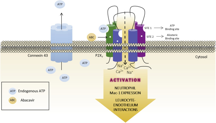
The cardiovascular toxicity of Abacavir is related to its purinergic structure. Purinergic P2X7-receptors (P2X7R), characterized by activation by high concentrations of ATP and with high plasticity, seem implicated. We appraise the nature of the interplay between Abacavir and P2X7R in generating vascular inflammation. The effects of Abacavir on leukocyte-endothelium interactions were compared with those of its metabolite carbovir triphosphate (CBV-TP) or ATP in the presence of apyrase (ATP-ase) or A804598 (P2X7R-antagonist). CBV-TP and ATP levels were evaluated by HPLC, while binding of Abacavir, CBV-TP and ATP to P2X7R was assessed by radioligand and docking studies. Hypersensitivity studies explored a potential allosteric action of Abacavir. Clinical concentrations of Abacavir (20 µmol/L) induced leukocyte-endothelial cell interactions by specifically activating P2X7R, but the drug did not show affinity for the P2X7R ATP-binding site (site 1). CBV-TP levels were undetectable in Abacavir-treated cells, while those of ATP were unaltered. The effects of Abacavir were Apyrase-dependent, implying dependence on endogenous ATP. Exogenous ATP induced a profile of proinflammatory actions similar to Abacavir, but was not entirely P2X7R-dependent. Docking calculations suggested ATP-binding to sites 1 and 2, and Abacavir-binding only to allosteric site 2. A combination of concentrations of Abacavir (1 µmol/L) and ATP (0.1 µmol/L) that had no effect when administered separately induced leukocyte-endothelium interactions mediated by P2X7R and involving Connexin43 channels. Therefore, Abacavir acts as a positive allosteric modulator of P2X7R, turning low concentrations of endogenous ATP themselves incapable of stimulating P2X7R into a functional proinflammatory agonist of the receptor.

Keywords: P2X7 receptor; abacavir; adenosine triphosphate; allosteric modulator; cardiovascular diseases; leukocyte-endothelium interactions.


Conflict of interest statement

JVE has received funds for speaking at symposia organized by Abbvie Farmaceutica, Astra Zeneca, Gilead Sciences and Pfizer. The remaining authors declare that the research was conducted in the absence of any commercial or financial relationships that could be construed as a potential conflict of interest.

Figures

FIGURE 1
FIGURE 1
Chemical structures of abacavir, carbovir triphosphate and ATP, and potential causes of the activation of P2X7R. (A) Abacavir (ABC), its active metabolite carbovir triphosphate (CBV-TP), and ATP share a close structural similarity. (B) The bottom part of the figure represents the four potential scenarios of the interaction of ABC with the P2X7R evaluated in the present study: (B1) direct activation of the P2X7R by ABC through its binding to ATP-binding sites; (B2) ABC is metabolized into its active metabolite CBV-TP, which subsequently stimulates the P2X7R by binding to ATP-binding sites; (B3) ABC indirectly stimulates the P2X7R, and ATP is the molecule that binds to its binding sites. ABC could induce an enhancement in the levels of extracellular ATP either by interfering with the enzymes implicated in its synthesis/degradation or by mobilizing intracellular ATP to the extracellular space; and (B4) ABC allosterically modulates the activation of P2X7R. ABC binds to site 2, rendering this receptor more susceptible to direct activation by endogenous ATP.
FIGURE 2
FIGURE 2
PMN-endothelial cell interactions. HUVEC and PMN were incubated (4 h) with ABC (20 µmol/L), ATP (0.05–10 µmol/L), CBV-TP (10–50 µmol/L), Bz-ATP (100 µmol/L) or vehicle (veh). In some cases, both cell types (HUVEC and PMN) were pretreated with suramin (Sur, non-selective P2R antagonist, 100 μmol/L, 60 min) or with A804598 (A80, P2X7R antagonist, 1 μmol/L, 30 min) prior to ABC (20 µmol/L), ATP (1 µmol/L) or CBV-TP (20 µmol/L). PMN rolling velocity (A, B), PMN rolling flux (C, D) and PMN adhesion (E, F) were quantified. Results are mean ± SEM, Veh: n ≥ 14, ABC: n ≥ 7, ATP 0.05: n ≥ 4, ATP 0.1: n ≥ 13, ATP 1: n ≥ 6, ATP 10: n ≥ 6, CBV-TP 10: n ≥ 6, CBV-TP 20: n ≥ 5, CBV-TP 30: n ≥ 5, CBV-TP 50: n ≥ 6, Bz-ATP 100: n ≥ 6, ABC 20 + Sur: n ≥ 4, ABC 20 + A80: n ≥ 4, ATP 1 + Sur: n ≥ 6, ATP 1 + A80: n ≥ 5, CBV-TP 20 + Sur: n ≥ 5, CBV-TP 20 + A80: n ≥ 5. *p < 0.05 or **p < 0.01 vs. corresponding value in vehicle-treated group. +p < 0.05 or ++p < 0.01 vs. corresponding value in ABC, ATP or CBV-TP-treated group (ANOVA followed by Newman-Keuls test).
FIGURE 3
FIGURE 3
Mac-1 expression and calcium influx in neutrophils. Whole blood (4 h) for Mac-1 expression or PMN (1 h) for calcium influx were treated with ABC (20 µmol/L), ATP (0.1–10 µmol/L) or CBV-TP (0.1–10 µmol/L), and the surface expression on neutrophils of the two subunits of Mac-1 CD11b (A, B) and CD18 (C, D), and calcium mobilization (E) were quantified. In some cases, blood or PMN were pretreated with suramin (Sur, nonselective P2R antagonist, 100 μmol/L, 60 min) or A804598 (A80, P2X7R antagonist, 1 μmol/L, 30 min) prior to treatment with ABC (20 µmol/L), ATP (1 µmol/L), ATP (5 µmol/L), CBV-TP (10 µmol/L) or Bz-ATP (100 µmol/L). Fluorescence values are expressed as percentage of mean of median fluorescence intensities of control cells (dotted line). Results are mean ± SEM, Veh: n ≥ 11, ABC 20: n ≥ 15, ATP 0.1: n ≥ 6, ATP 1: n ≥ 6, ATP 5: n = 10, ATP 10: n ≥ 7, CBV-TP 0.1: n ≥ 6, CBV-TP 1: n ≥ 10, CBV-TP 10: n ≥ 5, Bz-ATP 100: n ≥ 6, ABC 20 + Sur: n ≥ 9, ABC 20 + A80: n ≥ 5, ATP 1 + Sur: n ≥ 10, ATP 1 + A80: n ≥ 11, ATP 5 + A80: n ≥ 4, CBV-TP 10 + Sur: n ≥ 12, CBV-TP 10 + A80: n ≥ 4, Bz-ATP 100 + A80: n ≥ 4, *p < 0.05, **p < 0.01 or ****p < 0.0001 vs. corresponding value in vehicle-treated group. +p < 0.05 or ++p < 0.01 vs. corresponding value in ABC, ATP or CBV-TP-treated group (ANOVA followed by Newman-Keuls test), t p<0.05 vs. corresponding value in vehicle-treated group (t-test).
FIGURE 4
FIGURE 4
Effects of apyrase on PMN-endothelial cell interactions and on levels of ABC and ATP. (A) HUVEC and PMN were incubated (4 h) with ABC (20 µmol/L), ATP (1 µmol/L) or vehicle (veh). In some cases, both cell types (HUVEC and PMN) were pretreated with apyrase (Apy, ATPase, 100 μmol/L, 60 min) prior to ABC or ATP treatment. PMN rolling flux was quantified. (B) PMNs were incubated with ABC (20 µmol/L) or ATP (1 µmol/L). In some other experiments, cells were pretreated with apyrase (100 UI/L) and levels of ABC and ATP in the supernatants were quantified by HPLC (at 0, 1, 10, 60 and 240 min). Results are mean ± SEM, Veh: n = 15, ABC 20: n = 15, ABC 20 + Apy: n = 9, ATP 1: n = 6, ATP 1 + Apy: n = 4. **p < 0.01 vs. corresponding value in vehicle-treated group. ++p < 0.01 vs. corresponding value in ABC or ATP-treated group (ANOVA followed by Newman-Keuls test).
FIGURE 5
FIGURE 5
Role of endogenous ATP released by different channels on leukocyte-endothelial interactions induced by ABC. HUVEC and PMN or whole blood were incubated (4 h) with ABC (20 µmol/L). In some cases, they were pretreated with Carbenexolone (pannexin and connexin channels blocker, CBX, 100 µmol/L), Probenecid (Prob, pannexin and connexin channels and P2X7 receptor blocker, 150 µmol/L), Panx10 (mimetic inhibitory peptide that blocks pannexin-1 gap junctions, 300 µmol/L) or Gap19 (selective Connexin43 channel blocker Gap19, 300 µmol/L). PMN rolling velocity (A), PMN rolling flux (B) and PMN adhesion (C) were quantified after assembling the flow chamber and the expression of one of the two subunits of Mac-1 CD11b (D) on neutrophils was quantified by flow cytometry. Fluorescence values are expressed as percentage of mean of median fluorescence intensities of control cells (dotted line). Results are mean ± SEM, Veh: n ≥ 7, ABC 20: n ≥ 6, ABC 20 + CBX: n ≥ 6, ABC 20 + Prob: n ≥ 4, ABC 20 + Panx10: n ≥ 5, ABC 20 + Gap19: n ≥ 4. *p < 0.05 or **p < 0.01 vs. corresponding value in vehicle-treated group. +p < 0.05 or ++p < 0.01 vs. corresponding value in ABC-treated group (ANOVA followed by Newman-Keuls test).
FIGURE 6
FIGURE 6
Studies of radioligand-binding to P2X7R. (A) Displacement of [3H]A-804598 (P2X7 receptor antagonist, 2.5 nmol/L) by increasing the concentrations of Bz-ATP, ATP, CBV-TP and ABC (0.1 µmol/L-1 mmol/L) at membrane preparations of HUVEC. Results are mean ± SEM, Bz-ATP: n ≥ 3, ATP: n ≥ 6, CBV-TP: n ≥ 6, ABC: n ≥ 5; each experiment was performed in duplicate. (B) Affinities (pIC50 values) of Bz-ATP, ATP, ABC and CBV-TP for the human P2X7R.
FIGURE 7
FIGURE 7
Docking calculations of ABC and other ligands to P2X7 receptor. (A) Structure of human P2X7R built by homology modeling based on the X-ray crystal structure of panda P2X7R (5U2H). The P2X7R trimer is represented as a molecular surface. The three monomers are colored in deep purple, slate blue, and forest green. Each monomer is shaped like a dolphin (Hattori and Gouaux, 2012). Docked poses at ATP binding site (box 1) and A804598 (A80) binding site (box 2) of the different compounds studied. The P2X7R trimer is represented as a cartoon. ATP is depicted in yellow, A804598 in red, Bz-ATP in blue and CBV-TP in orange and ABC in lime green. (a1) Details of the ligands docked at box 1 showing some amino acid side chains at the interaction site. (a2) Details of the ligands docked at box 2 showing some amino acid side chain at the interaction site. (B) Superimposition of docked poses of P2X7R antagonist A804598 (A80, in red) and ABC (in lime green) at the binding site two of the P2X7R. The predicted binding energies are indicated under parenthesis (kcal mol−1).
FIGURE 8
FIGURE 8
Potentiation by ABC of the effects of ATP on PMN-endothelial cell interactions, the expression of CD11b and on calcium influx. HUVEC and PMN were treated (4 h) with ATP (0.001–20 µmol/L) in the presence or absence of ABC (1 µmol/L) and PMN rolling flux (A) was analyzed. Doses of ABC (1 µmol/L) and ATP (0.1 µmol/L) were selected to treat individually or in combination both PMN and HUVEC or whole blood. In some cases, both HUVEC and PMN or whole blood were pretreated with A804598 (A80, P2X7R antagonist, 1 μmol/L, 30 min) prior to ABC (1 µmol/L) and ATP (0.1 µmol/L). PMN rolling flux (B) was quantified after assembling the flow chamber and the expression of one of the two subunits of Mac-1 CD11b (C) on neutrophils was quantified by flow cytometry. (D) PMNs were treated (1 h) with Polymyxin B (Pol. 8 µmol/L), ABC (1 µmol/L) and ATP (0.1 µmol/L) individually or in combination and, in somes cases, PMN were pretreated with A804598 (A80, P2X7R antagonist, 1 μmol/L, 30 min) prior to Polymyxin B (Pol. 8 µmol/L) and ATP (0.1 µmol/L) or ABC (1 µmol/L) and ATP (0.1 µmol/L). Calcium mobilization was quantified by flow cytometry. Fluorescence values are expressed as a percentage of mean fluorescence intensities of control cells (dotted line). Results are mean ± SEM, Veh: n ≥ 5, Pol. 8: n = 3, ABC 1: n ≥ 5, ATP 0.1: n ≥ 5, ABC 1 + ATP 0.1: n ≥ 5, ABC 1 + ATP 0.1 + A80: n ≥ 5, ABC 1 + ATP 0.1 + Gap19: n ≥ 3 Pol. 8 + ATP 0.1: n = 3, Pol. 8 + ATP 0.1 + A80: n ≥ 3. *p < 0.05, **p < 0.01 or ***p < 0.001 vs. corresponding value in ABC-treated group. #p < 0.05, ##p < 0.01 or ###p < 0.001 vs. corresponding value in ATP-treated group. +p < 0.05 or ++p < 0.01 vs. corresponding value in the ABC + ATP-treated group (ANOVA followed by Newman-Keuls test).
FIGURE 9
FIGURE 9
Graphical abstract. Proposed role for ABC acting as an allosteric modulator of the P2X7 receptor. ABC binds to a specific site of the receptor (site 2, allosteric binding site), but not to ATP-binding site (site 1), facilitating the endogenous ATP-P2X7R binding to site one and, thus, the activation of the receptor with low and physiological concentrations of ATP. This will lead to an increase in the expression of the adhesion molecule Mac-1 in PMN, which interacts with ICAM-1 on endothelial cells, causing the recruitment of leukocytes seen in the vascular inflammatory response induced by ABC. The release of ATP to the extracellular milieu through Connexin43 channels is also implicated in this response.

References

    1. Abudara V., Bechberger J., Freitas-Andrade M., De Bock M., Wang N., Bultynck G., et al. (2014). The connexin43 mimetic peptide Gap19 inhibits hemichannels without altering gap junctional communication in astrocytes. Front. Cell. Neurosci. 8, 306. 10.3389/fncel.2014.00306 - DOI - PMC - PubMed
    1. Adinolfi E., Giuliani A. L., De Marchi E., Pegoraro A., Orioli E., Di Virgilio F. (2018). The P2X7 receptor: a main player in inflammation. Biochem. Pharmacol. 151, 234–244. 10.1016/j.bcp.2017.12.021 - DOI - PubMed
    1. Albalawi F., Lu W., Beckel J. M., Lim J. C., McCaughey S. A., Mitchell C. H. (2017). The P2X7 receptor primes IL-1β and the NLRP3 inflammasome in astrocytes exposed to mechanical strain. Front. Cell. Neurosci. 11, 227. 10.3389/fncel.2017.00227 - DOI - PMC - PubMed
    1. Alvarez A., Orden S., Andújar I., Collado-Diaz V., Núñez-Delgado S., Galindo M. J., et al. (2017). Cardiovascular toxicity of abacavir: a clinical controversy in need of a pharmacological explanation. AIDS 31, 1781–1795. 10.1097/QAD.0000000000001547 - DOI - PubMed
    1. Alvarez A., Rios-Navarro C., Blanch-Ruiz M. A., Collado-Diaz V., Andujar I., Martinez-Cuesta M. A., et al. (2017). Abacavir induces platelet-endothelium interactions by interfering with purinergic signalling: a step from inflammation to thrombosis. Antiviral Res. 141, 179–185. 10.1016/j.antiviral.2017.03.001 - DOI - PubMed