. 2021 Mar 29:12:653344.
doi: 10.3389/fimmu.2021.653344. eCollection 2021.

MicroLet-7b Regulates Neutrophil Function and Dampens Neutrophilic Inflammation by Suppressing the Canonical TLR4/NF-κB Pathway

Affiliations

MicroLet-7b Regulates Neutrophil Function and Dampens Neutrophilic Inflammation by Suppressing the Canonical TLR4/NF-κB Pathway

Binzhen Chen et al. Front Immunol. .

Abstract

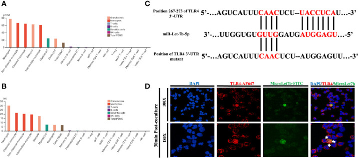
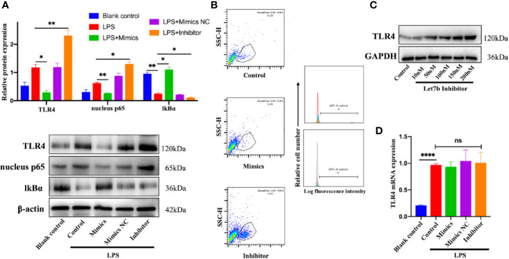
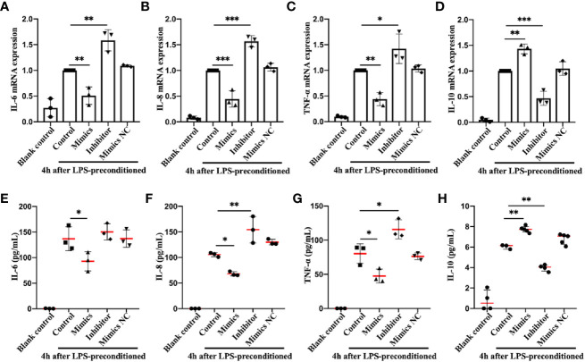
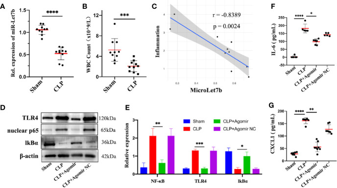
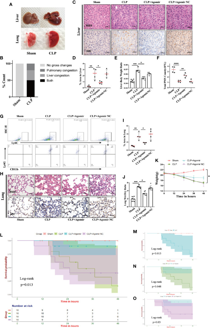
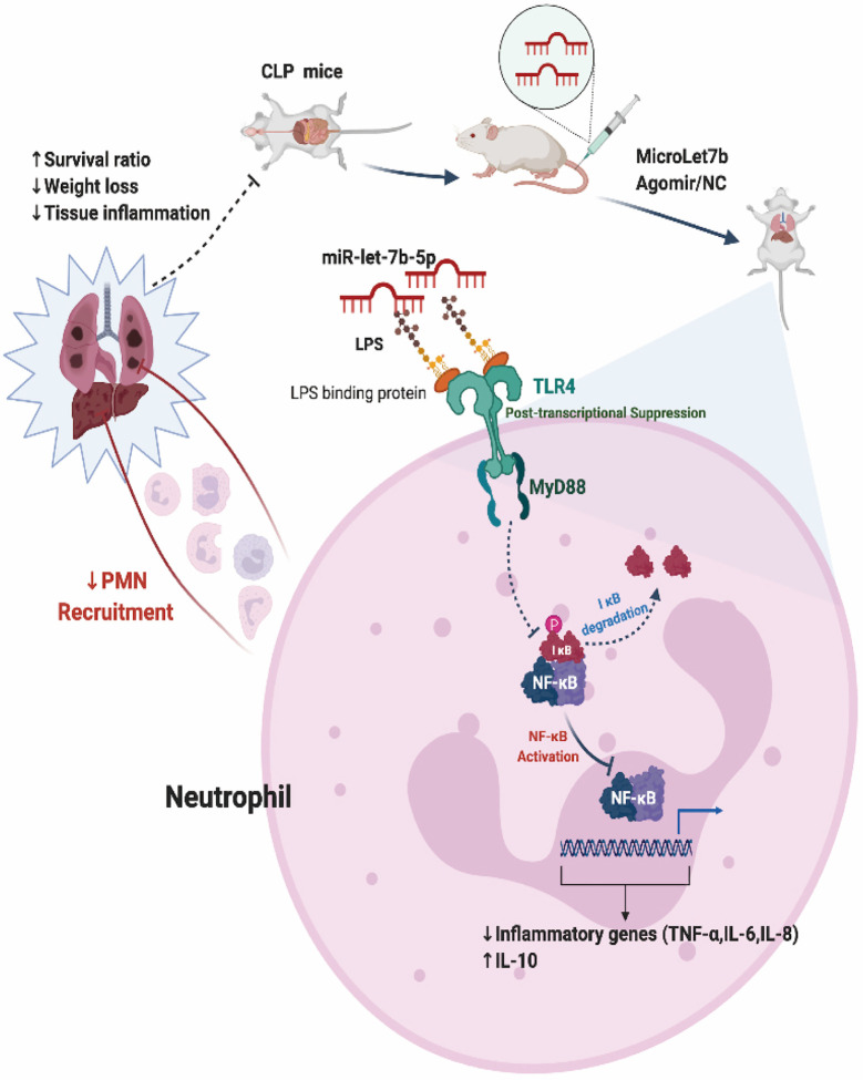
Sepsis is a heterogeneous syndrome caused by a dysregulated host response during the process of infection. Neutrophils are involved in the development of sepsis due to their essential role in host defense. COVID-19 is a viral sepsis. Disfunction of neutrophils in sepsis has been described in previous studies, however, little is known about the role of microRNA-let-7b (miR-let-7b), toll-like receptor 4 (TLR4), and nuclear factor kappa B (NF-κB) activity in neutrophils and how they participate in the development of sepsis. In this study, we investigated the regulatory pathway of miR-let-7b/TLR4/NF-κB in neutrophils. We also explored the downstream cytokines released by neutrophils following miR-let-7b treatment and its therapeutic effects in cecal ligation and puncture (CLP)-induced septic mice. Six-to-eight-week-old male C57BL/6 mice underwent CLP following treatment with miR-let-7b agomir. Survival (n=10), changes in liver and lungs histopathology (n=4), circulating neutrophil counts (n=4), the liver-body weight ratio (n=4-7), and the lung wet-to-dry ratio (n=5-6) were recorded. We found that overexpression of miR-let-7b could significantly down-regulate the expression of human-derived neutrophilic TLR4 at a post-transcriptional level, a decreased level of proinflammatory factors including interleukin-6 (IL-6), IL-8, tumor necrosis factor α (TNF-α), and an upregulation of anti-inflammatory factor IL-10 in vitro. After miR-let-7b agomir treatment in vivo, neutrophil recruitment was inhibited and thus the injuries of liver and lungs in CLP-induced septic mice were alleviated (p=0.01 and p=0.04, respectively), less weight loss was reduced, and survival in septic mice was also significantly improved (p=0.013). Our study suggested that miR-let-7b could be a potential target of sepsis.

Keywords: COVID-19; MicroLet-7b; TLR4/NF-κB; inflammation; neutrophil; sepsis.


Conflict of interest statement

The authors declare that the research was conducted in the absence of any commercial or financial relationships that could be construed as a potential conflict of interest.

Figures

Figure 1
Figure 1
miR-let-7b directly targets TLR4 in neutrophils. (A) Expression profiles based on within-sample normalized pTPM for TLR4 gene enriched in 18 blood cell types and total PBMC ranked according to protein transcript expression values (see www.proteinatlas.org for details). (B) Same as (A), but based on the between-sample NX values. (C) The sequence of human TLR4 3’-UTR (wild type and mutant) is predicted to be targeted by the mature sequence of miR-let-7b. Targeted nucleotides are labeled in red. (D) Confocal microscopy was performed on FITC-labeled miR-let-7b (green fluorescence) and cocultured for 30 minutes with AF647-labeled neutrophil TLR4 (red fluorescence) to demonstrate miR-let-7b internalization. Blue nuclear staining was performed with 4,6-diamidino-2-phenylindole (DAPI). One representative image of the immunostainings is shown. Scale bar denotes 100x and 180x objectives.
Figure 2
Figure 2
miR-let-7b activates the TLR4/NF-κB signaling and inhibits TLR4 expression via post-transcriptional regulation. (A) Neutrophils treated with 100 ng/mL LPS were then co-incubated with miR-let-7b mimics, miR-let-7b inhibitor, and miR-let-7b mimics NC (100 nM, respectively) for four hours followed by Western blotting to detect TLR4, nucleus p65, and cytosolic IkBα. *P < 0.05, **P < 0.01. (B) Isolated neutrophils were incubated with LPS alone or with indicated concentrations of miR-let-7b mimics and inhibitor (100 nM, each) for four hours followed by measurement of TLR4 binding (staining with AF647) by flow cytometry. The blue line is AF647-stained neutrophils cultured for four hours only (Blank), the red line is AF647-stained neutrophils pretreated with LPS and then cultured for four hours (Control), the brown line is AF647-stained neutrophils pretreated with LPS and then cocultured with miR-let-7b mimics for four hours (Let-7b mimics), and the gray line is AF647-stained neutrophils pretreated with LPS and then cocultured with miR-let-7b inhibitor for four hours (let-7b inhibitor). (C) A dose-dependent increase of TLR4 protein was determined with miR-let-7b inhibitor treatment. (D) RT-qPCR of miR-let-7b expression in human neutrophils. ****P < 0.0001, not significant (ns). All traces are representative of ≥3 independent experiments. Data represent mean ± SD (n=3).
Figure 3
Figure 3
Identification and expression of inflammation-related cytokines in neutrophils in vitro. RT-qPCR (A–D) and ELISA (E–H) detection in the supernatant of IL-6, IL-8, TNF-α, and IL-10 in human freshly isolated neutrophils. Cells were incubated with miR-let-7b mimics, miR-let-7b inhibitor, and miR-let-7b mimics NC (100 nM, respectively), and the supernatant was collected at 10 hours after stimulation with 100 ng/mL LPS for 4 h. Data represent mean ± SD (n=3–5). Tukey’s multiple comparison tests were used to generate the P-values indicated in the figure. *P < 0.05, **P < 0.01, ***P < 0.001.
Figure 4
Figure 4
Overexpression of miR-let-7b suppresses neutrophilic inflammation in vivo. (A) Expression of miR-let-7b in sham and CLP-induced septic mice from the peripheral blood using RT-qPCR (n=10, ****P < 0.0001). (B) Total WBC count in sham and CLP-induced septic mice from the peripheral blood (***P < 0.001). (C) Correlation analysis between the level of miR-let-7b and the degree of septic inflammation (r=-0.8389, p=0.0024). (D, E) Effects of miR-let-7b agomir and its NC on neutrophilic TLR4, nuclear NF-κB p65 and IkB expression in CLP-induced septic mice were assessed by western blotting. *P < 0.05, **P < 0.01, ***P < 0.001. (F, G) Serum levels of IL-6 (F) and CXCL1 (G) were measured by ELISA in mice (n = 5-8 mice per group, *P < 0.05, **P < 0.01, ****P < 0.0001).
Figure 5
Figure 5
Targeting miR-let-7b alleviates tissue injury and improves overall survival in CLP murine models. (A, B) Gross pathologic changes in mouse livers and lungs showing pulmonary/liver congestion in sham and CLP-treated mice groups. (C) Histological assessment of livers harvested at 48 hours. Shown are representative H&E stain (upper panel) and IHC images (lower panel). Infiltrating neutrophils were identified by CD11b (brown), Scale bars, 50 μm. (D) Area of IHC positive for CD11b expressed as a percentage and assessed in five aleatory images. *P < 0.05; **P < 0.01, one-way ANOVA, n= 4/group. (E) The whole liver was dissected and weighed for determination of liver-body weight ratio (liver to body ratio=liver mass/body mass). n=4–7/group. (F) Total neutrophil counts in the blood. **P < 0.01, ****P < 0.0001, one-way ANOVA, n= 4/group. (G) Percentage of neutrophil infiltration in mice lung was detected in BALF. Representative flow cytometry plots of CD11b+ and Ly6G+ frequency (bottom row) among Ly6Cint cells (top rows). At the end of the experiment (48 h), the lower two-thirds of the right lung were dissected, of which one-third was for lung histology (H, I, same as liver) and the other one-third was weighed for the lung wet-to-dry weight ratio analyses (J) (n=5–6, one-way ANOVA, *P < 0.05, **P < 0.01). (K) The weight of C57BL/6 mice was monitored every 12 hours (*P < 0.05). (L) Survival rate was performed by Kaplan-Meier analysis among four groups. p = 0.013. Survival rates were also performed between sham and CLP group (M), CLP and CLP + miR-let-7b agomir group (N), and CLP + miR-let-7b agomir and CLP + miR-let-7b agomir NC group (O). n=10 mice/group, Log-rank test, p = 0.013, p = 0.048 and p = 0.03, respectively.
Figure 6
Figure 6
Proposed model for miR-let-7b-mediated regulation of neutrophilic functions that target TLR4/NF-κB axis in vitro and in vivo.

Similar articles

Cited by

References

    1. Wang YX, Wang YY, Chen Y, Qin QS. Unique epidemiological and clinical features of the emerging 2019 novel coronavirus pneumonia (COVID-19) implicate special control measures. J Med Virol (2020) 92:568–76. 10.1002/jmv.25748 - DOI - PMC - PubMed
    1. Singer M, Deutschman CS, Seymour CW, Shankar-Hari M, Annane D, Bauer M, et al. . The Third International Consensus Definitions for Sepsis and Septic Shock (Sepsis-3). JAMA (2016) 315:801–10. 10.1001/jama.2016.0287 - DOI - PMC - PubMed
    1. Rhodes A, Evans LE, Alhazzani W, Levy MM, Antonelli M, Ferrer R, et al. . Surviving Sepsis Campaign: International Guidelines for Management of Sepsis and Septic Shock: 2016. Intensive Care Med (2017) 43:304–77. 10.1007/s00134-017-4683-6 - DOI - PubMed
    1. Fan Z, McArdle S, Marki A, Mikulski Z, Gutierrez E, Engelhardt B, et al. . Neutrophil recruitment limited by high-affinity bent beta2 integrin binding ligand in cis. Nat Commun (2016) 7:12658. 10.1038/ncomms12658 - DOI - PMC - PubMed
    1. Campbell EL, Bruyninckx WJ, Kelly CJ, Glover LE, McNamee EN, Bowers BE, et al. . Transmigrating Neutrophils Shape the Mucosal Microenvironment through Localized Oxygen Depletion to Influence Resolution of Inflammation. Immunity (2014) 40:66–77. 10.1016/j.immuni.2013.11.020 - DOI - PMC - PubMed

Publication types