. 2021 Feb;15(2):189-202.
doi: 10.1002/term.3165. Epub 2020 Dec 8.

Targeting HIF-α for robust prevascularization of human cardiac organoids

Affiliations

Targeting HIF-α for robust prevascularization of human cardiac organoids

Robert C Coyle et al. J Tissue Eng Regen Med. 2021 Feb.

Abstract

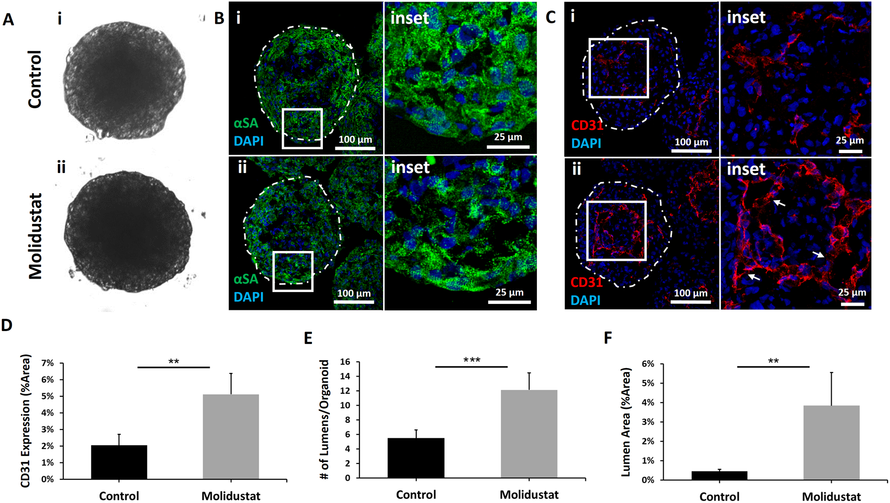
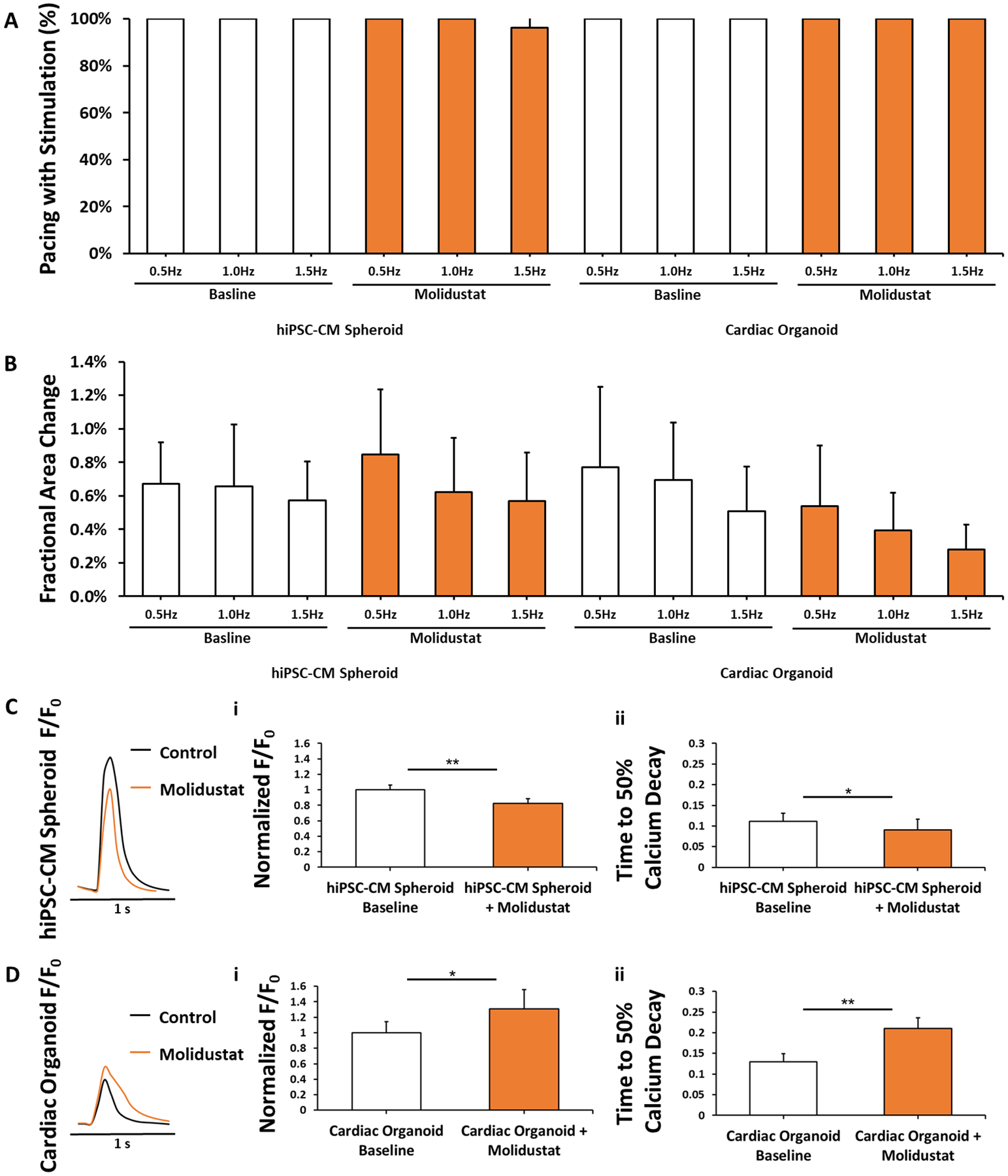
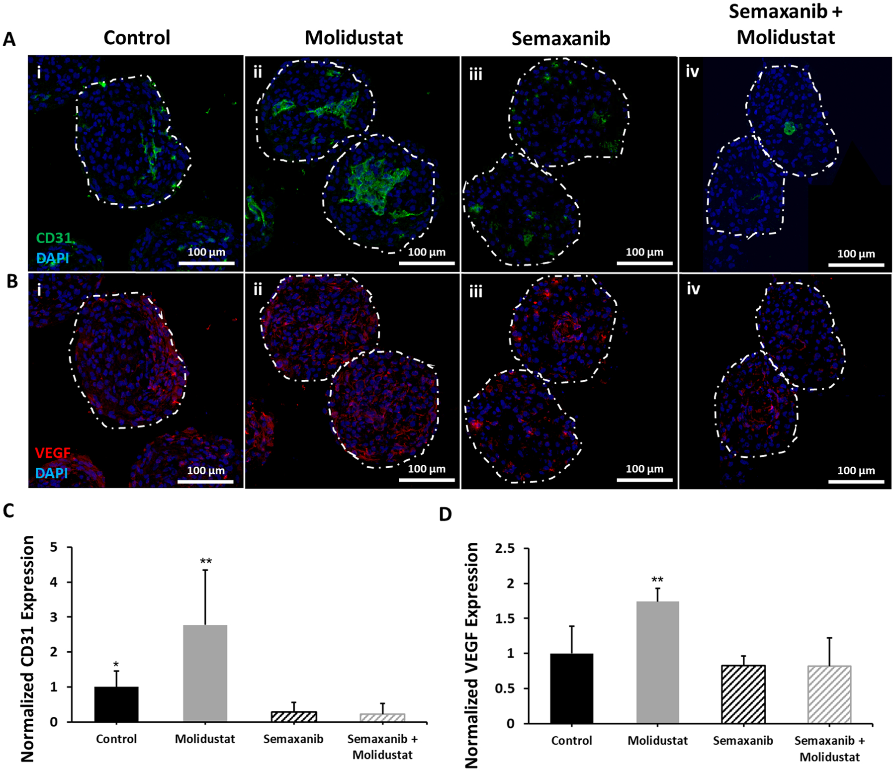
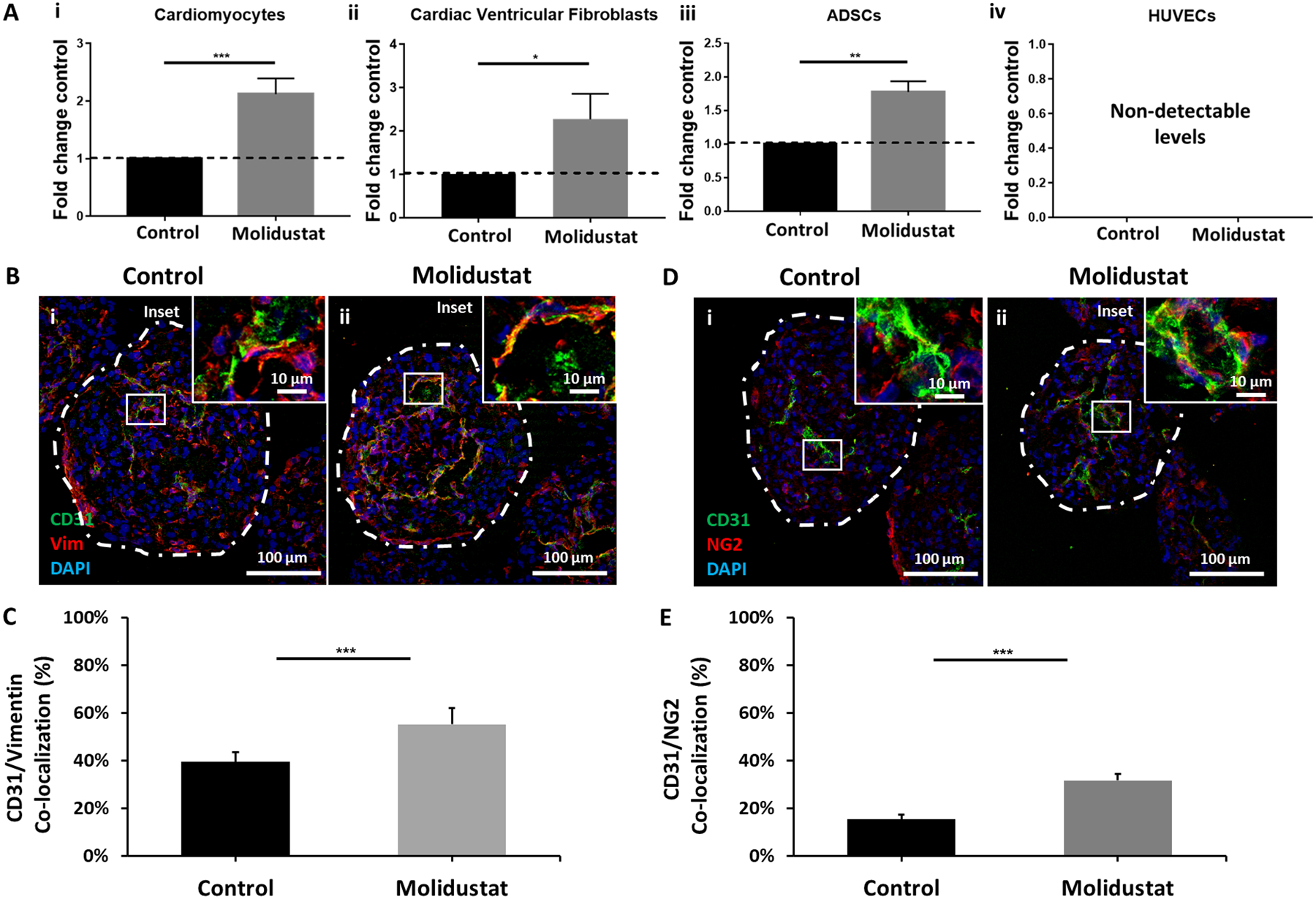
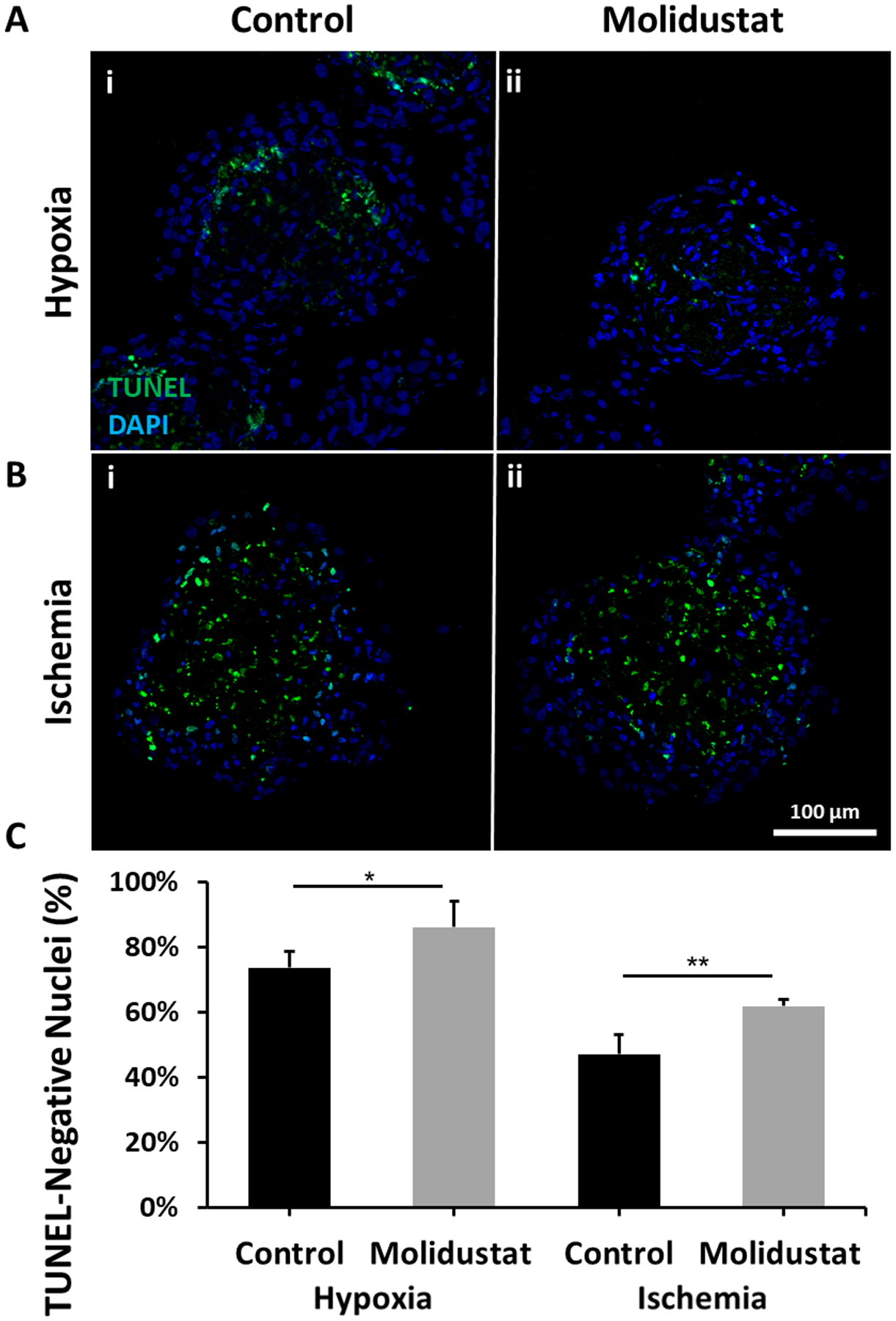
Prevascularized 3D microtissues have been shown to be an effective cell delivery vehicle for cardiac repair. To this end, our lab has explored the development of self-organizing, prevascularized human cardiac organoids by co-seeding human cardiomyocytes with cardiac fibroblasts, endothelial cells, and stromal cells into agarose microwells. We hypothesized that this prevascularization process is facilitated by the endogenous upregulation of hypoxia-inducible factor (HIF) pathway in the avascular 3D microtissues. In this study, we used Molidustat, a selective PHD (prolyl hydroxylase domain enzymes) inhibitor that stabilizes HIF-α, to treat human cardiac organoids, which resulted in 150 ± 61% improvement in endothelial expression (CD31) and 220 ± 20% improvement in the number of lumens per organoids. We hypothesized that the improved endothelial expression seen in Molidustat treated human cardiac organoids was dependent upon upregulation of VEGF, a well-known downstream target of HIF pathway. Through the use of immunofluorescent staining and ELISA assays, we determined that Molidustat treatment improved VEGF expression of non-endothelial cells and resulted in improved co-localization of supporting cell types and endothelial structures. We further demonstrated that Molidustat treated human cardiac organoids maintain cardiac functionality. Lastly, we showed that Molidustat treatment improves survival of cardiac organoids when exposed to both hypoxic and ischemic conditions in vitro. For the first time, we demonstrate that targeted HIF-α stabilization provides a robust strategy to improve endothelial expression and lumen formation in cardiac microtissues, which will provide a powerful framework for prevascularization of various microtissues in developing successful cell transplantation therapies.

Keywords: HIF; HIF prolyl-hydroxylase inhibitor; human cardiac organoid; human induced stem cell-derived cardiomyocyte; prevascularization; tissue engineering.


Conflict of interest statement

Conflicts of Interest Statement The authors have no conflicts of interest to report.

Figures

Figure 1.
Figure 1.. Human cardiac organoids treated with Molidustat result in improved endothelial expression and lumen formation.
Representative images of Control and Molidustat treated organoids after 10 days of culture: (A) brightfield, (B) immunofluorescent staining of cardiac marker α-sarcomeric actinin (αSA) showing comparable development of sarcomeric structures, and (C) immunofluorescent staining of endothelial marker CD31 showing improved organization of endothelial cells in Molidustat treated organoids. The white arrows in the inset (ii) point to the lumen structures. Green- αSA Red- CD31, Blue-DAPI. (D) Percent area expression of CD31 comparing Control and Molidustat treated organoids. (E) Total number of lumens per cardiac organoid when comparing Control and Molidustat treated groups. (F) Total sum of lumen area as a percentage of cardiac organoid area when comparing Control and Molidustat treated groups. n=at least 5 biological replicates. Mann-Whitney nonparametric test and two tailed confidence interval. (**) represents p<0.005, (***) represents p<0.0005.
Figure 2.
Figure 2.. Human cardiac organoid diameter influences results of Molidustat treatment on endothelial organization.
Human cardiac organoids with increased diameter result in varied expression of endothelial markers for (A) Control and (B) Molidustat treatment. Cardiac organoid diameter= (i) 275 μm, (ii) 325 μm, and (iii) 400 μm. Staining for cardiac-specific markers show consistent organization of hiPSC-CM development. Green- αSA Red- vWF, Blue-DAPI. (C) Diameter measurements show consistent size groupings of 275 μm, 325 μm, and 400 μm, with no significant difference in diameter between control and treatment groups. (D) Endothelial-specific marker vWF expression showed significant improvement for Molidustat treated organoids when compared to control for both 275 μm and 325 μm diameter organoids, but not between treatment groups of 400 μm diameter. (E) Total number of lumens per cardiac organoid when comparing Control and Molidustat treated groups of 400 μm diameter. (F) Total sum of lumen area as a percentage of cardiac organoid area when comparing Control and Molidustat treated groups of 400 μm diameter. n=at least 5 biological replicates. Mann-Whitney nonparametric test and two tailed confidence interval. (*) represents p<0.05, (**) represents p<0.005, (***) represents p<0.0005.
Figure 3.
Figure 3.. Molidustat treated organoids maintain cardiac functionality.
(A) Electrical pacing of hiPSC-CM Spheroids, hiPSC-CM Spheroids + Molidustat, Cardiac Organoids, and Cardiac Organoids + Molidustat at 0.5, 1.0, and 1.5Hz. n=15 biological replicates. (B) Fractional area change (FAC) of hiPSC-CMs, hiPSC-CMs + Molidustat, Cardiac Organoids, and Cardiac Organoids + Molidustat at 0.5, 1.0, and 1.5Hz. n=15 biological replicates. (C) Molidustat treated hiPSC-CM spheroids showed a decrease in both (i) peak calcium fluorescence and (ii) time to 50% calcium decay when compared to control. n=5 biological replicates. (D) Molidustat treated cardiac organoids showed a significant increase in both (i) peak calcium fluorescence and (ii) time to 50% calcium decay. n=5 biological replicates. Mann-Whitney nonparametric test and two tailed confidence interval. (*) represents p<0.05, (**) represents p<0.005.
Figure 4.
Figure 4.. Molidustat improves endothelial expression via upregulation of VEGF.
Representative images comparing (i) Control, (ii) Molidustat, (iii) Semaxanib, and (iv) Semaxanib + Molidustat treated organoids after 10 days of culture for (A) immunofluorescent staining of endothelial marker CD31 showing increased endothelial expression for Molidustat treated organoids and a decrease in endothelial expression for Semaxanib and Semaxanib + Molidustat treated organoids, and (B) immunofluorescent staining of VEGF showing increased expression for Molidustat treated organoids. Green- CD31, Red- VEGF, Blue- DAPI. Normalized expression of (C) CD31 and (D) VEGF as a function of organoid area relative to Control. n=5 biological replicates. Mann-Whitney nonparametric test and two tailed confidence interval. (*) represents p<0.05, (**) represents p<0.005.
Figure 5.
Figure 5.. Molidustat upregulates VEGF secretion of non-endothelial cell types.
(A) ELISA results show significant increase of VEGF secretion in (i) hiPSC-CMs, (ii) cardiac ventricular fibroblasts, and (iii) adipose-derived stem cells (ADSCs); (iv) Endothelial cells (HUVECS) did not express detectable concentration of VEGF in either group. (B) Vimentin expression showed (C) improved co-localization with endothelial cell-specific marker CD31 for (ii) Molidustat treated organoids when compared to (i) Control. n=at least 5 biological replicates. (D) NG2 expression showed (E) improved co-localization with endothelial cell-specific marker CD31 for (ii) Molidustat treated organoids when compared to (i) Control. n=at least 5 biological replicates.Mann-Whitney nonparametric test and two tailed confidence interval. (*) represents p<0.05, (**) represents p<0.005, (***) represents p<0.0005.
Figure 6.
Figure 6.. Molidustat treatment improves survival of cardiac organoids when exposed to hypoxic and ischemic conditions.
Viability assay shows that after 24 hours of exposure to (A) Hypoxia (1% O2) and (B) Ischemia (1% O2 and 1/10th media dilution), there is a (C) significant reduction in the number of TUNEL positive cells in (ii) Molidustat treated organoids when compared to (i) Control. n= at least 5 biological replicates. Mann-Whitney nonparametric test and two tailed confidence interval. (*) represents p<0.05, (**) represents p<0.005.

References

    1. Barad L, Schick R, Zeevi-Levin N, Itskovitz-Eldor J, & Binah O (2014). Human embryonic stem cells vs human induced pluripotent stem cells for cardiac repair. Can J Cardiol, 30(11), 1279–1287. doi: 10.1016/j.cjca.2014.06.023 - DOI - PubMed
    1. Burridge PW, Matsa E, Shukla P, Lin ZC, Churko JM, Ebert AD, … Wu JC (2014). Chemically defined generation of human cardiomyocytes. Nat Methods, 11(8), 855–860. doi: 10.1038/nmeth.2999 - DOI - PMC - PubMed
    1. Cheema U, Brown RA, Alp B, & MacRobert AJ (2008). Spatially defined oxygen gradients and vascular endothelial growth factor expression in an engineered 3D cell model. Cell Mol Life Sci, 65(1), 177–186. doi: 10.1007/s00018-007-7356-8 - DOI - PMC - PubMed
    1. Chong JJ, Yang X, Don CW, Minami E, Liu YW, Weyers JJ, … Murry CE (2014). Human embryonic-stem-cell-derived cardiomyocytes regenerate non-human primate hearts. Nature, 510(7504), 273–277. doi: 10.1038/nature13233 - DOI - PMC - PubMed
    1. Coyle R, Yao J, Richards D, & Mei Y (2019). The Effects of Metabolic Substrate Availability on Human Adipose-Derived Stem Cell Spheroid Survival. Tissue Eng Part A, 25(7–8), 620–631. doi: 10.1089/ten.TEA.2018.0163 - DOI - PMC - PubMed

Publication types

MeSH terms

LinkOut - more resources