Review
. 2021 Mar 30:12:20417314211004211.
doi: 10.1177/20417314211004211. eCollection 2021 Jan-Dec.

Bone defect reconstruction via endochondral ossification: A developmental engineering strategy

Affiliations
Review

Bone defect reconstruction via endochondral ossification: A developmental engineering strategy

Rao Fu et al. J Tissue Eng. .

Abstract

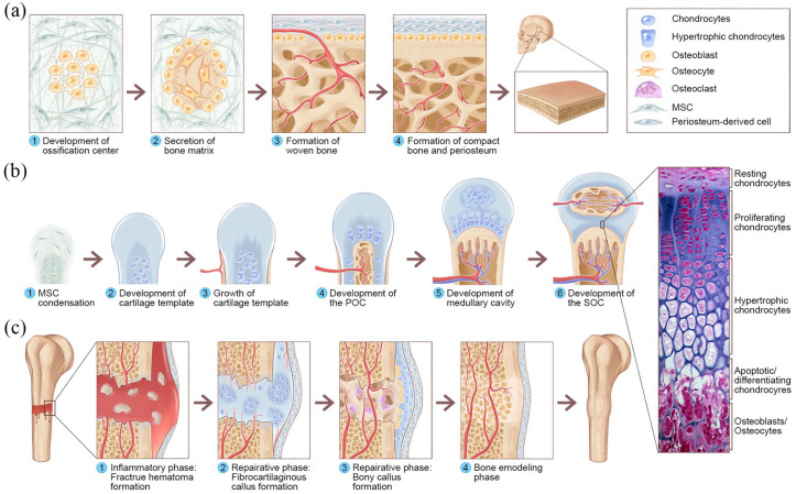
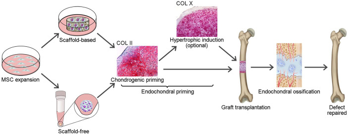
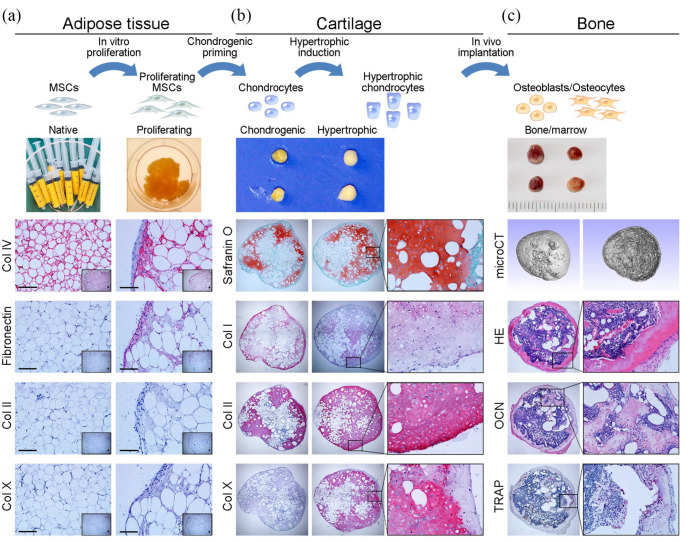
Traditional bone tissue engineering (BTE) strategies induce direct bone-like matrix formation by mimicking the embryological process of intramembranous ossification. However, the clinical translation of these clinical strategies for bone repair is hampered by limited vascularization and poor bone regeneration after implantation in vivo. An alternative strategy for overcoming these drawbacks is engineering cartilaginous constructs by recapitulating the embryonic processes of endochondral ossification (ECO); these constructs have shown a unique ability to survive under hypoxic conditions as well as induce neovascularization and ossification. Such developmentally engineered constructs can act as transient biomimetic templates to facilitate bone regeneration in critical-sized defects. This review introduces the concept and mechanism of developmental BTE, explores the routes of endochondral bone graft engineering, highlights the current state of the art in large bone defect reconstruction via ECO-based strategies, and offers perspectives on the challenges and future directions of translating current knowledge from the bench to the bedside.

Keywords: Developmental engineering; bone defect reconstruction; bone tissue engineering; endochondral ossification; hypertrophic cartilage.


Conflict of interest statement

Declaration of conflicting interests: The author(s) declared no potential conflicts of interest with respect to the research, authorship, and/or publication of this article.

Figures

Figure 1.
Figure 1.
Overview of IMO and ECO during embryonic bone development and fracture healing: (a) IMO follows four steps. Step 1: MSCs undergo condensation and form ossification centers. MSCs within the areas of condensation lead to the development of capillaries and osteoblasts. Step 2: Osteoblasts secrete osteoid, which then entraps the osteoblasts, and the osteoblasts transform into osteocytes. Step 3: Osteoid secreted around the capillaries result in trabecular matrix formation, while osteoblasts on the surface of the spongy bone become the periosteum. Step 4: The periosteum then creates compact bone superficial to the trabecular bone. The trabecular bone crowds blood vessels, which eventually condense into red marrow. (b) ECO follows six steps during embryonic bone development. Step 1: MSC condensation. Step 2: MSCs within the areas of condensation differentiate into chondrocytes to form the cartilage template of the future long bone, and MSCs in the periphery form the perichondrium. Step 3: Chondrocytes in the center of the template undergo hypertrophy, while cells in the periphery undergo direct osteogenic differentiation to form a periosteal collar of compact bone around the cartilage template. Step 4: Hypertrophic chondrocytes secrete osteogenic and angiogenic factors that initiate cartilage matrix mineralization and blood vessel invasion, resulting in POC formation. Step 5: The diaphysis elongates, and a medullary cavity forms as ossification continues. Step 6: After this initial bone formation, the same sequence of events occurs in the epiphyseal regions, leading to SOC formation, and (c) The healing of fractures follows three consecutive and overlapping phases. Inflammatory phase: Approximately 6–8 h after the fracture, a hematoma is formed at the fracture site. Reparative phase: Within approximately 48 h after the fracture, chondrocytes from the periosteum and marrow create an internal callus between the two ends of the broken bone and an external callus around the outside of the break. MSCs from the periosteum directly differentiate into osteoblasts, thereby stimulating appositional bone growth and enveloping the defect. Over the next several weeks, the cartilage in the calli is replaced by woven bone via ECO. Remodeling phase: The woven bone remodels into lamellar bone through osteoclast-osteoblast coupling, and the healing process is complete. The histological image of the epiphyseal plate of a growing long bone was adapted from Human Anatomy, sixth edition (Copyright © 2011 Pearson Education, Inc., Figure 6.12).
Figure 2.
Figure 2.
Schematic illustration of ECO-based strategies for large bone defect reconstruction.
Figure 3.
Figure 3.
Engineering hypertrophic cartilaginous tissue directly from human adipose tissue: (a) Human adipose tissue was harvested during liposuction surgery; the human adipose tissue was positive for COL IV but negative for fibronectin, COL II, and COL X. After 3 weeks of culture in proliferation medium, the adipose tissue was positive for COL IV and fibronectin but negative for COL II and COL X, indicating that proliferative culture results in more stromal cells in the adipose tissue. (b) Then, the cultured adipose tissue was subjected to endochondral priming. After 4 weeks of chondrogenic priming, the engineered constructs showed a cartilaginous phenotype, which was characterized by positive safranin O staining for GAG, weakly positive staining for COL I and COL X, and strongly positive staining for COL II. The chondrogenically primed constructs were cultured in HYM for 2 weeks, which resulted in strong positive staining for COL X, (c) The endochondrally primed constructs were subcutaneously implanted into nude mice for 12 weeks. MicroCT scanning of the retrieved constructs showed a bony shell around bone trabeculae inside. Bone tissue formation and morphological evidence of bone marrow in the retrieved constructs were identified histologically by hematoxylin and eosin (H&E) staining and osteocalcin (OCN) staining. Intensive bone resorption by osteoclasts, characterized as tartrate-resistant acid phosphatase (TRAP)-positive multinucleated cells, was observed in the inner margins of the bone marrow-like cavity surrounded by newly formed bone tissue.
Figure 4.
Figure 4.
Flow diagram for study selection. The inclusion criteria were as follows: (1) the constructs were engineered via chondrogenic and/or hypertrophic priming in vitro; (2) bone defect reconstruction in animal model. The included studies must meet all the above criteria at the same time. The excluded criteria were: (1) not an original article; (2) full text was not available; (3) not English language; (4) duplicate publications. Reports meet any of the above criteria were excluded.

References

    1. Fernandez de Grado G, Keller L, Idoux-Gillet Y, et al. Bone substitutes: a review of their characteristics, clinical use, and perspectives for large bone defects management. J Tissue Eng 2018; 9: 2041731418776819. - PMC - PubMed
    1. Wang W, Yeung KWK. Bone grafts and biomaterials substitutes for bone defect repair: A review. Bioact Mater 2017; 2: 224–247. - PMC - PubMed
    1. Buser Z, Brodke DS, Youssef JA, et al. Synthetic bone graft versus autograft or allograft for spinal fusion: a systematic review. J Neurosurg Spine 2016; 25: 509–516. - PubMed
    1. Roseti L, Parisi V, Petretta M, et al. Scaffolds for bone tissue engineering: state of the art and new perspectives. Mater Sci Eng C Mater Biol Appl 2017; 78: 1246–1262. - PubMed
    1. Wubneh A, Tsekoura EK, Ayranci C, et al. Current state of fabrication technologies and materials for bone tissue engineering. Acta Biomater 2018; 80: 1–30. - PubMed

LinkOut - more resources