. 2021 Apr 13:13:109.
doi: 10.1007/s40820-021-00620-8. eCollection 2021 Dec.

Human ACE2-Functionalized Gold "Virus-Trap" Nanostructures for Accurate Capture of SARS-CoV-2 and Single-Virus SERS Detection

Affiliations

Human ACE2-Functionalized Gold "Virus-Trap" Nanostructures for Accurate Capture of SARS-CoV-2 and Single-Virus SERS Detection

Yong Yang et al. Nanomicro Lett. .

Abstract

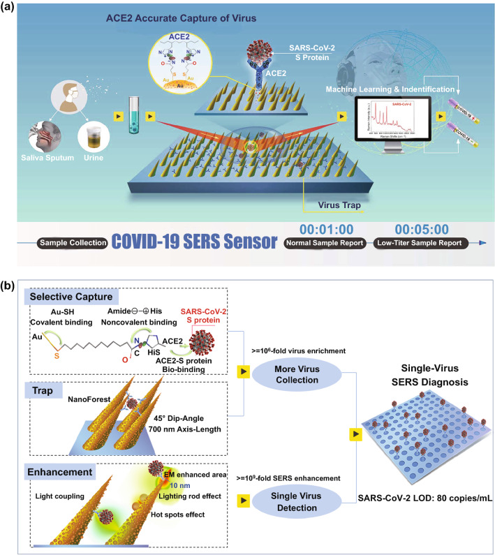
The current COVID-19 pandemic urges the extremely sensitive and prompt detection of SARS-CoV-2 virus. Here, we present a Human Angiotensin-converting-enzyme 2 (ACE2)-functionalized gold "virus traps" nanostructure as an extremely sensitive SERS biosensor, to selectively capture and rapidly detect S-protein expressed coronavirus, such as the current SARS-CoV-2 in the contaminated water, down to the single-virus level. Such a SERS sensor features extraordinary 106-fold virus enrichment originating from high-affinity of ACE2 with S protein as well as "virus-traps" composed of oblique gold nanoneedles, and 109-fold enhancement of Raman signals originating from multi-component SERS effects. Furthermore, the identification standard of virus signals is established by machine-learning and identification techniques, resulting in an especially low detection limit of 80 copies mL-1 for the simulated contaminated water by SARS-CoV-2 virus with complex circumstance as short as 5 min, which is of great significance for achieving real-time monitoring and early warning of coronavirus. Moreover, here-developed method can be used to establish the identification standard for future unknown coronavirus, and immediately enable extremely sensitive and rapid detection of novel virus.

Supplementary information: The online version contains supplementary material available at 10.1007/s40820-021-00620-8.

Keywords: Human ACE2; SARS-CoV-2; SERS; Single-virus detection; “Virus-trap” nanostructure.


Figures

Fig. 1
Fig. 1
Schematic diagram of COVID-19 SERS sensor design and single-virus detection mechanism. a Schematic diagram of COVID-19 SERS sensor design and operation procedure. SARS-CoV-2 can be localized by “virus-traps” nanoforest composed of oblique gold-nanoneedles array (GNAs), and be captured by ACE2 anchored on amide-modified GNAs from virus-containing urines even with complex multi-proteins circumstance. Through machine-learning and identification techniques, the identification standard of virus signals are established, and utilized for virus diagnoses. b Schematic diagrams of single-virus detection by selectively capturing and trapping virus, and the multi-SERS enhancement mechanism
Fig. 2
Fig. 2
Morphology analysis of gold-nanoneedles array and SERS spectra of viral protein. a SEM images of Au nanoneedles array fabricated by Ar+ ions irradiation at a tilted angle of 45° on an Au film of 500 nm in thickness. b Schematics of “virus-traps” nanoforest composed of tilted gold-nanoneedles array. c Calculated intensity distribution (|E|2) at 785 nm for a tilted Au nanoneedle array with the polarization of the incident laser along the x-axis. d Structure schematics of SARS-CoV-2 (left), and SERS spectra (right) of SARS-CoV-2 S protein and nucleocapsid protein, SARS-CoV S protein, and Human ACE2 protein at 100 nM level. e Calculated static Raman spectra of 4 types of main individual amino acids Tyr, Trp, His, Phe encoded in SARS-CoV-2 S on Au cluster
Fig. 3
Fig. 3
Affinity analysis of SARS-CoV-2 S, SARS-CoV S with ACE2. a Schematic illustration of S proteins’ binding on ACE2-functionalized GNAs of SARS-CoV-2 and SARS-CoV. SERS spectra of b SARS-CoV and c SARS-CoV-2 S proteins at the same concentration of 177 nM after bound to ACE2-functionalized GNAS for different binding time durations. d Intensity of detected SERS signals of SARS-CoV-2 S and SARS-CoV S after bound to ACE2-functionalized GNAs with different binding times. e Intensity of Raman bands (1027 cm−1) of SARS-CoV-2 S with different concentration detected with ACE2-functionalized GNAs by immersing in the diluted protein solution, and without ACE2-functionalized GNAs by dropping the corresponding concentration of diluted protein solution. It is worth noting that the application of dropping method is because GNAs without ACE2 modification have little affinity with SARS-CoV-2 S protein. The value marked on the line represents the number of S proteins in one Raman-focused window. η represents enrichment multiple by ACE2
Fig. 4
Fig. 4
Establishing and validating the SARS-CoV-2 virus identification standard based on machine-learning method. a SERS spectra of inactivated SARS-CoV-2, VN and VS in PBS solution and urine of healthy 8-year-old girl patient with the viral load of 2200 copies/mL. Navy blue, red, black, purple and blue lines represent inactivated SARS-CoV-2, VS, VN, VS in urine and VN in urine, respectively. b Schematic of SARS-CoV-2 S localized within the 10 nm EM enhancement area, and calculated intensity distribution (|E|2) at 785 nm for an oblique Au nanoneedle’s tip. c Key features of SERS patterns to classify the urine samples infected by VS (simulated contaminated water by SARS-CoV-2 virus), VN and healthy people via PCA. d DA results to identify the urines for chronic nephritis and VS-containing chronic nephritis. The green, red, blue balls represent the standard of negative urine, the standard of VS-positive urine, the standard of VN-positive urine. The green and red stars represent identified negative urine and identified VS-positive urine. e DA results to identify VS and VN virus mixed in the adult’s urine (2200 copies mL−1). The green, red, blue balls represent the standard of VS-negative urine, the standard of VS-positive urine, the standard of VN-positive urine. The red star represents identified VS-positive urine. f SERS mapping (40 × 30 μm2) of 300 measuring area for one urine sample, 42 dot-measuring-area can be identified as VS-positive

Similar articles

Cited by

References

    1. Huang CL, Wang YM, Li XW, Ren LL, Zhao JP, et al. Clinical features of patients infected with 2019 novel coronavirus in Wuhan. China. Lancet. 2020;395:497–506. doi: 10.1016/S0140-6736(20)30183-5. - DOI - PMC - PubMed
    1. Wu F, Zhao S, Yu B, Chen YM, Wang W, et al. A new coronavirus associated with human respiratory disease in China. Nature. 2020;579:265–269. doi: 10.1038/s41586-020-2008-3. - DOI - PMC - PubMed
    1. WHO Coronavirus Disease (COVID-19) Dashboard, data last updated: 2020/12/1, 7:08pm CEST. https://covid19.who.int/ (2020)
    1. Zheng S, Fan J, Yu F, Feng BH, Lou B, et al. Viral load dynamics and disease severity in patients infected with SARS-CoV-2 in Zhejiang province, China, January-March 2020: retrospective cohort study. BMJ. 2020;369:m1443. doi: 10.1136/bmj.m1443. - DOI - PMC - PubMed
    1. Peng L, Liu J, Xu WX, Luo QM, Chen DB, et al. SARS-CoV-2 can be detected in urine, blood, anal swabs, and oropharyngeal swabs specimens. J. Med. Virol. 2020 doi: 10.1002/jmv.25936. - DOI - PMC - PubMed