. 2021 Mar 19:24:462-476.
doi: 10.1016/j.omtn.2021.03.013. eCollection 2021 Jun 4.

Advancing peptide siRNA-carrier designs through L/D-amino acid stereochemical modifications to enhance gene silencing

Affiliations

Advancing peptide siRNA-carrier designs through L/D-amino acid stereochemical modifications to enhance gene silencing

Charles E Holjencin et al. Mol Ther Nucleic Acids. .

Abstract

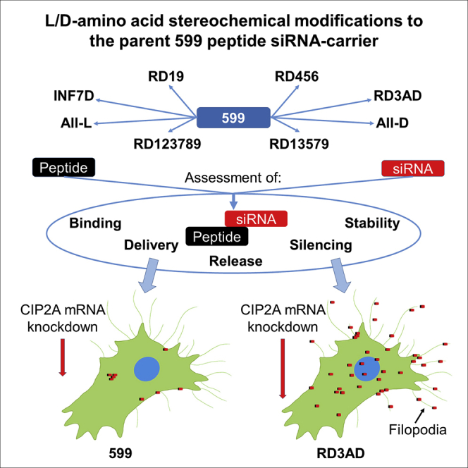
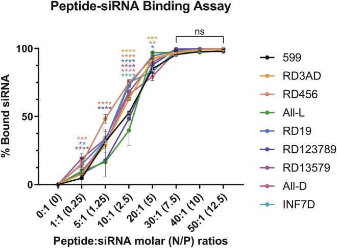
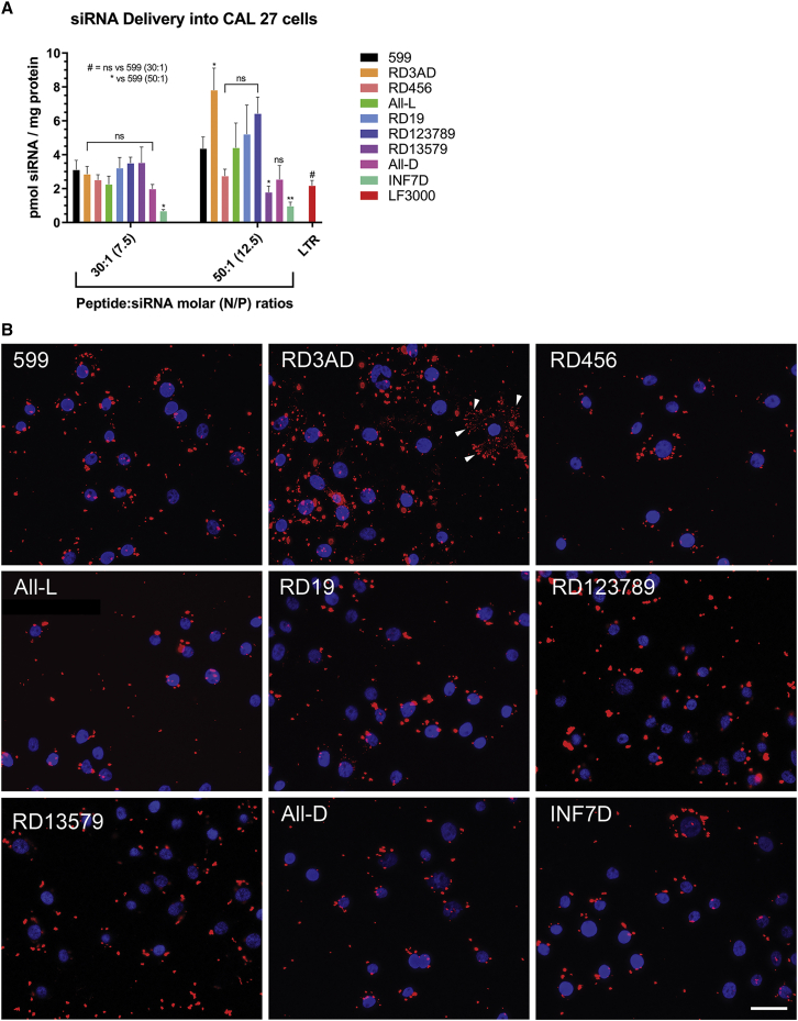
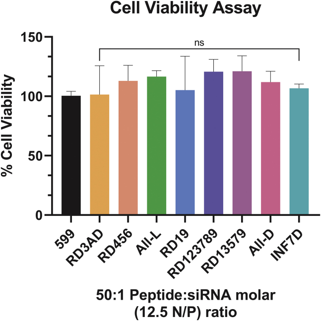
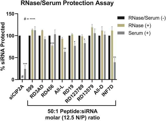
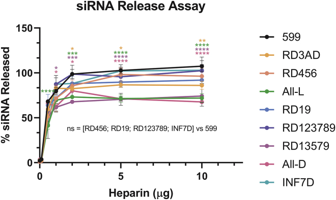
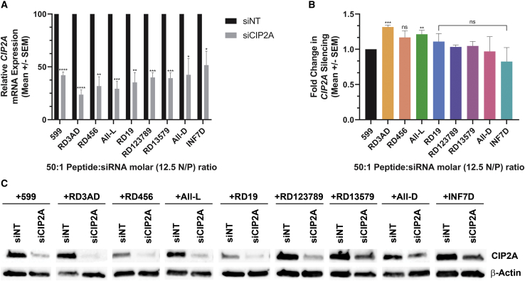
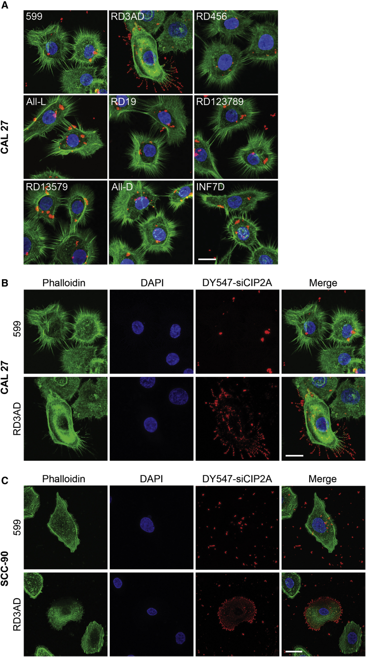
The 599 peptide has been previously shown to effectively deliver small interfering RNAs (siRNAs) to cancer cells, inducing targeted-oncogene silencing, with a consequent inhibition of tumor growth. Although effective, this study was undertaken to advance the 599 peptide siRNA-carrier design through L/D-amino acid stereochemical modifications. Consequently, 599 was modified to generate eight different peptide variants, incorporating either different stereochemical patterns of L/D-amino acids or a specific D-amino acid substitution. Upon analysis of the variants, it was observed that these modifications could, in some instances, increase/decrease the binding, nuclease/serum stability, and complex release of siRNAs, as well as influence the gene-silencing efficiencies of the complex. These modifications were also found to affect cellular uptake and intracellular localization patterns of siRNA cargo, with one particular variant capable of mediating binding of siRNAs to specific cellular projections, identified as filopodia. Interestingly, this variant also exhibited the most enhanced gene silencing in comparison to the parent 599 peptide, thus suggesting a possible connection between filopodia binding and enhanced gene silencing. Together, these data demonstrate the utility of peptide stereochemistry, as well as the importance of a key D-amino acid modification, in advancing the 599 carrier design for the enhancement of gene silencing in cancer cells.

Keywords: CIP2A; L/D-amino acid; RNAi; cancer; chirality; filopodia; peptide; siRNA; stereochemistry.


Conflict of interest statement

The authors declare no competing interests.

Figures

None
Graphical abstract
Figure 1
Figure 1
siRNA binding analysis of 599 and its peptide variants Densitometric quantitation of siCIP2A levels complexed to 599 or its peptide variants at increasing Peptide:siRNA molar (nitrogen:phosphate [N/P]) ratios. Data are mean ± SEM of three independent samples, where ∗p < 0.05, ∗∗p < 0.01, ∗∗∗p < 0.001, ∗∗∗∗p < 0.0001, and p ≥ 0.05 is not significant (ns) compared to 599 (2-way ANOVA).
Figure 2
Figure 2
Intracellular delivery of siRNAs in complex with 599 or its peptide variants (A) Quantitative siRNA uptake into CAL 27 cancer cells 2 h post-treatment with DY547-siCIP2A in complex with 599 or its peptide variants at either the 30:1 or 50:1 Peptide:siRNA molar (7.5 or 12.5 N/P) ratios. For comparison, the cells were also transfected using the commercial lipid-based transfection reagent (LTR), Lipofectamine 3000 (LF3000). The amount of siRNA delivered into cells in pmol per mg of protein is reported with each treatment normalized to DY547-siCIP2A alone. Data are mean ± SEM of three independent samples, where ∗p < 0.05, ∗∗p < 0.01, and p ≥ 0.05 is not significant (ns) compared to 599 (2-way ANOVA). (B) Confocal fluorescence microscopy analysis of CAL 27 cancer cells 2 h post-treatment with DY547-CIP2A (red) in complex with 599 or its peptide variants at 50:1 Peptide:siRNA molar (12.5 N/P) ratios. Nuclei (blue) were counterstained with DAPI. The arrowheads indicate highly ordered linear punctate uptake patterns of siRNAs in the RD3AD panel. Scale bar: 35 μm.
Figure 3
Figure 3
Assessment of cytotoxicity after treatment of cancer cells with 599 or its peptide variants in complex with siRNA The long-term cellular toxicity (as measured by a cell proliferation assay) of CAL 27 cancer cells was assessed 48 h post-treatment with 599 or its peptide variants in complex with a non-targeting siRNA (siNT) at 50:1 Peptide:siRNA molar (12.5 N/P) ratios. Data are mean ± SEM of three independent samples, where p ≥ 0.05 is not significant (ns) compared to 599 (1-way ANOVA).
Figure 4
Figure 4
Analyses of 599 and its peptide variants in their ability to protect siRNAs from RNase and serum-mediated degradation Naked and peptide-complexed siCIP2As at a 0:1 and 50:1 Peptide:siRNA molar (0 and 12.5 N/P) ratio, respectively, were incubated in the absence or presence of either RNase A or 50% human serum for 1 h at 37°C, after which densitometric quantitation was performed to determine the levels of protected siRNAs. Data are mean ± SEM of three independent samples, where ∗p < 0.05, ∗∗p < 0.01, ∗∗∗p < 0.001, ∗∗∗∗p < 0.0001, and p ≥ 0.05 is not significant compared to 599 (Student’s t test).
Figure 5
Figure 5
Analysis of siRNA release from 599 and its peptide variant complexes Densitometric quantitation of released siRNAs from 599 and its peptide variants complexed at the 50:1 Peptide:siRNA molar (12.5 N/P) ratio upon addition of increasing amounts of heparin (0–10 μg). Data are mean ± SEM of three independent samples, where ∗p < 0.05, ∗∗p < 0.01, ∗∗∗p < 0.001, ∗∗∗∗p < 0.0001, and p ≥ 0.05 is not significant (ns) compared to 599 (2-way ANOVA).
Figure 6
Figure 6
CIP2A gene silencing mediated by 599 and its peptide variants in complex with siCIP2A (A) Real-time PCR analysis of CIP2A mRNA levels in CAL 27 cancer cells 48 h post-treatment with siNT or siCIP2A in complex with either 599 or its peptide variants at 50:1 Peptide:siRNA molar (12.5 N/P) ratios. The CIP2A mRNA levels were normalized to endogenous 18S rRNA. Data are mean ± SEM of three independent samples, where ∗p < 0.05, ∗∗p < 0.01, ∗∗∗p < 0.001, ∗∗∗∗p < 0.0001, and p ≥ 0.05 is not significant compared to 599 (Student’s t test). (B) Fold change in CIP2A silencing mediated by the 599 peptide variants normalized to 599. Data are mean ± SEM of three independent samples, where ∗∗p < 0.01, ∗∗∗p < 0.001, and p ≥ 0.05 is not significant (ns) compared to 599 (Student’s t test). (C) Western blot analyses of CIP2A protein expression levels in CAL 27 cancer cells 48 h post-treatment with siNT or siCIP2A in complex with either 599 or its peptide variants at 50:1 Peptide:siRNA molar (12.5 N/P) ratios. β-actin protein levels were monitored to ensure equal loading of samples.
Figure 7
Figure 7
Cellular localization of siRNAs mediated by 599 and its peptide variants (A) Confocal fluorescence microscopy analysis of CAL 27 cancer cells 2 h post-treatment with DY547-siCIP2A (red) in complex with 599 or its peptide variants at 50:1 Peptide:siRNA molar (12.5 N/P) ratios. Filopodia (green) were stained with the selective F-actin label Alexa Fluor 488 phalloidin and nuclei (blue) were counterstained with DAPI. Scale bar: 17 μm. (B) Confocal fluorescence microscopy images of 599 and RD3AD-mediated localization of DY547-siCIP2A in CAL 27 cancer cells from (A) separated into their individual fluorophore signals. The merged images are also presented. Scale bar: 17 μm. (C) Confocal fluorescence microscopy analysis of SCC-90 cancer cells 2 h post-treatment with DY547-siCIP2A (red) in complex with 599 or RD3AD at a 50:1 Peptide:siRNA molar (12.5 N/P) ratio. Filopodia (green) were stained with the selective F-actin label Alexa Fluor 488 phalloidin and nuclei (blue) were counterstained with DAPI. Scale bar: 27 μm.
Figure 8
Figure 8
Time-course analyses of siRNA cellular localization mediated by RD3AD in comparison to 599 Confocal fluorescence microscopy analyses of CAL 27 cancer cells 0.5, 1, 2, and 4 h post-treatment with DY547-siCIP2A (red) in complex with 599 or RD3AD at a 50:1 Peptide:siRNA molar (12.5 N/P) ratio. Alexa Fluor 488 phalloidin was used to stain the F-actin (green) in filopodia, as well as the cytoplasm of cells, so as to help demarcate the cell bodies. Nuclei (blue) were counterstained with DAPI. The upper-half panels represent merged images of the red and blue fluorophore signals only, while the lower-half panels represent all three (red, blue, and green) fluorophore signals combined. Cross-sectional views comprising z sections along the x and y planes are also presented within each panel. Yellow fluorescence reveals the potential colocalization between siRNA and F-actin. Scale bar: 70 μm.

References

    1. Fire A., Xu S., Montgomery M.K., Kostas S.A., Driver S.E., Mello C.C. Potent and specific genetic interference by double-stranded RNA in Caenorhabditis elegans. Nature. 1998;391:806–811. - PubMed
    1. Rana T.M. Illuminating the silence: understanding the structure and function of small RNAs. Nat. Rev. Mol. Cell Biol. 2007;8:23–36. - PubMed
    1. Elbashir S.M., Harborth J., Lendeckel W., Yalcin A., Weber K., Tuschl T. Duplexes of 21-nucleotide RNAs mediate RNA interference in cultured mammalian cells. Nature. 2001;411:494–498. - PubMed
    1. Cummings J.C., Zhang H., Jakymiw A. Peptide carriers to the rescue: overcoming the barriers to siRNA delivery for cancer treatment. Transl. Res. 2019;214:92–104. - PMC - PubMed
    1. Bobbin M.L., Rossi J.J. RNA Interference (RNAi)-Based Therapeutics: Delivering on the Promise? Annu. Rev. Pharmacol. Toxicol. 2016;56:103–122. - PubMed