Case Reports
. 2021 Mar 31:11:638905.
doi: 10.3389/fonc.2021.638905. eCollection 2021.

Pancreatic Leiomyosarcoma With Schistosomiasis Hematobia: A Case Report and Literature Review

Affiliations
Case Reports

Pancreatic Leiomyosarcoma With Schistosomiasis Hematobia: A Case Report and Literature Review

Qiang Li et al. Front Oncol. .

Abstract

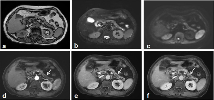
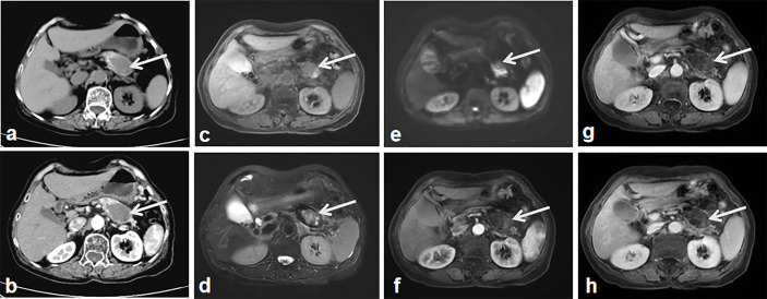
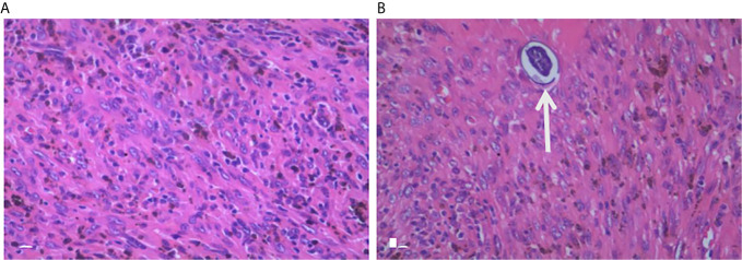
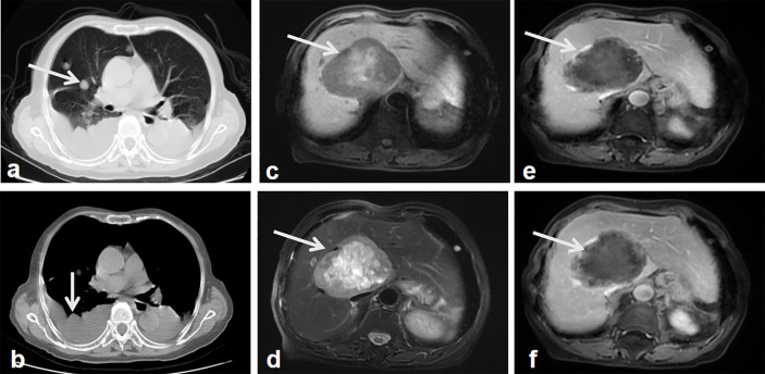
Pancreatic leiomyosarcoma (PL) is a very rare, malignant neoplasm with a very poor prognosis. Here, we examine a novel case of PL with schistosomiasis hematobia. The patient had been initially misdiagnosed by the first magnetic resonance imaging (MRI). The second imaging examination demonstrated an enlarged heterogeneous tumor mass in the body-tail of pancreas. Following image analysis, the patient underwent a pancreatectomy, splenectomy and lymph node dissections. Sixteen months after the tumor resection, follow-up computed tomography (CT) and MRI revealed tumor metastasis in the liver and lung. PL has non-specific clinical manifestations and imaging characteristics, making early diagnosis very challenging. When it is difficult to distinguish between benign and malignant pancreatic lesions, short-term imaging follow-up is preferred. In this case report, we discuss the relationship between PL and schistosomiasis hematobia.

Keywords: differential diagnosis; imaging; metastasis; pancreatic leiomyosarcoma; schistosomiasis hematobia.


Conflict of interest statement

The authors declare that the research was conducted in the absence of any commercial or financial relationships that could be construed as a potential conflict of interest.

Figures

Figure 1
Figure 1
The first abdominal MRI. Both MR T1 (A) and T2 weighted image (B) showed the atrophy of the pancreas and inhomogeneous signal change in the body and tail of the pancreas. Diffusion weighted image didn’t show the apparent tumor mass-related signal change (C). Gadolinium-enhanced T1 weighted images including early arterial phase (D), late arterial phase (E) and portal phase images (F), depicted multiple irregular hypointense signals without demonstrating a typical tumor imaging characteristics, which was regarded as an indeterminate lesion in the pancreas. A recommendation of imaging follow-up was made for the patient.
Figure 2
Figure 2
The second imaging. CT without contrast administration show the significantly enlarged tumor mass with heterogeneous components of central cystic low attenuation, peripheral solid isointensity and small patchy calcification (A, arrow). Contrast-enhanced CT shows the tumor mass with intermediate enhancement of the peripheral component and the non-enhanced cyst-like central area (B, arrow). MR T2 (C) and T2 weighted images (D) show heterogeneous hyper-signal intensity and hypo-signal intensity in the central area of the tumor (arrow). DWI shows the apparent diffusion restriction-induced heterogeneous hypersignal intensity (E, arrow). Contrast-enhanced MR weighted images of arterial (F), portal (G) and equilibrium phase (H) show the tumor mass with intermediate enhancement of the peripheral component, the non-enhanced cyst-like central area (arrows).
Figure 3
Figure 3
Pathological examination. Histology with H&E staining of the excised pancreatic tumor mass shows large pleomorphic, spindle-shaped cells, fibrosis, which indicate a stromal malignancy (A). Another slide of the tumor shows schistosome in the tumor tissue (B, arrow).
Figure 4
Figure 4
Follow-up images after surgery. Lung CT shows multiple nodules in the lung and thoracic effusion, which indicate the lung and possible pleural metastasis (A, B, arrow). MR T1 weighted images show a well-demarcated, heterogeneous hypointense signal tumor in the right lobe of the liver with patchy hypersignal intensities in the central of the tumor (C, arrow). MR weighted images show the tumor mass with heterogeneous hypersignal intensities (D, arrow). Contrast-enhanced MR weighted images of arterial (E), portal phase (F) show the tumor mass with intermediate enhancement of the peripheral component, the non-enhanced hypointense signal central area (arrows).

References

    1. Bacelar A, Castro LG, de Queiroz AC, Cafe E. Association between prostate cancer and schistosomiasis in young patients: a case report and literature review. Braz J Infect Dis (2007) 11:520–2. 10.1590/S1413-86702007000500014 - DOI - PubMed
    1. Ross CF. Leiomyosarcoma of the pancreas. Br J Surg (1951) 39:53–6. 10.1002/bjs.18003915311 - DOI - PubMed
    1. Reyes MC, Huang X, Bain A, Ylagan L. Primary pancreatic leiomyosarcoma with metastasis to the liver diagnosed by endoscopic ultrasound-guided fine needle aspiration and fine needle biopsy: A Case Report and Review of Literature. Diagn Cytopathol (2016) 44:1070–3. 10.1002/dc.23540 - DOI - PubMed
    1. Hur YH, Kim HH, Park EK, Seoung JS, Kim JW, Jeong YY, et al. . Primary leiomyosarcoma of the pancreas. J Korean Surg Soc (2011) 81 Suppl 1:S69–73. 10.4174/jkss.2011.81.Suppl1.S69 - DOI - PMC - PubMed
    1. Muhammad SU, Azam F, Zuzana S. Primary pancreatic leiomyosarcoma: a case report. cases J (2008) 1:280. 10.1186/1757-1626-1-280 - DOI - PMC - PubMed

Publication types

LinkOut - more resources