Review
. 2021 Mar 25:9:595914.
doi: 10.3389/fcell.2021.595914. eCollection 2021.

Ghrelin-Mediated Regeneration and Plasticity After Nervous System Injury

Affiliations
Review

Ghrelin-Mediated Regeneration and Plasticity After Nervous System Injury

Irina Stoyanova et al. Front Cell Dev Biol. .

Abstract

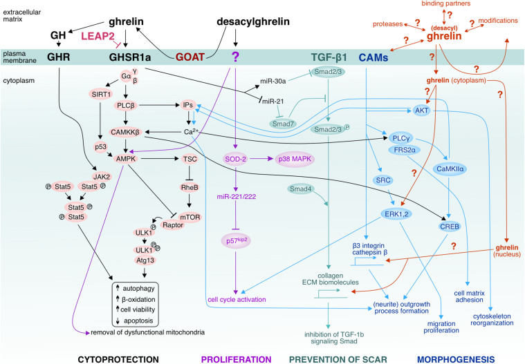
The nervous system is highly vulnerable to different factors which may cause injury followed by an acute or chronic neurodegeneration. Injury involves a loss of extracellular matrix integrity, neuronal circuitry disintegration, and impairment of synaptic activity and plasticity. Application of pleiotropic molecules initiating extracellular matrix reorganization and stimulating neuronal plasticity could prevent propagation of the degeneration into the tissue surrounding the injury. To find an omnipotent therapeutic molecule, however, seems to be a fairly ambitious task, given the complex demands of the regenerating nervous system that need to be fulfilled. Among the vast number of candidates examined so far, the neuropeptide and hormone ghrelin holds within a very promising therapeutic potential with its ability to cross the blood-brain barrier, to balance metabolic processes, and to stimulate neurorepair and neuroactivity. Compared with its well-established systemic effects in treatment of metabolism-related disorders, the therapeutic potential of ghrelin on neuroregeneration upon injury has received lesser appreciation though. Here, we discuss emerging concepts of ghrelin as an omnipotent player unleashing developmentally related molecular cues and morphogenic cascades, which could attenuate and/or counteract acute and chronic neurodegeneration.

Keywords: GHSR; brain and spinal cord injury; ischemia; neurogenesis; stroke; synaptic activity.


Conflict of interest statement

The authors declare that the research was conducted in the absence of any commercial or financial relationships that could be construed as a potential conflict of interest.

Figures

FIGURE 1
FIGURE 1
Ghrelin-induced effector pathways and unavowed signaling hotspots. Ghrelin activates the cascade via GHSR1a/CaMKKβ/AMPK to suppress endoplasmic reticulum stress. LEAP2 has been suggested to inhibit GHSR1a. Ghrelin-activated AMPK inhibits mTOR via activation of TSC and inactivation of Raptor, leading to reduced phosphorylation of ULK1 and therefore enhanced ULK1 kinase activity which triggers autophagy (Kim et al., 2011). Ghrelin’s pro-autophagic effect has been shown to improve hepatosteatosis by increasing abundance of mtDNA and inducing mitochondrial free fatty acid β-oxidation. Not only CaMKKβ but also SIRT1-p53 modulates AMPK in the setting of autophagy (e.g., in hypothalamic ghrelin signaling). Ghrelin can act pro-autophagically via the growth hormone (GH) under fat-depleted famine conditions by activating GH receptor-mediated JKA2-pStat cascades. Furthermore, ghrelin has been shown to upregulate antifibrotic (miR-30a) microRNA and downregulate profibrotic (miR-21) microRNA, thus affecting the TGF-β1-Smad pathway and ameliorating skeletal muscle fibrosis upon injury. Plasma membrane-associated GOAT has been proposed to locally convert desacylghrelin to ghrelin. Also desacylghrelin can stimulate the AMPK activity in order to induce autophagy by decreasing reactive oxygen species accumulation and apoptosis, thereby protecting e.g., cardiomyocytes from ischemic injury. Furthermore, desacylghrelin has been shown to stimulate SOD-2 which leads to increased expression of miR-221 and miR-222. In turn, these miRs suppress p57kip2 expression in satellite cells of skeletal muscle, thereby accelerating cell cycle re-entry and proliferation. These events facilitate muscle regeneration. Desacylghrelin-mediated SOD-2 upregulation also increases myogenesis and decreases reactive oxygen species generation, thus promoting tissue regeneration after injury (Yanagi et al., 2018). It has remained unclear whether desacylghrelin/ghrelin can be internalized by the target cells and may act in the cytoplasm, e.g., on AKT or ERK1,2 or by far, whether ghrelin can translocate to the nucleus and affect gene expression. Does ghrelin also associate with cell adhesion molecules (CAMs) or other binding partners at the plasma membrane and/or undergo recycling? It is possible that the desacylghrelin/ghrelin-mediated signaling cascades might be highly intertwined with the signaling cascades mediated by the CAMs. CAMs, such as cadherins, form signaling units with TGF-β1. The interplay between CAMs with their binding partners leads to recruitment of catenins and junction plakoglobin to the nucleus and regulate transcription [e.g., α-catenin can regulate actin bundling (AKT)]. CAMs convey also signals to kinases (for example, SRC family kinases and the Tyr-protein kinase) and phosphatases (PLCγ). Given that the CAM-mediated cascades govern morphogenic events such as cytoskeletal reorganization, process formation, neurite outgrowth and dynamic events like proliferation and migration (Cavallaro and Dejana, 2011), it is conceivable that one possible action of ghrelin to stimulate regeneration could be via affecting these CAM-cascades. AKT (actin); AMPK (5’ adenosine monophosphate-activated protein kinase); Atg13 (autophagy-related); CaMKIIα (Ca2+-calmodulin-dependent kinase); CAMKKβ (Calcium/calmodulin-dependent protein kinase kinase 2); CREB (cAMP response element-binding protein); ECM (extracellular matrix); ERK1,2 (extracellular regulated kinase); Frs2α (substrate of fibroblast growth factor receptor); GHSR1a (growth hormone secretagogue receptor 1a); Gα,β,γ (G protein alpha, beta and gamma subunits); IP3 (inositol triphosphate); JAK2 (Janus kinase 2); LEAP (Liver-expressed antimicrobial peptide 2); mTOR (mechanistic target of rapamycin); P38 MAPK (p38 mitogen-activated protein kinases); P53 (protein p53); P57kip2 (Cyclin-dependent kinase inhibitor 1C); PLCβ (phospholipase C); PLCγ (phospholipase); Raptor (Regulatory-associated protein of mTOR); RheB (Ras homolog enriched in brain); SIRT1 (NAD-dependent deacetylase sirtuin-1); Smad2/3, Smad4, Smad7 (Mothers against decapentaplegic homolog 2/3/4/7); SOD-2 (Superoxide dismutase 2); SRC (Src kinase); Stat5 (Signal transducer and activator of transcription 5); TSC (tuberous sclerosis complex); ULK1 (Unc-51 like kinase-1).
FIGURE 2
FIGURE 2
Effect of ghrelin on network activity. Raster plots of the neuronal activity recorded over 20 min (x-axis) in cultures incubated with ghrelin and controls at age 6 days in vitro (DIV). The top rows of the panels depict only the electrodes in contact with active neurons (electrode numbers are on y-axis). Each tick represents a recorded action potential. The bottom rows of the two panels represent the summed network activity. (A) Network activity in a sister culture under control conditions, 47 spikes recorded in 1 min, immature activity pattern. (B) Network activity in a culture chronically treated with ghrelin, 3,740 spikes recorded in 1 min, showing mature activity pattern. With permission from BMC Neuroscience (Stoyanova and le Feber, 2014).

References

    1. Abizaid A., Hougland J. L. (2020). Ghrelin signaling: GOAT and GHS-R1a take a LEAP in complexity. Trends Endocrinol. Metab. 31 107–117. 10.1016/j.tem.2019.09.006 - DOI - PMC - PubMed
    1. Akalu Y., Molla M. D., Dessie G., Ayelign B. (2020). Physiological effect of ghrelin on body systems. Int. J. Endocrinol. 2020:1385138. 10.1155/2020/1385138 - DOI - PMC - PubMed
    1. Al Massadi O., López M., Tschöp M., Diéguez C., Nogueiras R. (2017). Current understanding of the hypothalamic ghrelin pathways inducing appetite and adiposity. Trends Neurosci. 40 167–180. 10.1016/j.tins.2016.12.003 - DOI - PubMed
    1. Allbrand M., Åman J., Lodefalk M. (2018). Placental ghrelin and leptin expression and cord blood ghrelin, adiponectin, leptin, and C-peptide levels in severe maternal obesity. J. Matern. Fetal. Neonatal. Med. 31 2839–2846. 10.1080/14767058.2017.1358262 - DOI - PubMed
    1. Andrews Z. B. (2011). The extra-hypothalamic actions of ghrelin on neuronal function. Trends Neurosci. 34 31–40. 10.1016/j.tins.2010.10.001 - DOI - PubMed

LinkOut - more resources