. 2021 Apr 1:9:663037.
doi: 10.3389/fcell.2021.663037. eCollection 2021.

Periodontal Inflammation-Triggered by Periodontal Ligament Stem Cell Pyroptosis Exacerbates Periodontitis

Affiliations

Periodontal Inflammation-Triggered by Periodontal Ligament Stem Cell Pyroptosis Exacerbates Periodontitis

Qin Chen et al. Front Cell Dev Biol. .

Abstract

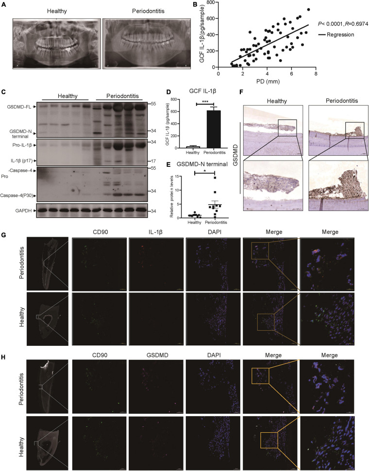
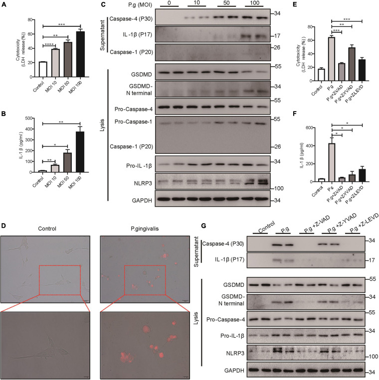
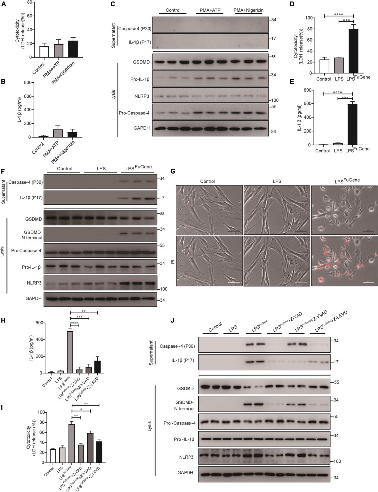
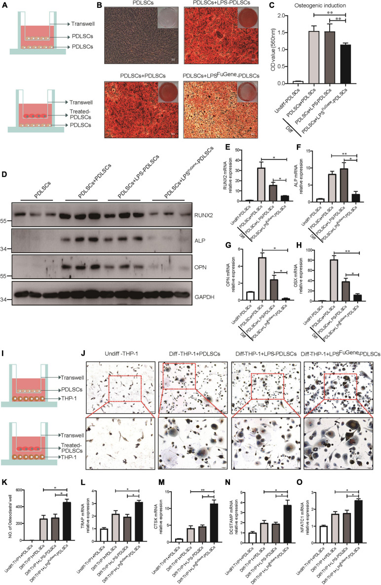
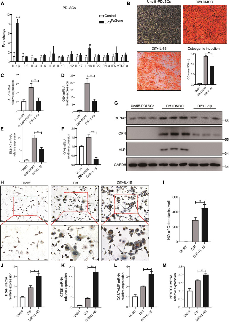
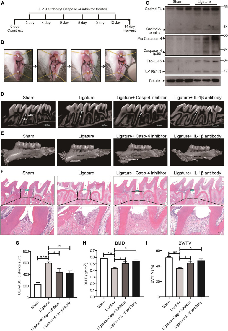
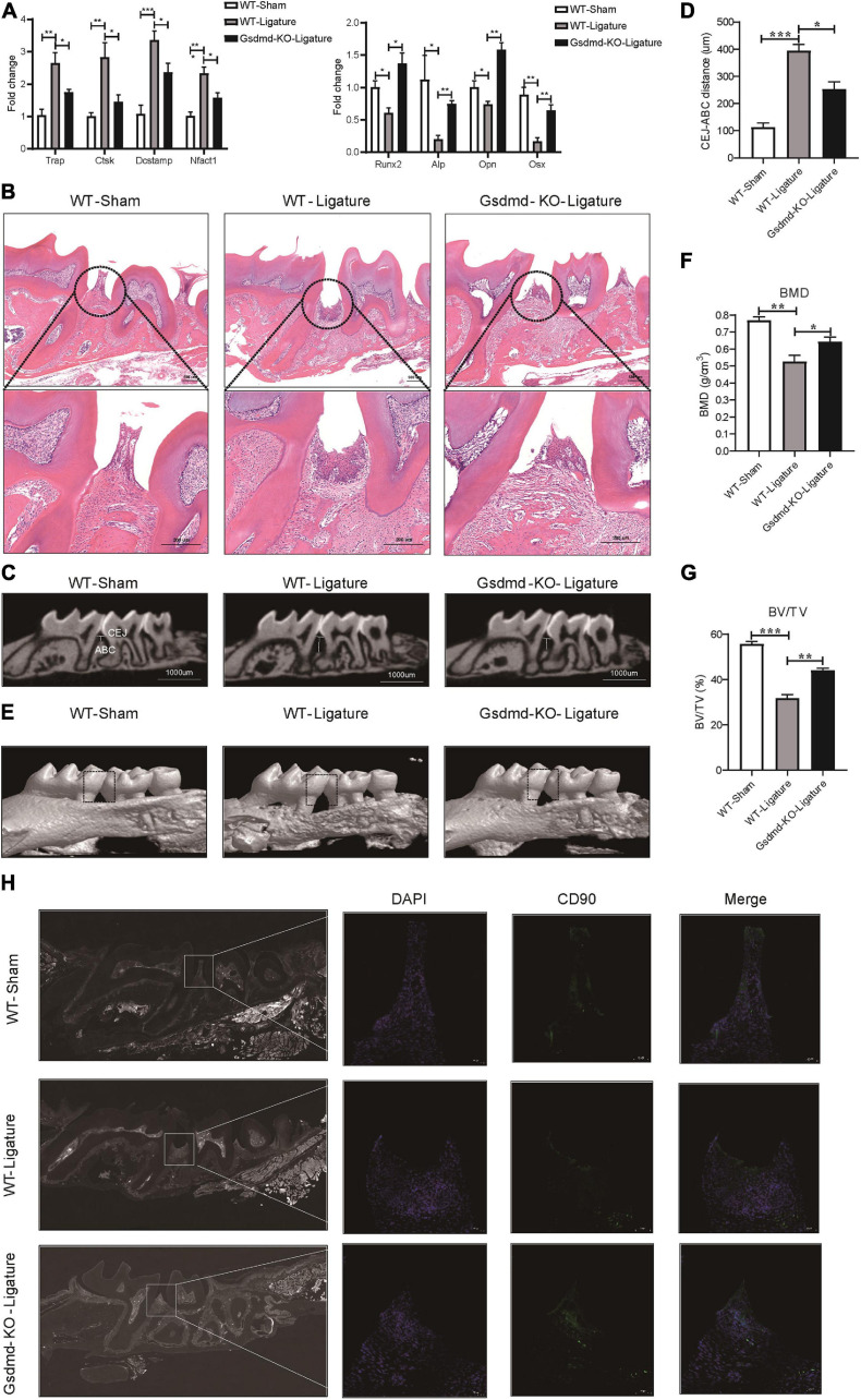
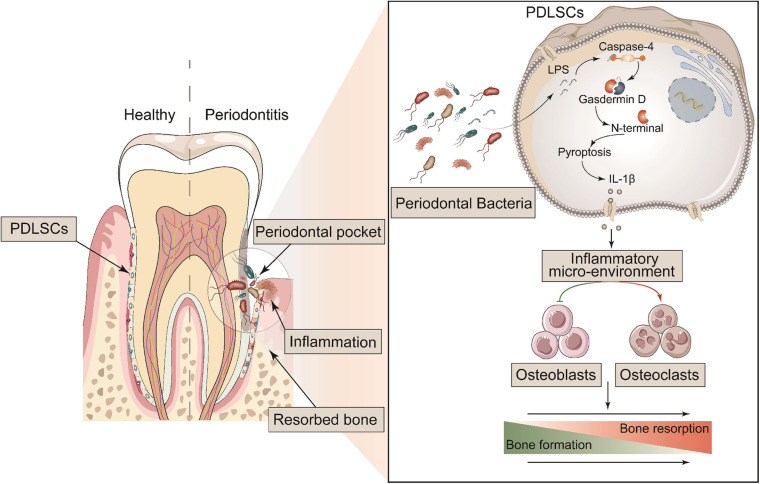
Periodontitis is an immune inflammatory disease that leads to progressive destruction of bone and connective tissue, accompanied by the dysfunction and even loss of periodontal ligament stem cells (PDLSCs). Pyroptosis mediated by gasdermin-D (GSDMD) participates in the pathogenesis of inflammatory diseases. However, whether pyroptosis mediates PDLSC loss, and inflammation triggered by pyroptosis is involved in the pathological progression of periodontitis remain unclear. Here, we found that PDLSCs suffered GSDMD-dependent pyroptosis to release interleukin-1β (IL-1β) during human periodontitis. Importantly, the increased IL-1β level in gingival crevicular fluid was significantly correlated with periodontitis severity. The caspase-4/GSDMD-mediated pyroptosis caused by periodontal bacteria and cytoplasmic lipopolysaccharide (LPS) dominantly contributed to PDLSC loss. By releasing IL-1β into the tissue microenvironment, pyroptotic PDLSCs inhibited osteoblastogenesis and promoted osteoclastogenesis, which exacerbated the pathological damage of periodontitis. Pharmacological inhibition of caspase-4 or IL-1β antibody blockade in a rat periodontitis model lead to the significantly reduced loss of alveolar bone and periodontal ligament damage. Furthermore, Gsdmd deficiency alleviated periodontal inflammation and bone loss in mouse experimental periodontitis. These findings indicate that GSDMD-driven PDLSC pyroptosis and loss plays a pivotal role in the pathogenesis of periodontitis by increasing IL-1β release, enhancing inflammation, and promoting osteoclastogenesis.

Keywords: GSDMD; IL-1β; PDLSC; periodontitis; pyroptosis.


Conflict of interest statement

The authors declare that the research was conducted in the absence of any commercial or financial relationships that could be construed as a potential conflict of interest.

Figures

FIGURE 1
FIGURE 1
The inflammatory lesions in periodontitis were correlated with the pyroptosis of PDLSCs. (A) Panoramic radiograph from healthy and periodontitis patients. (B) Scatter plot and regression line for the regression between GCF IL-1b level and probing depth (PD) from periodontitis patients (n = 65). (C) Periodontium tissues were collected from periodontitis patients and healthy individuals, and the expression of GSDMD, IL-1β, and Caspase-4 was analyzed by western blot. GAPDH was used as the loading control. (D) GCF from healthy subjects and periodontitis patients was analyzed to determine the concentrations of human IL-1β by ELISA. (E) The intensities of the GSDMD-N terminus were quantified using imaging software and normalized to that of GAPDH. Ten periodontium tissue specimens from periodontitis patients and healthy individuals were examined. (F) Representative immunohistochemical staining of GSDMD in human periodontal ligament sections from healthy and periodontitis patients. The signals of GSDMD appear brown in sections counterstained with hematoxylin (blue). Experiments were repeated independently more than three times. (G) Immunofluorescence staining of CD90 (green) and IL-1β (red) in human tooth longitudinal sections from severe periodontitis patients and healthy (orthodontic) patients. (H) Immunofluorescence staining of CD90 (green) and GSDMD (purple) in human tooth longitudinal sections from severe periodontitis patients and healthy patients. Nuclei were identified by staining with DAPI. Scale bars, 50 μm. The data are presented as the mean ± SEM. *P < 0.05, ***P < 0.001.
FIGURE 2
FIGURE 2
P. gingivalis induced pyroptosis in PDLSCs dependent on caspase-4. (A–C) PDLSCs were infected with P. gingivalis (MOI, 10, 50, or 100) for 24 h. (A) The amount of cytoplasmic LDH released into the culture supernatant was measured by an LDH Cytotoxicity Assay Kit. (B) Cell culture supernatants were assayed for human IL-1β by ELISA. (C) The IL-1β, Caspase-4 and Caspase-1 secreted into the culture supernatants and the GSDMD cleavage, Pro-IL-1β, Pro-Caspase-4, Pro-Caspase-1, NLRP3, and GAPDH in the cell lysates (lysis) were detected by immunoblotting. (D) The morphology of PDLSCs with P. gingivalis (MOI, 100) infection. Cells were stained with propidium iodide (PI, red) and analyzed under a microscope. (E–G) PDLSCs were pretreated with the pan-caspase inhibitor Z-VAD-FMK (20 μM), the caspase-1 inhibitor Z-YVAD-FMK (20 μM), or the capase-4 inhibitor Z-LEVD-FMK (20 μM) for 1 h before P. gingivalis (MOI, 100) infection for 24 h. (E,F) Cell culture supernatant was collected and assayed for LDH release (E) and IL-1β secretion (F). (G) The IL-1β and Caspase-4 secreted into the culture supernatants and the GSDMD cleavage, Pro-IL-1β, Pro-Caspase-4, NLRP3, and GAPDH in the cell lysates (lysis) were detected by immunoblotting. The data are presented as the mean ± SEM. *P < 0.05, **P < 0.01, ***P < 0.001, ****P < 0.0001.
FIGURE 3
FIGURE 3
PDLSC pyroptosis was mediated by the caspase-4-dependent non-canonical pyroptosis pathway. (A–C) PDLSCs were pretreated with PMA for 1 h and stimulated with ATP or nigericin for 4 h. (A,B) Cell culture supernatant was collected and assayed for LDH release (A) and IL-1β secretion (B). (C) The IL-1β and Caspase-4 secreted into the culture supernatants and the GSDMD cleavage, Pro-IL-1β, Pro-Caspase-4, NLRP3, and GAPDH in the cell lysates (lysis) were detected by immunoblotting. (D–G) PDLSCs were treated with LPS or transfected with LPS using FuGene HD for 16 h. Cells without treatment were used as controls. (D,E) Cell culture supernatant was collected and assayed for LDH release (D) and IL-1β secretion (E). (F) The IL-1β and Caspase-4 secreted into the culture supernatants and the GSDMD cleavage, Pro-IL-1β, Pro-Caspase-4, NLRP3, and GAPDH in the cell lysates (lysis) were detected by immunoblotting. (G) Representative images of propidium iodide (PI, red) uptake. (H–J) PDLSCs were pretreated with the pan-caspase inhibitor Z-VAD-FMK (20 μM), the caspase-1 inhibitor Z-YVAD-FMK (20 μM), or the capase-4 inhibitor Z-LEVD-FMK (20 μM) for 1 h before transfection with LPS (LPSFuGene) for 16 h. (H) IL-1β ELISA in supernatants. (I) Percentage of LDH release in supernatants. (J) The IL-1β and Caspase-4 secreted into the culture supernatants and the GSDMD cleavage, Pro-IL-1β, Pro-Caspase-4, NLRP3, and GAPDH in the cell lysates (lysis) were detected by immunoblotting. The data are presented as the mean ± SEM. *P < 0.05, **P < 0.01, ***P < 0.001, ****P < 0.0001.
FIGURE 4
FIGURE 4
The paracrine effect of PDLSC pyroptosis on osteoblast differentiation and osteoclast differentiation. (A) Schematic diagram of coculture design indicating the placement of PDLSCs with LPS, with LPSFuGene treatment, or without treatment on the transwell insert with the lower chamber containing healthy PDLSCs. (B–H) Cells in the cocultured transwell system were maintained in osteogenic differentiation medium for 21 days. PDLSCs in a single culture (without coculture) were maintained in MEM-α medium as a control. (B) Entire plate views and micrographs of alizarin red staining after culturing in osteogenic differentiation medium. (C) Quantification of the alizarin red staining results. (D) Immunoblots for RUNX2, ALP, and OPN in whole-cell lysates. GAPDH was used as a loading control. RUNX2 (E), ALP (F), OPN (G), and OSX (H) mRNAs were subjected to real-time PCR analysis, and the expression levels were normalized to that of 36B4. (I) Schematic diagram of coculture design indicating the placement of PDLSCs with LPS treatment, with LPSFuGene treatment, or without treatment on transwell insert with the lower chamber containing THP-1 cells. (J–O) The THP-1 cells in the coculture system were cultured in osteoclast differentiation medium for 14 days. THP-1 cells in a single culture were maintained in RPMI-1640 medium as a control. (J) Micrographs of TRAP staining after culturing in osteoclast differentiation medium. (K) Quantification of TRAP-positive multinucleated cells (nuclei > 3). TRAP (L), CTSK (M), DCSTAMP (N), and NFATC1 (O) mRNAs were subjected to real-time PCR analysis, and the expression levels were normalized to that of 36B4. The data are presented as the mean ± SEM. *P < 0.05, **P < 0.01.
FIGURE 5
FIGURE 5
IL-1β was the key factor in PDLSC pyroptosis to regulate osteoblast and osteoclast differentiation. (A) Expression profiles of several inflammatory cytokines in PDLSCs under LPSFuGene treatment for 16 h. (B–G) PDLSCs were incubated with osteogenic differentiation medium for 21 days with or without 500 pg/mL IL-1β treatment. (B) Representative images of alizarin red staining and quantification of the staining results. ALP (C), OSX (D), RUNX2 (E), and OPN (F) mRNAs were subjected to real-time PCR analysis, and the expression levels were normalized to that of 36B4. (G) Immunoblots for RUNX2, ALP, and OPN in whole-cell lysates. GAPDH was used as a loading control. (H–M) THP-1 cells were incubated with osteoclast differentiation medium for 14 days with or without 500 pg/mL IL-1β treatment. (H) Micrographs of TRAP staining after culturing in osteoclast differentiation medium. (I) Quantification of TRAP-positive multinucleated cells (nuclei > 3). TRAP (J), CTSK (K), DCSTAMP (L), and NFATC1 (M) mRNAs were subjected to real-time PCR analysis, and the expression levels were normalized to that of 36B4. The data are presented as the mean ± SEM. *P < 0.05, **P < 0.01.
FIGURE 6
FIGURE 6
Caspase-4-mediated non-canonical pyroptosis was involved in ligature-induced rat periodontitis. (A–I) Rats were divided randomly into a sham group, ligature-induced rat periodontitis group (ligature group), ligature with caspase-4 inhibitor injection into the subperiosteum at the left buccal and palatal gingivae of the first maxillary molars group (Ligature + Casp-4 inhibitor group), and ligature with IL-1b antibody injection into the subperiosteum at the first maxillary molar group (Ligature + IL-1b antibody group). (A) Timeline of the animal experiment. (B) Technical procedures of ligature-induced rat periodontitis. A 4-0 silk ligature was looped around the first molar of the rat maxilla under anesthesia and maintained for 14 days. The red arrow indicated the drugs injection site. (C) Periodontium tissues were collected from ligature-induced rat periodontitis group and sham group, and the expression of Gsdmd, IL-1β, and caspase-4 was analyzed by western blot. Tubulin was used as the loading control. (D) Representative micro-CT sagittal images of maxillary molars after insertion of ligature. CEJ, cement-enamel junction; ABC, alveolar bone crest. The line indicates the distance from the CEJ to the ABC on the buccal side of the ligature site. (E) The images show the reconstructed sagittal 3D images from the computerized tomography of the maxillary molars. The squares formed by the continuous dotted line show visual differences in alveolar bone from different animal groups. (F) Tissue sections from rats were prepared after 14 days of periodontitis induction and processed for hematoxylin and eosin (H&E) staining. (G) The distances (um) between CEJ and ABC were measured after periodontitis induction. The bone mineral density (H) and bone/tissue volume (I) of the selected squares regions on the ligature site were calculated. The data are presented as the mean ± SEM. *P < 0.05, **P < 0.01, ***P < 0.001.
FIGURE 7
FIGURE 7
Gsdmd–/– mice presented with reduced periodontal inflammation and bone loss in experimental periodontitis. (A–H) Mice were divided into the WT-sham group, WT-ligature group, and Gsdmd-KO-ligature group. Silk thread was used to form a ligature around the left maxillary first molar for 2 weeks to induce periodontitis. (A) Periodontium tissues were collected from each group, and the mRNA expression of osteoblast markers (including Runx2, Alp, Opn, and Osx) and osteoclast markers (including Trap, Ctsk, Dcstamp, and Nfact1) was analyzed by real-time PCR. 36b4 was used as an internal control. (B) Tissue sections from mice were prepared after 14 days of periodontitis induction and processed for hematoxylin and eosin (H&E) staining. (C) Representative micro-CT sagittal images of maxillary molars after insertion of ligature. CEJ, cement-enamel junction; ABC, alveolar bone crest. The line indicates the distance from the CEJ to the ABC on the buccal side of the ligature site. (D) The distances (μm) between CEJ and ABC were measured after periodontitis induction. (E) The images of bone surrounding first molars were analyzed by 3D micro-computed tomography. The squares formed by the continuous dotted line show visual differences in alveolar bone from animal groups. The bone mineral density (F) and bone/tissue volume (G) of the selected square regions on the ligature site were calculated. (H) Immunofluorescence staining of CD90 (green) in mouse maxillary molar longitudinal sections from the periodontitis model and sham group. Nuclei were identified by staining with DAPI. Scale bars, 50 μm. The data are presented as the mean ± SEM. *P < 0.05, **P < 0.01, ***P < 0.001.
FIGURE 8
FIGURE 8
Schematic diagram of the mechanism of GSDMD-mediated PDLSC pyroptosis in the promotion of periodontitis. PDLSCs commit Caspase-4/GSDMD-mediated non-canonical pyroptosis in periodontopathogen-induced periodontitis. The pyroptotic PDLSCs express IL-1β and release it to the cellular microenvironment following membrane rupture. IL-1β disrupts the homeostatic balance of bone formation and resorption by inhibiting osteoblastogenesis and promoting osteoclastogenesis, jointly aggravating the process of periodontitis.

Similar articles

Cited by

References

    1. Aglietti R. A., Dueber E. C. (2017). Recent insights into the molecular mechanisms underlying pyroptosis and gasdermin family functions. Trends Immunol. 38 261–271. 10.1016/j.it.2017.01.003 - DOI - PubMed
    1. Al-Habib M., Huang G. T.-J. (2019). Dental mesenchymal stem cells: dental pulp stem cells, periodontal ligament stem cells, apical papilla stem cells, and primary teeth stem cells—isolation, characterization, and expansion for tissue engineering. Methods Mol. Biol. 1922 59–76. 10.1007/978-1-4939-9012-2_7 - DOI - PubMed
    1. American Academy of Periodontology (2011). Comprehensive periodontal therapy: a statement by the American Academy of Periodontology. J. Periodontol. 82 943–949. 10.1902/jop.2011.117001 - DOI - PubMed
    1. Aoyama N., Kure K., Minabe M., Izumi Y. (2019). Increased heart failure prevalence in patients with a high antibody level against periodontal pathogen. Int. Heart J. 60 1142–1146. 10.1536/ihj.19-010 - DOI - PubMed
    1. Aral K., Milward M. R., Kapila Y., Berdeli A., Cooper P. R. (2020). Inflammasomes and their regulation in periodontal disease: a review. J. Periodontal Res. 55 473–487. 10.1111/jre.12733 - DOI - PubMed

LinkOut - more resources