Review
. 2021 Apr 1:9:668171.
doi: 10.3389/fcell.2021.668171. eCollection 2021.

Telomere Replication: Solving Multiple End Replication Problems

Affiliations
Review

Telomere Replication: Solving Multiple End Replication Problems

Erin Bonnell et al. Front Cell Dev Biol. .

Abstract

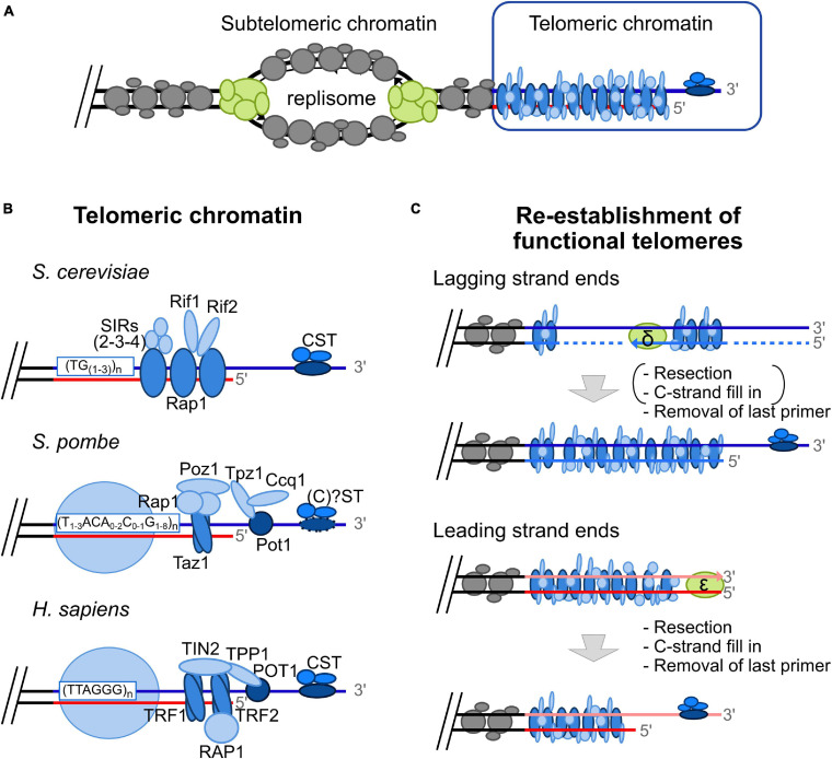
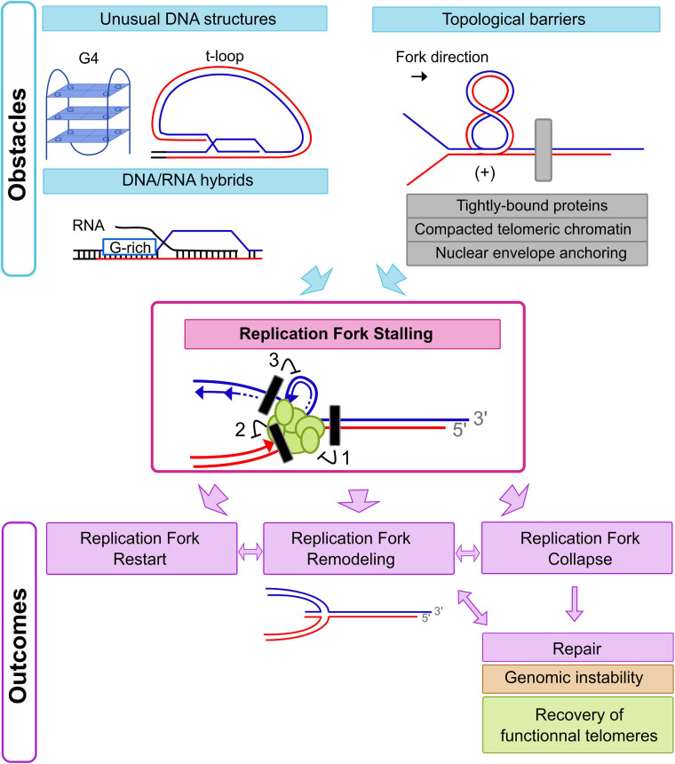
Eukaryotic genomes are highly complex and divided into linear chromosomes that require end protection from unwarranted fusions, recombination, and degradation in order to maintain genomic stability. This is accomplished through the conserved specialized nucleoprotein structure of telomeres. Due to the repetitive nature of telomeric DNA, and the unusual terminal structure, namely a protruding single stranded 3' DNA end, completing telomeric DNA replication in a timely and efficient manner is a challenge. For example, the end replication problem causes a progressive shortening of telomeric DNA at each round of DNA replication, thus telomeres eventually lose their protective capacity. This phenomenon is counteracted by the recruitment and the activation at telomeres of the specialized reverse transcriptase telomerase. Despite the importance of telomerase in providing a mechanism for complete replication of telomeric ends, the majority of telomere replication is in fact carried out by the conventional DNA replication machinery. There is significant evidence demonstrating that progression of replication forks is hampered at chromosomal ends due to telomeric sequences prone to form secondary structures, tightly DNA-bound proteins, and the heterochromatic nature of telomeres. The telomeric loop (t-loop) formed by invasion of the 3'-end into telomeric duplex sequences may also impede the passage of replication fork. Replication fork stalling can lead to fork collapse and DNA breaks, a major cause of genomic instability triggered notably by unwanted repair events. Moreover, at chromosomal ends, unreplicated DNA distal to a stalled fork cannot be rescued by a fork coming from the opposite direction. This highlights the importance of the multiple mechanisms involved in overcoming fork progression obstacles at telomeres. Consequently, numerous factors participate in efficient telomeric DNA duplication by preventing replication fork stalling or promoting the restart of a stalled replication fork at telomeres. In this review, we will discuss difficulties associated with the passage of the replication fork through telomeres in both fission and budding yeasts as well as mammals, highlighting conserved mechanisms implicated in maintaining telomere integrity during replication, thus preserving a stable genome.

Keywords: DNA replication; genome stability; replication fork stability; telomeres; telomeric chromatin.


Conflict of interest statement

The authors declare that the research was conducted in the absence of any commercial or financial relationships that could be construed as a potential conflict of interest.

Figures

FIGURE 1
FIGURE 1
The “Unusual” telomeric chromatin and the “classical” End Replication Problem. (A) Replication origins in subtelomeric areas fire in S-phase (humans) or in late S-phase (yeasts). At each fork, the replisome, a protein complex schematized here in green, allows DNA duplication. At the leading strand, DNA is synthesized by DNA polymerase ε in a continuous fashion, whereas at lagging strand, DNA synthesis by DNA polymerase δ occurs in a discontinuous fashion, i.e., in the form of Okazaki fragments. Subtelomeric chromatin is displayed in gray and the unusual telomeric chromatin is represented in blue. (B) Telomeric chromatin is unusual due to the binding of specific proteins in a sequence specific manner and lack of classical nucleosomes. Whereas telomeric chromatin in S. cerevisiae is devoid of nucleosomes (Wright et al., 1992), histones are present over telomeric repeats in S. pombe and humans in a non-canonical fashion (Greenwood et al., 2018). Rap1 recognizes dsDNA budding yeast telomeric repeats [(TG1-3) n] whereas Cdc13p binds the ssDNA telomeric overhang (Wellinger and Zakian, 2012). Telomere-bound Rap1 recruits several proteins such as the SIR complex (Sir2/Sir3/Sir4), and Rif1/Rif2. Cdc13 recruits Stn1 and Ten1, forming the CST complex. In S. pombe, Taz1 binds as homodimer on duplex telomeric DNA, whereas Pot1 recognizes single strand telomeric DNA. These two telomere-bound proteins recruit several proteins: Rap1, Poz1, Tpz1, and Ccq1 (Shelterin-like complex) (Moser and Nakamura, 2009). Whereas the homolog of Cdc13 has not been identified in this model organism, Stn1, and Ten1 are known to bind to telomeric ssDNA without forming a complex with the other ssDNA-binding protein Pot1 (Martín et al., 2007). Contrary to the heterogeneous telomeric repeats found in S. cerevisiae and S. pombe, TTAGGG repeats are found in most vertebrate species, including humans. The Shelterin complex is associated with human telomeric DNA and is comprised of TRF1 and TRF2 bound as homodimers on duplex DNA, POT1 on ssDNA, and associated proteins: RAP1, TIN2 and TPP1 (De Lange, 2005). (C) The “classical” End Replication Problem leading to progressive telomere shortening is the consequence of the unusual DNA structure of telomeres, i.e., the constitutive 3′ overhang, that has to be reformed after conventional replication, and the unidirectionality of DNA synthesis by conventional replicative DNA polymerase (from 5′ to 3′). Indeed, the G-rich strand (blue line) is used as DNA template by lagging strand machinery (primase-DNA polymerase α, synthesizing a RNA-DNA primer (dotted line) followed by extension by DNA polymerase δ). Removal of the last primer is expected to be sufficient to reform functional telomeres, at least in yeast. The leading strand machinery (DNA polymerase ε) allows complementary synthesis of the C-rich strand leading to a blunt end. 5′ resection followed by C-strand fill in and removal of the last primer allows re-establishment of functional telomeres. It should be noted that resection and C-strand fill in occur at lagging strand ends in humans [mentioned under parentheses in the scheme; (Wu et al., 2012)].
FIGURE 2
FIGURE 2
Initiation and outcomes of Replication Fork Stalling at chromosomal ends. Replication forks could stall just upstream to or on telomeric repeat tracts due to different obstacles. Hampering of replication fork progression may be caused by an incapacity of DNA unwinding by replicative helicases (block 1), a situation expected in the context of topological barriers (gray rectangle on the figure). Tightly bound proteins, compacted telomeric chromatin, and nuclear envelope anchoring are strong topological barriers at chromosomal ends. In humans, the unusual DNA structure of the t-loop could also induce a topological stress in front of the replication fork. At least two other situations could induce replication fork stalling with lesions inhibiting only leading strand synthesis (block 2) or lagging strand synthesis (block 3). Given that G4s could be formed on the G-rich strand (blue line) during lagging strand synthesis, a lagging strand specific defect could be expected with this kind of replication stress. In contrast, t-loops or DNA/RNA hybrids could lead to leading strand synthesis defects. Depending on the kind of replication stress encountered, there are various pathways to deal with the consequences of a stalled replication fork. Replication restart can occur by alleviation of the replication stress and repriming events. Replication fork remodeling with fork reversal could also follow replication fork stalling. In addition, complete collapse of the replication fork could occur, resulting in DSBs or one-sided DSBs that initiate appropriate or inappropriate repair pathways.

References

    1. Abdallah P., Luciano P., Runge K. W., Lisby M., Géli V., Gilson E., et al. (2009). A two-step model for senescence triggered by a single critically short telomere. Nat. Cell Biol. 11 988–993. 10.1038/ncb1911 - DOI - PMC - PubMed
    1. Amiard S., Doudeau M., Pinte S., Poulet A., Lenain C., Faivre-Moskalenko C., et al. (2007). A topological mechanism for TRF2-enhanced strand invasion. Nat. Struct. Mol. Biol. 14 147–154. 10.1038/nsmb1192 - DOI - PubMed
    1. Anderson B. H., Kasher P. R., Mayer J., Szynkiewicz M., Jenkinson E. M., Bhaskar S. S., et al. (2012). Mutations in CTC1, encoding conserved telomere maintenance component 1, cause Coats plus. Nat. Genet. 44 338–342. 10.1038/ng.1084 - DOI - PubMed
    1. Arora R., Lee Y., Wischnewski H., Brun C. M., Schwarz T., Azzalin C. M. (2014). RNaseH1 regulates TERRA-telomeric DNA hybrids and telomere maintenance in ALT tumour cells. Nat. Commun. 5:5220. 10.1038/ncomms6220 - DOI - PMC - PubMed
    1. Audry J., Maestroni L., Delagoutte E., Gauthier T., Nakamura T. M., Gachet Y., et al. (2015). RPA prevents G−rich structure formation at lagging−strand telomeres to allow maintenance of chromosome ends. EMBO J. 34 1942–1958. 10.15252/embj.201490773 - DOI - PMC - PubMed

LinkOut - more resources