

Primary intramedullary melanoma of lumbar spinal cord: A case report
- PMID: 33869613
- PMCID: PMC8026835
- DOI: 10.12998/wjcc.v9.i10.2352
Primary intramedullary melanoma of lumbar spinal cord: A case report
Abstract
Background: Primary intramedullary melanoma is a very rare tumor, most frequently occurring in the cervical and thoracic spinal cord.
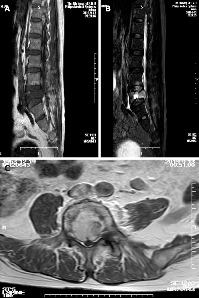
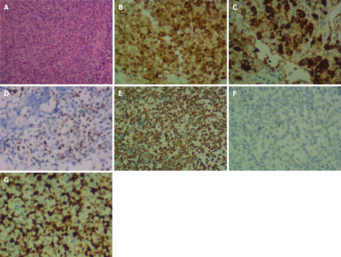
Case summary: We present a rare case in which the primary intramedullary melanoma was located in the lumbar spine. A 56-year-old man complained of progressive intermittent pain in the lumbar area. Thoracic magnetic resonance imaging showed a spinal intramedullary tumor between the L3 and S1 levels. The tumor was resected entirely, and the diagnosis of malignant melanoma was confirmed by histopathology.
Conclusion: Primary melanoma of the spinal cord, particularly intramedullary localization, has rarely been reported in the previous literature. We describe a primary malignant melanoma of the lumbar spinal cord and discuss the challenges associated with the diagnosis.
Keywords: Case report; Histopathological examination; Immunohistochemical staining; Intramedullary melanoma; Lumbar; Magnetic resonance imaging; Spinal cord.
©The Author(s) 2021. Published by Baishideng Publishing Group Inc. All rights reserved.
Conflict of interest statement
Conflict-of-interest statement: The authors have no conflicts of interest to report.
Figures
Similar articles
-
An uncommon intramedullary tumor: primary spinal cord melanoma.Asian Spine J. 2014 Aug;8(4):512-5. doi: 10.4184/asj.2014.8.4.512. Epub 2014 Aug 19. Asian Spine J. 2014. PMID: 25187871 Free PMC article.
-
Primary intramedullary malignant melanoma: can imaging lead to the correct diagnosis?J Int Med Res. 2020 Oct;48(10):300060520966152. doi: 10.1177/0300060520966152. J Int Med Res. 2020. PMID: 33103574 Free PMC article.
-
Easily misdiagnosed delayed metastatic intraspinal extradural melanoma of the lumbar spine: A case report and review of the literature.Oncol Lett. 2013 Jun;5(6):1799-1802. doi: 10.3892/ol.2013.1299. Epub 2013 Apr 10. Oncol Lett. 2013. PMID: 23833644 Free PMC article.
-
Intramedullary spinal cord metastases of malignant melanoma: an autopsy case report and review of the literature.Clin Neuropathol. 2010 Sep-Oct;29(5):334-40. doi: 10.5414/npp29334. Clin Neuropathol. 2010. PMID: 20860898 Review.
-
Primary spinal melanoma: a case report and literature review.Chin Med J (Engl). 2012 Nov;125(22):4138-41. Chin Med J (Engl). 2012. PMID: 23158158 Review.
Cited by
-
Intramedullary primary spinal cord melanoma: illustrative case.J Neurosurg Case Lessons. 2025 Mar 10;9(10):CASE24732. doi: 10.3171/CASE24732. Print 2025 Mar 10. J Neurosurg Case Lessons. 2025. PMID: 40063999 Free PMC article.
-
Primary thoracolumbar intraspinal malignant melanoma: A case report.World J Clin Cases. 2024 Jun 6;12(16):2904-2910. doi: 10.12998/wjcc.v12.i16.2904. World J Clin Cases. 2024. PMID: 38899297 Free PMC article.
References
-
- Liubinas SV, Maartens N, Drummond KJ. Primary melanocytic neoplasms of the central nervous system. J Clin Neurosci. 2010;17:1227–1232. - PubMed
Publication types
LinkOut - more resources
Full Text Sources
Other Literature Sources
