. 2021 Mar 29:21:399-412.
doi: 10.1016/j.omtm.2021.03.020. eCollection 2021 Jun 11.

Improvement of HSV-1 based amplicon vectors for a safe and long-lasting gene therapy in non-replicating cells

Affiliations

Improvement of HSV-1 based amplicon vectors for a safe and long-lasting gene therapy in non-replicating cells

Marie Soukupová et al. Mol Ther Methods Clin Dev. .

Abstract

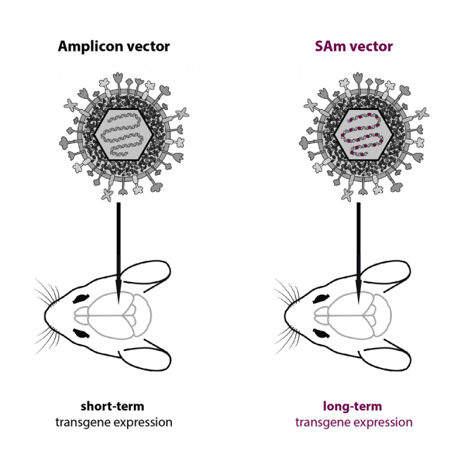
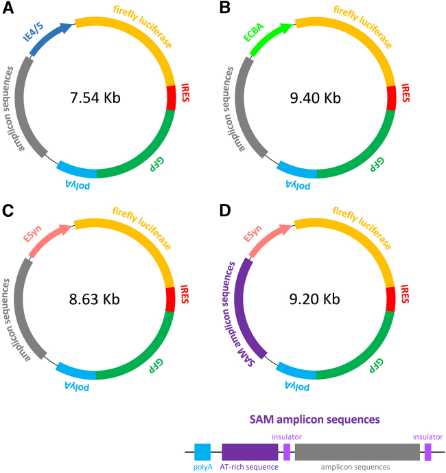
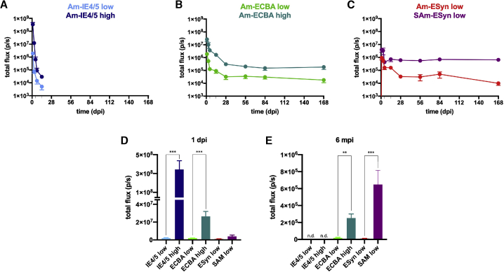
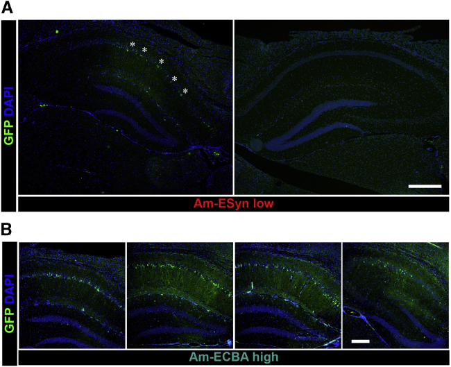
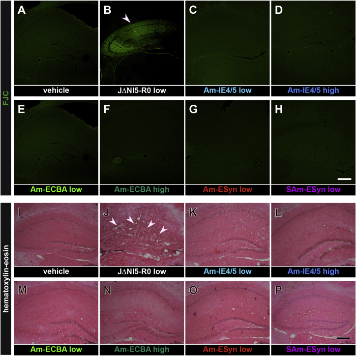
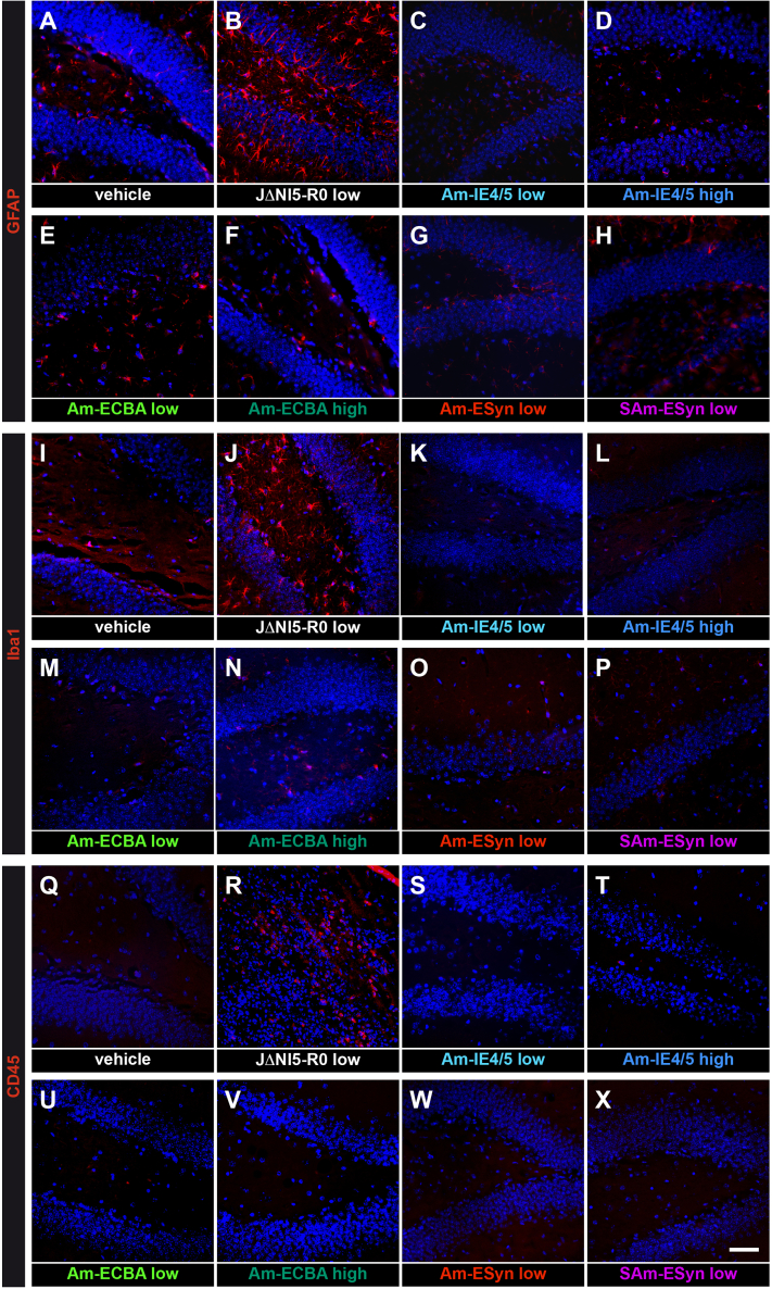
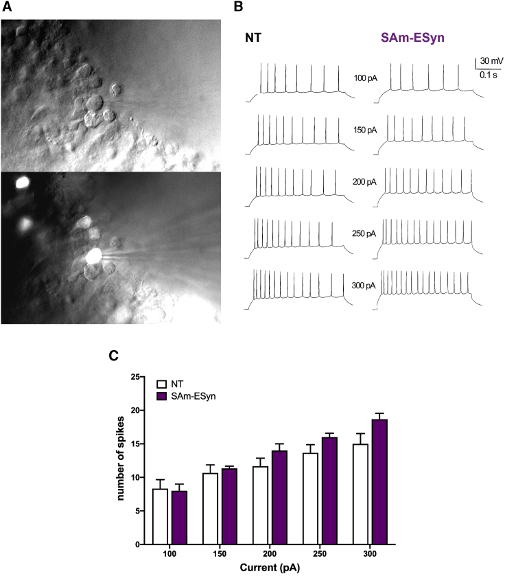
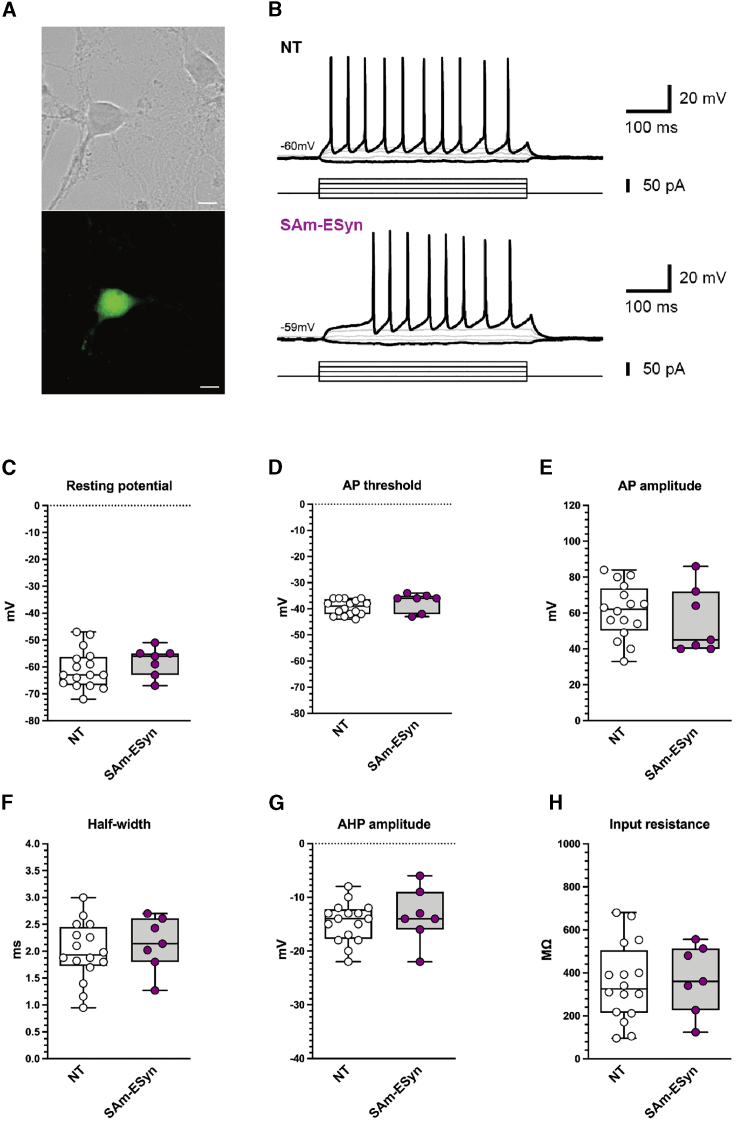
A key factor for developing gene therapy strategies for neurological disorders is the availability of suitable vectors. Currently, the most advanced are adeno-associated vectors that, while being safe and ensuring long-lasting transgene expression, have a very limited cargo capacity. In contrast, herpes simplex virus-based amplicon vectors can host huge amounts of foreign DNA, but concerns exist about their safety and ability to express transgenes long-term. We aimed at modulating and prolonging amplicon-induced transgene expression kinetics in vivo using different promoters and preventing transgene silencing. To pursue the latter, we deleted bacterial DNA sequences derived from vector construction and shielded the transgene cassette using AT-rich and insulator-like sequences (SAm technology). We employed luciferase and GFP as reporter genes. To determine transgene expression kinetics, we injected vectors in the hippocampus of mice that were longitudinally scanned for bioluminescence for 6 months. To evaluate safety, we analyzed multiple markers of damage and performed patch clamp electrophysiology experiments. All vectors proved safe, and we managed to modulate the duration of transgene expression, up to obtaining a stable, long-lasting expression using the SAm technology. Therefore, these amplicon vectors represent a flexible, efficient, and safe tool for gene delivery in the brain.

Keywords: Central Nervous System; amplicon vector; herpes simplex virus; hippocampus.


Conflict of interest statement

The authors declare no competing interests.

Figures

None
Graphical abstract
Figure 1
Figure 1
Structure of the plasmids used for construction of amplicon vectors Genes encoding two reporter products, firefly luciferase (Luc) and green fluorescence protein (GFP), are linked with an internal ribosomal entry site (IRES) element, and the GFP gene is followed by a poly(A) tail. (A–D) Different promotor sequences were placed ahead of Luc to regulate transcription: the IE4/5 promoter in the pAm-IE4/5-luciferase-GFP plasmid (A); the cytomegalovirus enhancer chicken-β-actin (ECBA) promoter in the pAm-ECBA-luciferase-GFP plasmid (B); the neuron-specific enhanced synapsin (ESyn) promoter in the pAm-ESyn-luciferase-GFP plasmid (C), and the pSAm-ESyn-luciferase-GFP plasmid (D). The size of each plasmid is reported in the respective scheme. Based on these sizes and considering that each plasmid will produce a concatemer closely matching the size of the HSV genome (about 150 kb), it can be estimated that pAm-IE4/5-LiG2 will be represented 20 times in the respective amplicon vector, pAm-ECBA-LiG2 16 times, pAm-ESyn-LiG2 17 times, and pSAm-ESyn-LiG2 16 times. “Steady” amplicon (SAm) sequences, i.e., insulator sequences of about 0.5 Kb and an approximately 2 Kb AT-rich sequence, were cloned in the pSAm-ESyn-luciferase-GFP plasmid (D). Amplicons vectors were assembled from these plasmids as described in the Materials and methods.
Figure 2
Figure 2
Bioluminescence (BLI) imaging experimental plan (Top) Experimental flow chart illustrating the time points when BLI imaging was longitudinally performed. (Bottom) Representative BLI images of the head of mice injected with the two vectors in which transgenes are driven by the ESyn promoter (vAm-ESyn-LiG2, vSAm-ESyn-LiG2). Animals were anesthetized and BLI scanned 15–20 min after i.p. injection of D-luciferin (150 mg/kg).
Figure 3
Figure 3
Kinetics of luciferase expression in animals injected with the different amplicon vectors (A–C) Time course of the BLI signal in mice injected with low titer (light blue) or high titer (dark blue) vAm-IE4/5-LiG2 (A); low titer (light green) or high titer (dark green) vAm-ECBA-LiG2 (B); low titer vAm-ESyn-LiG2 (red) or low titer vSAm-ESyn-LiG2 (purple) (C). (D and E) A comparison of the BLI signals produced by injection of the different amplicon vectors at the earliest (1 dpi, D) and latest (6 mpi, E) time points. 14 animals were injected with either low or high titer vAm-IE4/5-LiG2 vector, of which 8 were killed at 4 dpi, and 6 at 14 dpi. 28 animals per group were injected with other vectors, of which 16 were killed after at 4 dpi, 6 at 2 mpi, and 6 at 6 mpi. Therefore, data in (D) are the means ± SEM of 14 low or high titer vAm-IE4/5-LiG2 injected-mice and 28 animals of the other groups; data in (E) are the means ± SEM of 6 animals per group (but vAm-IE4/5-LiG2 injected-mice were not determined, n.d.). ∗∗p < 0.01; ∗∗∗p < 0.001; Mann-Whitney U test.
Figure 4
Figure 4
GFP expression and vector diffusion in the vAm-ESyn-LiG2 injected mouse hippocampus, at 4 dpi (A) Representative coronal section of a mouse hippocampus of low titer vAm-ESyn-LiG2, near the level of amplicon vector injection. Note GFP-positive pyramidal cells in the injected hippocampus (asterisks), and absence of GFP signal in the contralateral hippocampus. (B) Representative images of serial coronal sections immunostained for GFP (green) and counterstained with DAPI (blue) covering about 900 μm (anterior to posterior) of an injected hippocampus, across the site of inoculation of high titer vAm-ECBA-LiG2. Horizontal bar in (A), 500 μm; horizontal bar in (B), 250 μm.
Figure 5
Figure 5
Absence of detectable neurodegeneration and alterations in cytoarchitecture in the hippocampus of amplicon-injected animals (A–H) Dorsal hippocampal, FJC-stained sections prepared from a negative control, vehicle-injected (A), a positive control, JΔNI R0 injected (B), and amplicon vector injected animals (C–H, as indicated), at 4 dpi. Note numerous FCJ-positive in the positive control, but not in other groups. (I–P) As above, hematoxylin and eosin stained. Obvious cell loss, cytoarchitecture damage, and cell infiltration in (J), but not in other groups. These images are representative of 5 section per animal, 6 animals per group except 4 animals per group in (C), (D) (K), and (L). Horizontal bar in (H) (for A–H panels), 500 μm; horizontal bar in (P) (for I–P panels), 320 μm.
Figure 6
Figure 6
Absence of detectable neuroinflammatory reaction in the hippocampus of amplicon-injected animals (A–H) Dorsal hippocampal, GFAP-stained sections prepared from a negative control, vehicle-injected (A), a positive control, JΔNI R0 injected (B), and various amplicon vector injected animals (C–H, as indicated), at 4 dpi. Note numerous GFAP-positive cells in the positive control, but not in other groups. (I–P) As above, IBA-1-stained. Note numerous IBA-1-positive cells in the positive control, but not in other groups. (Q–X) As above, CD45-stained. Again, note numerous CD45-positive cells in the positive control, but not in other groups. These images are representative of 5 section per animal, 6 animals per group except 4 animals per group in (C), (D), (K), (L), (S), and (T). Horizontal bar in (X) (for all panels), 70 μm.
Figure 7
Figure 7
Whole-cell patch clamp recordings in pyramidal neurons from hippocampal slices (A) Representative image of GFP-expressing neurons from a hippocampal slice prepared 4 days after inoculation of the vSAm-ESyn-LiG2 amplicon vector (top, brightfield image; bottom, GFP fluorescence overlaid on the brightfield image). The recording pipette can be seen in the bottom image. (B) Representative traces from a naive, not transduced (NT) pyramidal neuron and a pyramidal neuron infected by vSAm-ESyn-LiG2, showing the AP firing pattern as response to square-wave depolarizing current injections (100 to 300 nA). (C) Number of APs fired in response to current injected steps. Data are the means ± SEM of 4 animals per group (value for each animal was the average of 2–3 cells).
Figure 8
Figure 8
Whole-cell patch clamp recordings from primary hippocampal neuronal cultures (A) Representative image of native GFP signal in a vSAm-ESyn-LiG2 infected hippocampal neuron. Scale bar, 10 μm. (B) Current-clamp recordings of a hippocampal neuron showing voltage response to step current injections at various amplitudes in mock (NT) and vSAm-ESyn-LiG2 infected cells. (C–H) Basic electrophysiological parameters of mock (white circles; n = 16) and vSAm-ESyn-LiG2 infected neurons (purple circles; n = 7). AP, action potential; AHP, afterhyperpolarization.

Similar articles

Cited by

References

    1. Simonato M., Manservigi R., Marconi P., Glorioso J. Gene transfer into neurones for the molecular analysis of behaviour: focus on herpes simplex vectors. Trends Neurosci. 2000;23:183–190. - PubMed
    1. Simonato M., Bennett J., Boulis N.M., Castro M.G., Fink D.J., Goins W.F., Gray S.J., Lowenstein P.R., Vandenberghe L.H., Wilson T.J. Progress in gene therapy for neurological disorders. Nat. Rev. Neurol. 2013;9:277–291. - PMC - PubMed
    1. Chen S.H., Haam J., Walker M., Scappini E., Naughton J., Martin N.P. Recombinant Viral Vectors as Neuroscience Tools. Curr. Protoc. Neurosci. 2019;87:e67. - PMC - PubMed
    1. Choudhury S.R., Hudry E., Maguire C.A., Sena-Esteves M., Breakefield X.O., Grandi P. Viral vectors for therapy of neurologic diseases. Neuropharmacology. 2017;120:63–80. - PMC - PubMed
    1. Duan D. Systemic delivery of adeno-associated viral vectors. Curr. Opin. Virol. 2016;21:16–25. - PMC - PubMed

LinkOut - more resources