

Primary evisceration for neonatal endogenous endophthalmitis: A report of two cases
- PMID: 33869894
- PMCID: PMC8042423
- DOI: 10.1016/j.ajoc.2021.101081
Primary evisceration for neonatal endogenous endophthalmitis: A report of two cases
Abstract
Purpose: To present two cases of neonatal endophthalmitis with poor prognosis that were managed with primary evisceration.
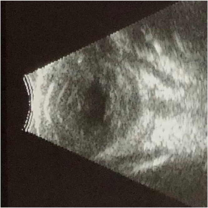
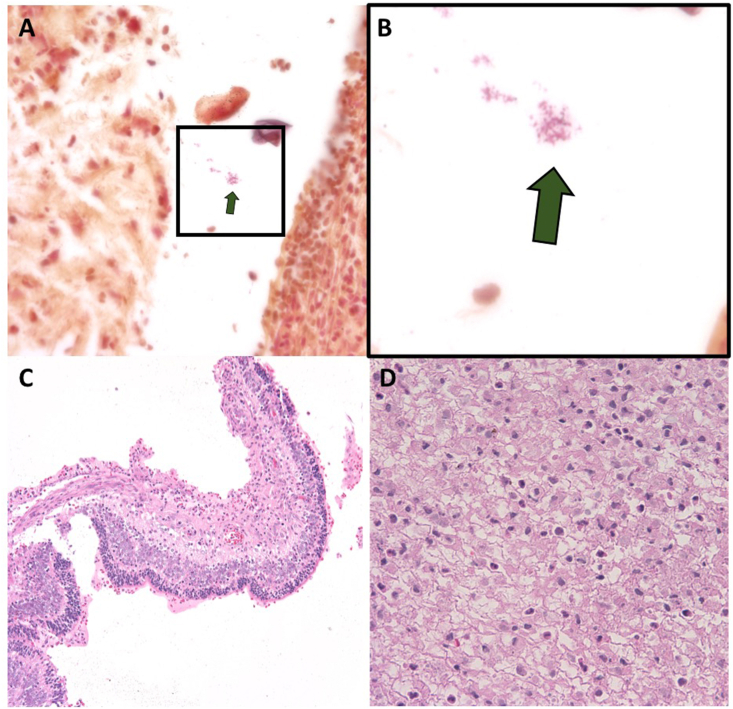
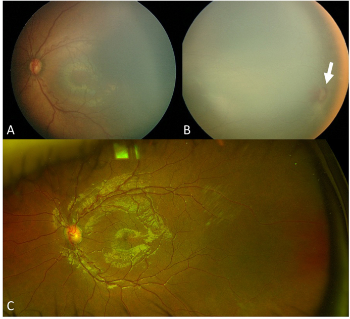
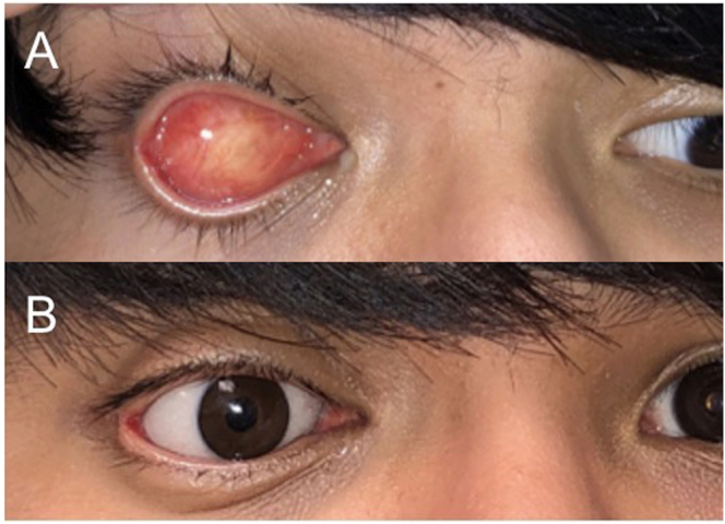
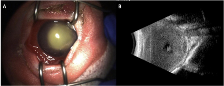
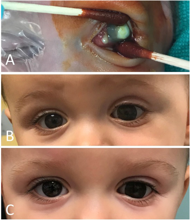
Observations: Case 1 is a 27-weeks' gestation neonate who developed Pseudomonas aeruginosa endophthalmitis complicated by globe rupture. Case 2 describes a 34-weeks' gestation neonate with Serratia marcescens endophthalmitis. Both patients had poor prognosis and thus underwent primary evisceration with good long-term cosmetic outcomes at 15 years and 17 months, respectively.
Conclusions and importance: Primary evisceration should be considered in neonates with endophthalmitis with a poor prognosis and can result in good long-term cosmesis.
Keywords: Endophthalmitis; Evisceration; Oculoplastics; Retina.
© 2021 The Authors.
Conflict of interest statement
The authors have no relevant financial disclosures to report.
Figures
Similar articles
-
The results of evisceration with primary porous implant placement in patients with endophthalmitis.Korean J Ophthalmol. 2010 Oct;24(5):279-83. doi: 10.3341/kjo.2010.24.5.279. Epub 2010 Oct 5. Korean J Ophthalmol. 2010. PMID: 21052507 Free PMC article.
-
Primary implant placement with evisceration in patients with endophthalmitis.Ophthalmology. 2000 Sep;107(9):1661-4; discussion 1664-5. doi: 10.1016/s0161-6420(00)00262-1. Ophthalmology. 2000. PMID: 10964824
-
Primary implant placement with evisceration in patients with endophthalmitis.Am J Ophthalmol. 2007 May;143(5):902-4. doi: 10.1016/j.ajo.2006.11.029. Epub 2006 Dec 20. Am J Ophthalmol. 2007. PMID: 17452189
-
Endogenous Serratia marcescens endophthalmitis with dark hypopyon: case report and review.Surv Ophthalmol. 2001 Nov-Dec;46(3):259-68. doi: 10.1016/s0039-6257(01)00263-6. Surv Ophthalmol. 2001. PMID: 11738433 Review.
-
Outcomes of orbital implants after evisceration and enucleation in patients with endophthalmitis.Curr Opin Ophthalmol. 2010 Sep;21(5):375-9. doi: 10.1097/ICU.0b013e32833b7a56. Curr Opin Ophthalmol. 2010. PMID: 20489621 Review.
Cited by
-
Neonatal Endogenous Endophthalmitis: A Case Report.Cureus. 2022 Feb 15;14(2):e22256. doi: 10.7759/cureus.22256. eCollection 2022 Feb. Cureus. 2022. PMID: 35228981 Free PMC article.
References
-
- Jalali S., Pehere N., Rani P.K. Treatment outcomes and clinicomicrobiological characteristics of a protocol-based approach for neonatal endogenous endophthalmitis. Eur J Ophthalmol. 2014;24:424–436. - PubMed
-
- Jackson T.L., Eykyn S.J., Graham E.M., Stanford M.R. Endogenous bacterial endophthalmitis: a 17-year prospective series and review of 267 reported cases. Surv Ophthalmol. 2003;48:403–423. - PubMed
-
- Stensvold H.J., Klingenberg C., Stoen R. Neonatal morbidity and 1-year survival of extremely preterm infants. Pediatrics. 2017;139 - PubMed
Publication types
LinkOut - more resources
Full Text Sources
Other Literature Sources
