. 2021 Jan 27;3(3):100250.
doi: 10.1016/j.jhepr.2021.100250. eCollection 2021 Jun.

Genetic or pharmacological reduction of cholangiocyte senescence improves inflammation and fibrosis in the Mdr2 -/- mouse

Affiliations

Genetic or pharmacological reduction of cholangiocyte senescence improves inflammation and fibrosis in the Mdr2 -/- mouse

Mohammed Alsuraih et al. JHEP Rep. .

Abstract

Background & aims: Cholangiocyte senescence is important in the pathogenesis of primary sclerosing cholangitis (PSC). We found that CDKN2A (p16), a cyclin-dependent kinase inhibitor and mediator of senescence, was increased in cholangiocytes of patients with PSC and from a PSC mouse model (multidrug resistance 2; Mdr2 -/-). Given that recent data suggest that a reduction of senescent cells is beneficial in different diseases, we hypothesised that inhibition of cholangiocyte senescence would ameliorate disease in Mdr2 -/- mice.

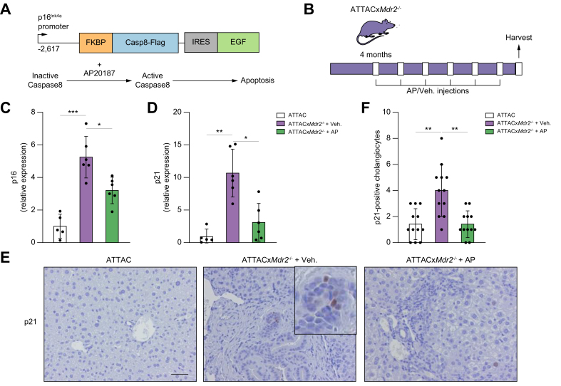
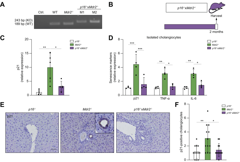
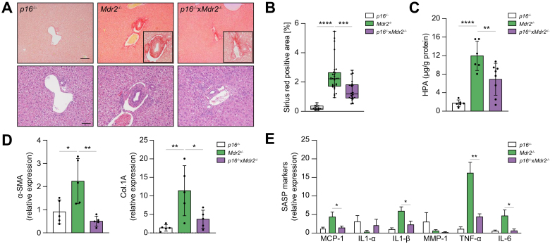
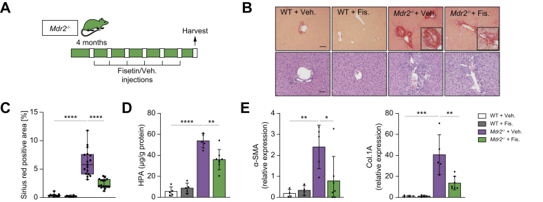
Methods: We used 2 novel genetic murine models to reduce cholangiocyte senescence: (i) p16Ink4a apoptosis through targeted activation of caspase (INK-ATTAC)xMdr2 -/-, in which the dimerizing molecule AP20187 promotes selective apoptotic removal of p16-expressing cells; and (ii) mice deficient in both p16 and Mdr2. Mdr2 -/- mice were also treated with fisetin, a flavonoid molecule that selectively kills senescent cells. p16, p21, and inflammatory markers (tumour necrosis factor [TNF]-α, IL-1β, and monocyte chemoattractant protein-1 [MCP-1]) were measured by PCR, and hepatic fibrosis via a hydroxyproline assay and Sirius red staining.

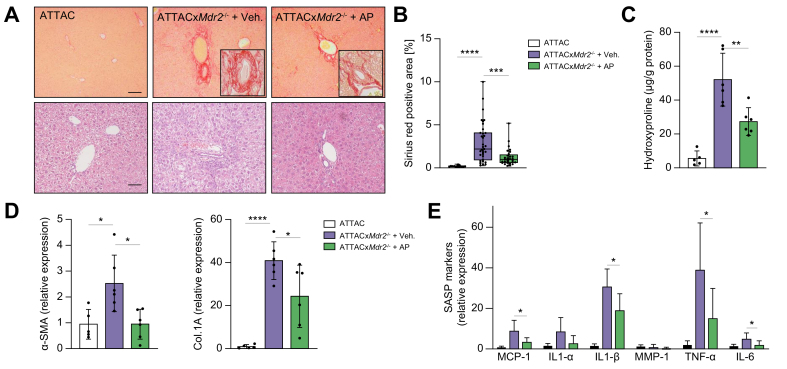
Results: AP20187 treatment reduced p16 and p21 expression by ~35% and ~70% (p >0.05), respectively. Expression of inflammatory markers (TNF-α, IL-1β, and MCP-1) decreased (by 60%, 40%, and 60%, respectively), and fibrosis was reduced by ~60% (p >0.05). Similarly, p16 -/- xMdr2 -/- mice exhibited reduced p21 expression (70%), decreased expression of TNF-α, IL-1β (60%), and MCP-1 (65%) and reduced fibrosis (~50%) (p >0.05) compared with Mdr2 -/- mice. Fisetin treatment reduced expression of p16 and p21 (80% and 90%, respectively), TNF-α (50%), IL-1β (50%), MCP-1 (70%), and fibrosis (60%) (p >0.05).

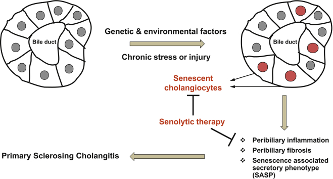
Conclusions: Our data support a pathophysiological role of cholangiocyte senescence in the progression of PSC, and that targeted removal of senescent cholangiocytes is a plausible therapeutic approach.

Lay summary: Primary sclerosing cholangitis is a fibroinflammatory, incurable biliary disease. We previously reported that biliary epithelial cell senescence (cell-cycle arrest and hypersecretion of profibrotic molecules) is an important phenotype in primary sclerosing cholangitis. Herein, we demonstrate that reducing the number of senescent cholangiocytes leads to a reduction in the expression of inflammatory, fibrotic, and senescence markers associated with the disease.

Keywords: ALP, alkaline phosphatase; AP, AP20187; Apoptosis resistance; BCL2, B cell lymphoma 2; Bcl-xL, B-cell lymphoma-extra large; Biliary epithelial cell; CCA, cholangiocarcinoma; CKI, cyclin-dependent kinase inhibitor; Cellular senescence; Cholestatic liver disease; Col.1A, collagen 1A; D, dasatinib; EVs, extracellular vesicles; FKBP-Casp8, FK506-binding-protein-caspase 8; IF, immunofluorescence; INK-ATTAC, p16Ink4a apoptosis through targeted activation of caspase; IR, irradiation; MCL1, myeloid cell leukemia 1; MCP-1, monocyte chemoattractant protein-1; MMP, matrix metalloproteinase; NHC, normal human cholangiocyte; PSC, primary sclerosing cholangitis; Primary sclerosing cholangitis; Q, quercetin; RT, reverse transcription; SA-β-gal, senescence-associated β-gal; SASP, senescence-associated secretory phenotype; Senescence-associated secretory phenotype; Senolytics; TNF, tumour necrosis factor; WT, wild-type; mdr2, multidrug-resistance 2; qPCR, quantitative PCR; α-SMA, α-smooth muscle actin; β-Gal, β-galactosidase.


Conflict of interest statement

The authors have no conflict of interest related to the manuscript. Please refer to the accompanying ICMJE disclosure forms for further details.

Figures

None
Graphical abstract
Fig. 1
Fig. 1
Genetic deletion of senescent cholangiocytes in the Mdr2-/- mouse model of PSC. (A) Schematic diagram of the INK-ATTAC suicide gene. AP, a synthetic drug that induces membrane-bound FKBP-Casp8 dimerization, resulted in apoptosis in cells expressing p16Ink4A. (B) Schematic of the experimental design. Crossbred ATTACxMdr2-/- mice at 4 months of age received AP and vehicle injections every 7 days. (C,D) p16 and p21 mRNA expression in whole-liver tissue, as assessed by RT-PCR, were significantly decreased in ATTACxMdr2-/- mice following AP treatment. (E) Representative images of p21 immunohistochemistry of WT (left), ATTACxMdr2-/- vehicle-treated (middle), and ATTACxMdr2-/- AP-treated (right) mice. Magnification: 20×. Thus, AP treatment reduced the number of p21-positive cholangiocytes. (F) Quantification of p21-positive cholangiocytes. Each dot represents an image with at least 1 bile duct. Bars represent mean ± SD; n = 5–7. Scale bar: 50 μm (E). ∗p <0.05, ∗∗p <0.001, ∗∗∗p <0.0001 (Tukey’s multiple comparisons test). AP, AP20187; FKBP-Casp8, FK506-binding-protein-caspase 8; (INK-)ATTAC, p16Ink4a apoptosis through targeted activation of caspase; Mdr2, multidrug-resistance 2; PSC, primary sclerosing cholangitis; RT, reverse transcription; WT, wild-type.
Fig. 2
Fig. 2
Genetic deletion of senescent cholangiocytes decreases inflammatory and fibrotic markers. (A) Representative images of Picrosirius-red-stained liver sections (magnification: 10×) showing deposition of collagen (upper panel) and H&E-stained liver sections (magnification: 20×) showing generalised visualisation of liver parenchyma (lower panel) from WT mice (left), ATTACxMdr2-/- vehicle-treated mice (middle), and ATTACxMdr2-/- AP-treated mice (right). Based on histopathological evaluation of Picrosirius-red-stained liver sections, a significant reduction in fibrosis was observed in the ATTACxMdr2-/- AP-treated mice. (B) Quantification of Picrosirius-red-stained liver sections; data presented as the percentage of red stain-positive areas. Each dot represents an image with at least 1 bile duct. (C) Biochemical assessment of hepatic fibrosis confirmed marked decreased hydroxyproline concentration upon AP treatment. (D,E) mRNA expression of fibrotic markers (α-SMA and Col.1A) and SASP markers (MCP-1, IL-1α, IL-1β, MMP-1, TNF-α, and IL-6) in whole-liver tissue was significantly reduced, except those marked ‘n.s.’, in the ATTACxMdr2-/- AP-treated mice, as assessed by RT-PCR. Bars represent mean ± SD; n = 5–7. Scale bars: 100 and 50 μm (A). ∗p <0.05, ∗∗p <0.01, ∗∗∗p <0.001, ∗∗∗∗p <0.0001 (Tukey’s multiple comparisons test). α-SMA, α-smooth muscle actin; AP, AP20187; Col.1A, collagen 1A; (INK-)ATTAC, p16Ink4a apoptosis through targeted activation of caspase; MCP-1, monocyte chemoattractant protein-1; Mdr2, multidrug-resistance 2; MMP-1, matrix metalloproteinase 1; RT, reverse transcription; SASP, senescence-associated secretory phenotype; TNF, tumour necrosis factor; WT, wild-type.
Fig. 3
Fig. 3
Genetic reduction of p16 reduces senescence markers in the Mdr2-/- mouse. (A) PCR genotyping showing the p16-/- DNA construct. (KO) band represents the neomycin-resistance cassette on exon-1α of the Ink4a/Arf/Ink4b locus. (B) Schematic of the experimental design. P16-/-, Mdr2-/-, and crossbred p16-/-xMdr2-/- mice were harvested at 2 months of age. (C) Hepatic p21 mRNA expression, as assessed by RT-PCR, was reduced in p16-/-xMdr2-/- mice. (D) mRNA expression of senescence markers in isolated cholangiocytes, as assessed by RT-PCR, was reduced in p16-/-xMdr2-/- mice. (E) Representative images of p21 immunohistochemistry of p16-/- (left), Mdr2-/- (middle), and p16-/-xMdr2-/- mice (right) (magnification: 20×). A significant reduction in the number of p21-positive cholangiocytes was observed in p16-/-xMdr2-/- mice compared with the control group. (F) Quantification of p21-positive cholangiocytes. Each dot represents an image with at least 1 bile duct. Bars represent mean ± SD; n = 5–7. Scale bar: 50 μm (D). ∗p <0.05, ∗∗p <0.01, (Tukey’s multiple comparisons test). Mdr2, multidrug-resistance 2; RT, reverse transcription; TNF, tumour necrosis factor; WT, wild-type.
Fig. 4
Fig. 4
Genetic reduction of p16 decreases hepatic inflammation and fibrosis. (A) Representative images of Picrosirius-red-stained liver sections (magnification: 10×) showing deposition of collagen (upper panel) and H&E-stained liver sections (magnification: 20×) showing generalized visualization of liver parenchyma (lower panel) of p16-/- (left), Mdr2-/- (middle), and p16-/-xMdr2-/- mice (right) (magnification: 20×). p16-/-xMdr2-/- mice exhibited a marked reduction in hepatic fibrosis. (B) Quantification of Picrosirius-red-stained liver sections; data presented as % red stain positive area. Each dot represents an image with at least 1 bile duct. (C) Biochemical assessment of hepatic fibrosis confirmed a markedly decreased hydroxyproline concentration in p16-/-xMdr2-/- mice. (D,E) Analysis of multiple fibrotic (a-SMA and Col.1A) and SASP genes (MCP1, IL1a, IL1b, MMP1, TNFa, and IL6) by RT-PCR showing significant reduction, except those marked ‘n.s.’, in whole-liver tissue from p16-/-xMdr2-/- mice. Bars represent mean ± SD; n = 5–7. Scale bars: 100 and 50 μm (A). ∗p <0.05, ∗∗p <0.01, ∗∗∗p <0.001, ∗∗∗∗p <0.0001 (Tukey’s multiple comparisons test). a-SMA, α-smooth muscle actin; Col.1A, collagen 1A; MCP1, monocyte chemoattractant protein-1; Mdr2, multidrug-resistance 2; MMP-1, matrix metalloproteinase 1; RT, reverse transcription; SASP, senescence-associated secretory phenotype; TNF, tumour necrosis factor; WT, wild-type.
Fig. 5
Fig. 5
Fisetin targets senescent cholangiocytes in vitro. (A) Senolytic compounds at various concentrations were applied to proliferating and senescent cholangiocytes (induced experimentally by irradiation). Fisetin and A1331 significantly reduced the number of senescent cholangiocytes as measured by Crystal violet. Bars represent mean ± SEM; n = 4. (B,C) Fisetin and A1331 induced apoptosis in senescent cholangiocytes. (D) Fisetin did not induce apoptosis in non-proliferating or non-senescent cholangiocytes. Proliferating and senescent cholangiocytes were treated with fisetin and A1331, respectively for 12 h and Caspase-3/7 activity was measured using a luminescent substrate. (F) Fisetin did not induce apoptosis in isolated hepatocytes from WT or Mdr2-/- mice. Data represent mean ± SD; n = 4 at each concentration. ∗p <0.01 (Student’s t test). Mdr2, multidrug-resistance 2; NHC, normal human cholangiocyte; WT, wild-type.
Fig. 6
Fig. 6
Fisetin-treatment reduces inflammation and fibrosis in the Mdr2-/- mouse. (A) Schematic of the experimental design. WT and Mdr2-/- mice were gavaged with fisetin and vehicle every 7 days for 2 months. (B) Representative images of Picrosirius-red-stained liver sections (10×) showing deposition of collagen (upper panel) and haematoxylin and eosin-stained liver sections (20×) showing generalized visualization of liver parenchyma (lower panel) of WT vehicle and fisetin-treated mice, and vehicle and fisetin-treated Mdr2-/- mice (20×). Based on histopathological evaluation of picrosirius-stained liver sections, a significant reduction was observed in fisetin-treated Mdr2-/- mice. (C) Quantification of Picrosirius-red-stained liver sections; data presented as the percentage of red stain-positive areas. Each dot represents an image with at least 1 bile duct. (D) Biochemical assessment of hepatic fibrosis confirmed the markedly decreased hydroxyproline concentration upon fisetin treatment. (E) Fibrotic marker assessment. The mRNA expression of α-SMA and Col.1A was reduced following fisetin treatment. Bars represent mean ± SD; n = 5–7. Scale bars: 100 and 50 μm (A). ∗p <0.05, ∗∗p <0.01, ∗∗∗p <0.001, ∗∗∗∗p <0.0001 (Tukey’s multiple comparisons test). α-SMA, α-smooth muscle actin; Col.1A, collagen 1A; HPA, hydroxyproline; Mdr2, multidrug-resistance 2; WT, wild-type.
Fig. 7
Fig. 7
Fisetin treatment decreases senescence markers in the liver of the Mdr2-/- mouse. (A) Representative images of p21 immunohistochemistry of WT vehicle and fisetin-treated Mdr2-/- vehicle and fisetin-treated mice (magnification: 20×). (B) Quantification of p21-positive cholangiocytes. Each dot represents an image with at least 1 bile duct. (C) mRNA expression of the cellular senescence mediators, p16 and p21, was decreased significantly following fisetin treatment. (D) Gene expression analysis of various SASP genes by RT-PCR showing a significant decrease, except those marked by ‘n.s.’, in whole-liver tissue of fisetin-treated Mdr2-/-mice. (E) mRNA expression of senescence markers in sampled cholangiocytes, as assessed by RT-PCR, was reduced in Mdr2-/- mice treated with fisetin. Bars represent mean ± SD; n = 5–7. Scale bar: 50 μm (A). ∗p <0.05, ∗∗p <0.001, ∗∗∗p <0.0001 (Tukey’s multiple comparisons test). MCP-1, monocyte chemoattractant protein-1; Mdr2, multidrug-resistance 2; MMP-1, matrix metalloproteinase 1; RT, reverse transcription; SASP, senescence-associated secretory phenotype; TNF, tumour necrosis factor; WT, wild-type.

References

    1. Lazaridis K.N., LaRusso N.F. Primary sclerosing cholangitis. N Engl J Med. 2016;375:1161–1170. - PMC - PubMed
    1. Karlsen T.H., Folseraas T., Thorburn D., Vesterhus M. Primary sclerosing cholangitis - a comprehensive review. J Hepatol. 2017;67:1298–1323. - PubMed
    1. Hirschfield G.M., Karlsen T.H., Lindor K.D., Adams D.H. Primary sclerosing cholangitis. Lancet. 2013;382:1587–1599. - PubMed
    1. Tabibian J.H., O'Hara S.P., Splinter P.L., Trussoni C.E., LaRusso N.F. Cholangiocyte senescence by way of N-ras activation is a characteristic of primary sclerosing cholangitis. Hepatology. 2014;59:2263–2275. - PMC - PubMed
    1. Tabibian J.H., Trussoni C.E., O'Hara S.P., Splinter P.L., Heimbach J.K., LaRusso N.F. Characterization of cultured cholangiocytes isolated from livers of patients with primary sclerosing cholangitis. Lab Invest. 2014;94:1126–1133. - PMC - PubMed