. 2020 Nov:1:100015.
doi: 10.1016/j.brain.2020.100015. Epub 2020 Sep 3.

In vivo estimates of axonal stretch and 3D brain deformation during mild head impact

Affiliations

In vivo estimates of axonal stretch and 3D brain deformation during mild head impact

Andrew K Knutsen et al. Brain Multiphys. 2020 Nov.

Abstract

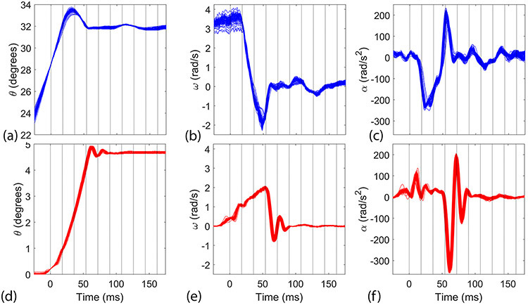
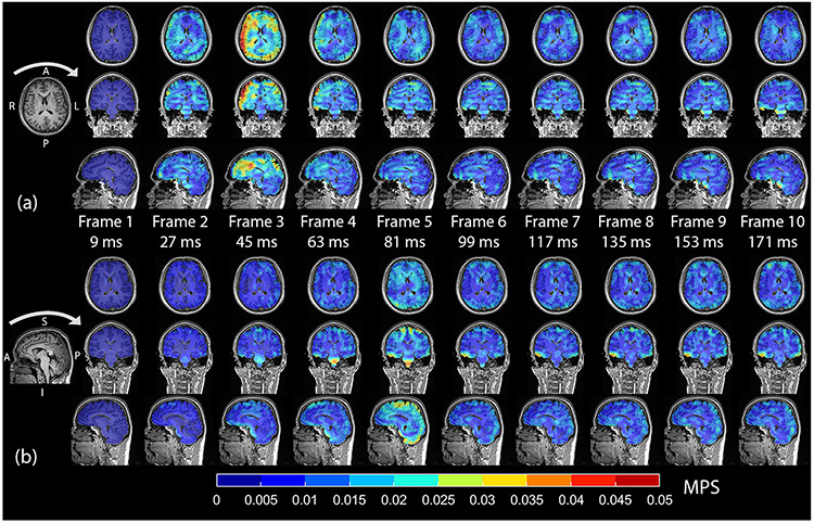
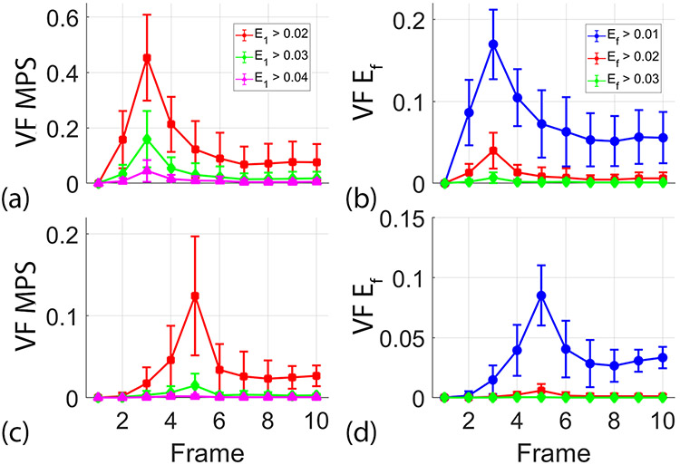
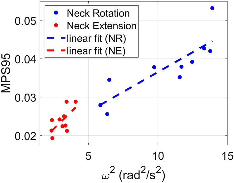
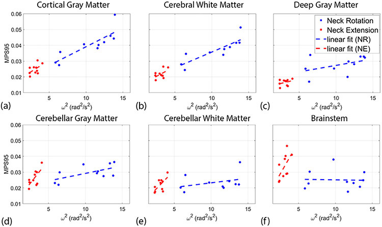
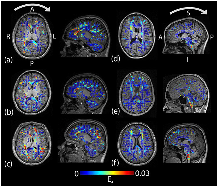
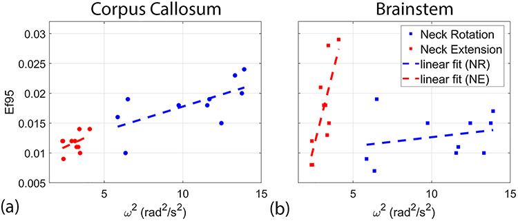
The rapid deformation of brain tissue in response to head impact can lead to traumatic brain injury. In vivo measurements of brain deformation during non-injurious head impacts are necessary to understand the underlying mechanisms of traumatic brain injury and compare to computational models of brain biomechanics. Using tagged magnetic resonance imaging (MRI), we obtained measurements of three-dimensional strain tensors that resulted from a mild head impact after neck rotation or neck extension. Measurements of maximum principal strain (MPS) peaked shortly after impact, with maximal values of 0.019-0.053 that correlated strongly with peak angular velocity. Subject-specific patterns of MPS were spatially heterogeneous and consistent across subjects for the same motion, though regions of high deformation differed between motions. The largest MPS values were seen in the cortical gray matter and cerebral white matter for neck rotation and the brainstem and cerebellum for neck extension. Axonal fiber strain (Ef) was estimated by combining the strain tensor with diffusion tensor imaging data. As with MPS, patterns of Ef varied spatially within subjects, were similar across subjects within each motion, and showed group differences between motions. Values were highest and most strongly correlated with peak angular velocity in the corpus callosum for neck rotation and in the brainstem for neck extension. The different patterns of brain deformation between head motions highlight potential areas of greater risk of injury between motions at higher loading conditions. Additionally, these experimental measurements can be directly compared to predictions of generic or subject-specific computational models of traumatic brain injury.

Keywords: axonal strain; brain biomechanics; tagged MRI; traumatic brain injury.


Figures

Figure 1:
Figure 1:
Plots of (a, d) angular position (θ), (b, e) angular velocity (ω), and (c, f) angular acceleration (α) vs time for each repetition during (a–c) neck rotation and (d–f) neck extension. The vertical lines indicate the temporal boundaries of the image frames.
Figure 2:
Figure 2:
Axial, coronal, and sagittal views of MPS (color) and MPRAGE (grayscale) for each frame/time as measured during (a) one neck rotation experiment and (b) one neck extension experiment. Impact occurred during the second frame for neck rotation and the fourth frame for neck extension. Peak angular deceleration occurred during frame 2 for neck rotation and frame 4 for neck extension. The arrows above the MPRAGE on the left-most portion of the figure indicate the direction of head motion prior to impact. L=left, R=right, A=anterior, P=posterior, I=inferior, S=superior.
Figure 3:
Figure 3:
Average (+/− standard error) of volume fraction (VF) of (a, c) maximum principal strain (MPS) and (b, d) axonal fiber strain (Ef) at multiple thresholds for each acquired image frame during (a–b) neck rotation and (c–d) neck extension. The temporal resolution between image frames is 18 ms.
Figure 4:
Figure 4:
Relationship between 95th percentile of maximum principal strain (MPS95) at the time of peak deformation and angular velocity squared (ω2) during neck rotation and neck extension. Each data point shows the result for a single subject. The dashed lines show linear fits of the data.
Figure 5:
Figure 5:
Relationship between 95th percentile of maximum principal strain (MPS95) at the time of peak deformation and angular velocity squared (ω2) in the (a) cortical gray matter, (b) cerebellar gray matter, (c) deep gray matter, (d) cerebral white matter, (e) cerebellar white matter, and (f) brainstem. Each data point shows the result for the single subject. The dashed lines show a linear fit of the data.
Figure 6:
Figure 6:
Axial and sagittal views of fiber strain (Ef) (color) and MPRAGE (grayscale) during (a–c) neck rotation and (d–f) neck extension in six subjects at the time of peak deformation (frame 3 for neck rotation and frame 5 for neck extension). Values of Ef were mapped to 3D ellipsoids that indicate the principal direction and degree of anisotropy from the diffusion tensors. Arrows above the images in the top row indicate the direction of head motion prior to impact. L=left, R=right, A=anterior, P=posterior, I=inferior, S=superior.
Figure 7:
Figure 7:
Relationships between the 95th percentile of axonal fiber strain (Ef95) and angular velocity squared (ω2) in the (a) corpus callosum and (b) brainstem. The dashed lines show linear fit of the data.

References

    1. Faul M and Coronado V, Epidemiology of traumatic brain injury. Handb Clin Neurol, 2015. 127: p. 3–13. - PubMed
    1. Gardner RC and Yaffe K, Epidemiology of mild traumatic brain injury and neurodegenerative disease. Mol Cell Neurosci, 2015. 66(Pt B): p. 75–80. - PMC - PubMed
    1. Carroll LJ, et al., Methodological issues and research recommendations for mild traumatic brain injury: the WHO Collaborating Centre Task Force on Mild Traumatic Brain Injury. J Rehabil Med, 2004(43 Suppl): p. 113–25. - PubMed
    1. Cassidy JD, et al., Incidence, risk factors and prevention of mild traumatic brain injury: results of the WHO Collaborating Centre Task Force on Mild Traumatic Brain Injury. J Rehabil Med, 2004(43 Suppl): p. 28–60. - PubMed
    1. Bell ED, Donato AJ, and Monson KL, Cerebrovascular dysfunction following subfailure axial stretch. J Mech Behav Biomed Mater, 2017. 65: p. 627–633. - PMC - PubMed