. 2021 May 14;2(5):575-590.e5.
doi: 10.1016/j.medj.2021.04.009. Epub 2021 Apr 13.

SARS-CoV-2 colonization of maternal and fetal cells of the human placenta promotes alteration of local renin-angiotensin system

Affiliations

SARS-CoV-2 colonization of maternal and fetal cells of the human placenta promotes alteration of local renin-angiotensin system

Sonam Verma et al. Med. .

Abstract

Background: Severe acute respiratory syndrome coronavirus 2 (SARS-CoV-2) infection appears to increase the risk of adverse pregnancy outcomes, such as pre-eclampsia in pregnant women. The mechanism(s) by which this occurs remains unclear.

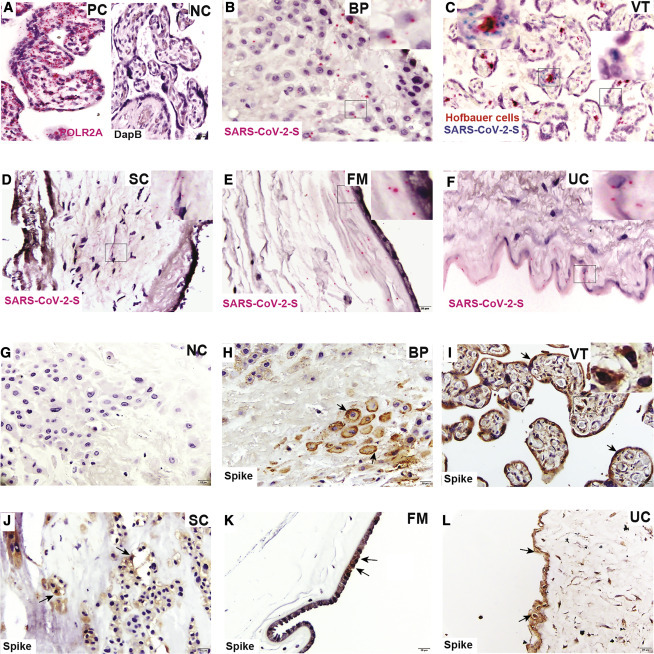
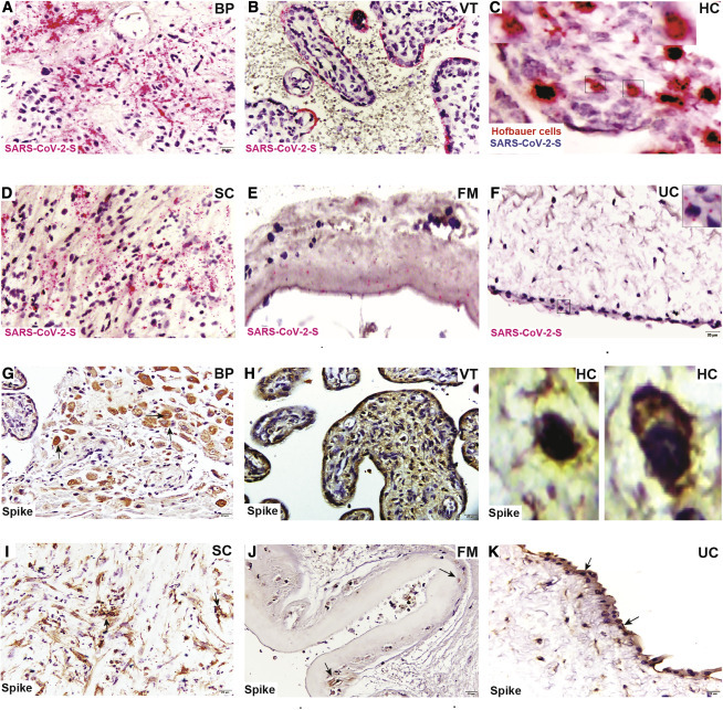
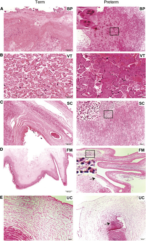
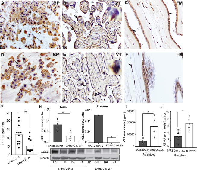
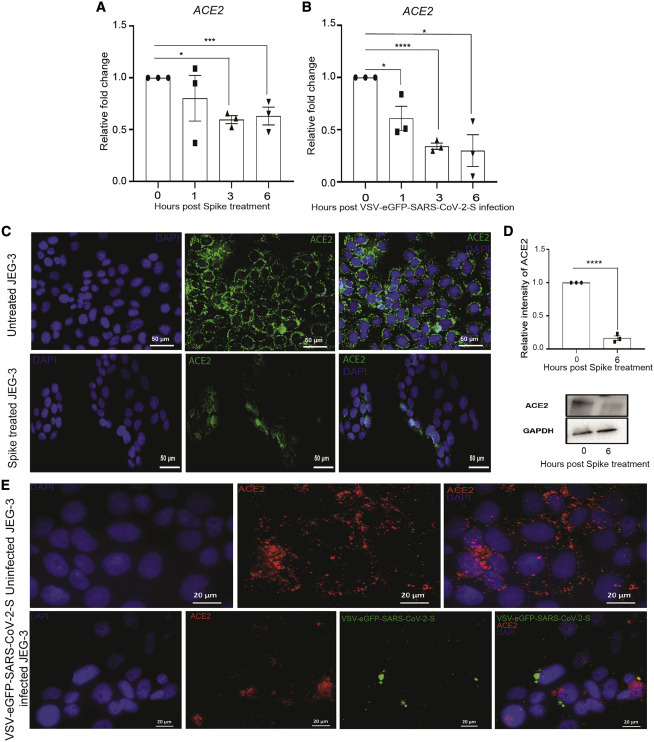
Methods: We investigated the pathophysiology of SARS-CoV-2 at maternal-fetal interface in pregnant women who tested positive for the virus using RNA in situ hybridization (viral RNA), immunohistochemistry, and hematoxylin and eosin staining. To investigate whether viral infection alters the renin angiotensin system (RAS) in placenta, which controls blood pressure, we treated human trophoblasts with recombinant spike protein or a live modified virus with a vesicular stomatitis viral backbone expressing spike protein (VSV-S).

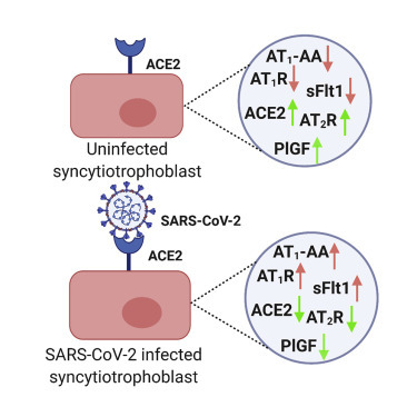
Findings: Viral colonization was highest in maternal decidua, fetal trophoblasts, Hofbauer cells, and in placentas delivered prematurely. We localized SARS-CoV-2 to cells expressing angiotensin-converting enzyme 2 (ACE2) and demonstrate that infected placentas had significantly reduced ACE2. In response to both spike protein and VSV-S, cellular ACE2 decreased although angiotensin II receptor type 1 (AT1R) increased with concomitant increase in soluble fms-like tyrosine kinase-1 (sFlt1). Viral infection decreased pro-angiogenic factors, AT2R, and placental growth factor, which competitively binds to sFlt1. Sera from infected pregnant women had elevated levels of sFlt1 and angiotensin II type 1-receptor autoantibodies prior to delivery, both signatory markers of pre-eclampsia.

Conclusions: SARS-CoV-2 colonizes ACE2-expressing maternal and fetal cells in the placenta. Infection in pregnant women correlates with alteration of placental RAS. As RAS regulates blood pressure, SARS-CoV-2 infection may thus increase adverse hemodynamic outcomes, such as pre-eclampsia in pregnant women.

Funding: NIH/NICHD grants R01 HD091218 and 3R01HD091218-04S1 (RADx-UP Supplement).

Keywords: ACE2; AT1-AA; AT1R; PlGF; decidua; extravillous trophoblasts; placenta; pre-eclampsia; pregnancy; sFlt1.


Conflict of interest statement

I.U.M. serves on the Scientific Advisory Board of Luca Biologics. The authors declare no competing interests.

Figures

None
Graphical abstract
Figure 1
Figure 1
Term placenta from SARS-CoV-2-infected women shows SARS-CoV-2 localization (A) RNAscope ISH positive control (PC) (RNA polymerase II subunit A [POLR2A]) and negative control (NC) (bacterial gene; DapB). (B–F) RNA-ISH images of SARS-CoV-2-S RNA (B) in the basal plate (BP) in extravillous trophoblast cells (EVTs); (C) villous tissue (VT); syncytiotrophoblasts (STBs) shown in right side inset and Hofbauer cells (HCs) in left side inset; (D) subchorion (SC); (E) fetal membrane (FM); and (F) umbilical cord (UC). (G) Immunohistochemical (IHC) NC. (H–L) IHC staining for the presence of viral spike protein in (H) BP; (I) VT, including STBs (black arrow) and HCs (right side inset); (J) SC; (K) FM; and (L) UC. Black arrows show the location of spike protein in respective tissues. Scale bar represents 20 μm.
Figure 2
Figure 2
Preterm placenta from SARS-CoV-2-positive woman shows higher SARS-CoV-2 viral presence (A–F) RNAscope ISH images depict presence of (A) SARS-CoV-2 spike RNA in BP (EVTs), (B) VT in STBs and (C) fetal HCs (insets), (D) SC, (E) FM, and (F) UC (inset). (G–K) Immunohistochemical staining for SARS-CoV-2 spike protein demonstrating virus localization in (G) BP, (H) VT (including STBs and fetal HCs in insets), (I) SC, (J) FM, and (K) UC. Scale bar represents 20 μm.
Figure 3
Figure 3
Histopathological findings in SARS-CoV-2-positive preterm placenta (A–E) (Left) H&E staining of term placental compartments does not indicate obvious histopathological changes. (Right) H&E staining of preterm placental compartments is shown. (A) Trophoblast giant cells (inset) BP. (B) No histopathological defects found in VT. (C) Acute chorioamnionitis, infiltration of neutrophils (inset) in SC. (D) Necrotic amniocyte (inset), edema of amnion (arrow), and pigmented macrophages in FM. (E) Acute funisitis or vasculitis in UC. Black arrows indicate defects in respective tissue. Scale bar represents 100 μm.
Figure 4
Figure 4
SARS-CoV-2 infection in pregnant women is associated with changes in RAS components (A–F) Immunohistochemical (IHC) staining for (A) ACE2 in BP, (B) VT (STBs and HC; inset), and (C) FM in uninfected and (D) BP, (E) VT (STBs and HC; inset), and (F) FM in SARS-CoV-2-infected placenta. Arrows indicate expression of ACE2. Scale bar represents 20 μm. (G) Semiquantitative analysis of ACE2 expression in uninfected and SARS-CoV-2-infected placenta. (H) Densitometric analysis of ACE2 protein expression in term (left panel) and preterm (right panel) placenta of SARS-CoV-2-negative and positive pregnant women. Absolute quantification was performed after normalization with β-actin. The respective blots of ACE2 expression and loading control β-actin are shown. (I and J) Pre-delivery soluble fms-like tyrosine kinase 1 (sFlt1) and angiotensin II type 1-receptor autoantibody (AT1-AA) levels in sera of uninfected and SARS-CoV-2-infected pregnant women. P1–P3, SARS-CoV-2-negative placenta; P4, SARS-CoV-2-negative preterm placenta; S1–S3, SARS-CoV-2-positive placenta; S4, SARS-CoV-2-positive preterm placenta. Values are expressed as mean ± SEM. ∗p < 0.05, ∗∗p < 0.01, ∗∗∗p < 0.001, and ∗∗∗∗p < 0.0001.
Figure 5
Figure 5
ACE2 expression decreases when treated or infected with spike or VSV-EGFP-SARS-CoV-2-S (A and B) Relative expression of ACE2 normalized with GAPDH in JEG-3 cells treated or infected with spike or VSV-EGFP-SARS-CoV-2-S at 0, 1, 3, and 6 h treatment or post-infection. (C and D) Expression of ACE2 in untreated and spike-protein-treated JEG-3 cells after 6 h of treatment by (C) immunofluorescence and (D) western blot, respectively (magnification scale 20×; scale bar represents 50 μm). (E) Immunofluorescence images of uninfected and VSV-EGFP-SARS-CoV-2-S-infected JEG-3 cells after 6 h post-infection, showing expression of ACE2 (magnification scale 63×; scale bar represents 20 μm). Values are expressed as mean ± SEM of three independent experiments. ∗p < 0.05, ∗∗p < 0.01, ∗∗∗p < 0.001, and ∗∗∗∗p < 0.0001.

References

    1. Nishiga M., Wang D.W., Han Y., Lewis D.B., Wu J.C. COVID-19 and cardiovascular disease: from basic mechanisms to clinical perspectives. Nat. Rev. Cardiol. 2020;17:543–558. - PMC - PubMed
    1. Napoli C., Tritto I., Benincasa G., Mansueto G., Ambrosio G. Cardiovascular involvement during COVID-19 and clinical implications in elderly patients. A review. Ann. Med. Surg. (Lond.) 2020;57:236–243. - PMC - PubMed
    1. Madjid M., Safavi-Naeini P., Solomon S.D., Vardeny O. Potential effects of coronaviruses on the cardiovascular system: a review. JAMA Cardiol. 2020;5:831–840. - PubMed
    1. Bonafè M., Prattichizzo F., Giuliani A., Storci G., Sabbatinelli J., Olivieri F. Inflamm-aging: Why older men are the most susceptible to SARS-CoV-2 complicated outcomes. Cytokine Growth Factor Rev. 2020;53:33–37. - PMC - PubMed
    1. Elshafeey F., Magdi R., Hindi N., Elshebiny M., Farrag N., Mahdy S., Sabbour M., Gebril S., Nasser M., Kamel M., et al. A systematic scoping review of COVID-19 during pregnancy and childbirth. Int. J. Gynaecol. Obstet. 2020;150:47–52. - PMC - PubMed

Publication types

Substances