Review
. 2021 Oct;100(11):1201-1209.
doi: 10.1177/00220345211004842. Epub 2021 Apr 17.

Disease-Induced Changes in Salivary Gland Function and the Composition of Saliva

Affiliations
Review

Disease-Induced Changes in Salivary Gland Function and the Composition of Saliva

G B Proctor et al. J Dent Res. 2021 Oct.

Abstract

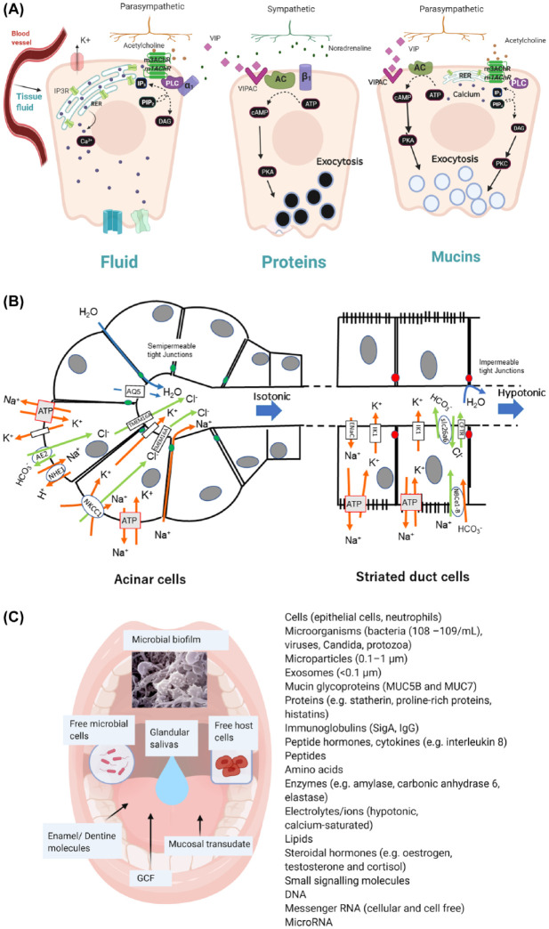
Although the physiological control of salivary secretion has been well studied, the impact of disease on salivary gland function and how this changes the composition and function of saliva is less well understood and is considered in this review. Secretion of saliva is dependent upon nerve-mediated stimuli, which activate glandular fluid and protein secretory mechanisms. The volume of saliva secreted by salivary glands depends upon the frequency and intensity of nerve-mediated stimuli, which increase dramatically with food intake and are subject to facilitatory or inhibitory influences within the central nervous system. Longer-term changes in saliva secretion have been found to occur in response to dietary change and aging, and these physiological influences can alter the composition and function of saliva in the mouth. Salivary gland dysfunction is associated with different diseases, including Sjögren syndrome, sialadenitis, and iatrogenic disease, due to radiotherapy and medications and is usually reported as a loss of secretory volume, which can range in severity. Defining salivary gland dysfunction by measuring salivary flow rates can be difficult since these vary widely in the healthy population. However, saliva can be sampled noninvasively and repeatedly, which facilitates longitudinal studies of subjects, providing a clearer picture of altered function. The application of omics technologies has revealed changes in saliva composition in many systemic diseases, offering disease biomarkers, but these compositional changes may not be related to salivary gland dysfunction. In Sjögren syndrome, there appears to be a change in the rheology of saliva due to altered mucin glycosylation. Analysis of glandular saliva in diseases or therapeutic interventions causing salivary gland inflammation frequently shows increased electrolyte concentrations and increased presence of innate immune proteins, most notably lactoferrin. Altering nerve-mediated signaling of salivary gland secretion contributes to medication-induced dysfunction and may also contribute to altered saliva composition in neurodegenerative disease.

Keywords: Sjögren syndrome; mucins; oral health; salivation; secretion; sialadenitis.


Conflict of interest statement

Declaration of Conflicting Interests: The authors declared no potential conflicts of interest with respect to the research, authorship, and/or publication of this article.

Figures

Figure 1.
Figure 1.
Nerve-regulated salivary secretion. Peripheral and central involvement in reflex-stimulated secretion by the major salivary glands.
Figure 2.
Figure 2.
Salivary gland secretion of fluid and protein. (A) Coupling of nerve-mediated stimuli to secretion of fluid, proteins, and mucins by salivary acinar cells. Release of acetylcholine from parasympathetic (ps) nerves activates muscarinic receptors (m3/m1 AChRs), leading to activation of phospholipase C (PLC) and generation of IP3 (inositol triphosphate) from PIP2 (polyinositol biphosphate), binding to IP3 receptor and release of Ca2+ from RER (rough endoplasmic reticulum), increasing intracellular calcium concentration. Noradrenaline (NA) release from sympathetic (sy) nerves can cause a minor secretion of fluid through α1 adrenoceptors. Protein exocytosis is evoked by activation of PKA (protein kinase A) by intracellular cAMP generated from ATP by AC (adenylate cyclase) activated following NA binding to β1-adrenoceptors or vasointesinal polypeptide (VIP) signaling from ps nerves. Exocytosis from mucous acinar cells appears to occur without involvement of sy nerves. Acetylcholine release from ps nerves activates exocytosis through increased intracellular Ca2+ and PKC (protein kinase C) activated by DAG (diacylglycerol). VIP also signals mucous cell exocytosis through PKA activation (see Appendix). (B) Cellular mechanisms of fluid secretion. Acinar cell secretion of isotonic NaCl and water is followed by striated duct cell absorption of NaCl and secretion of KHCO3 in a hypotonic fluid. The principal membrane transporter proteins involved are shown. Acinar cell secretion is dependent on an apical chloride channel (TMEM16A), basolateral sodium, potassium cotransporter (NKCC1), antiports (AE2, NHE1), potassium channels, water channel (AQ5), and paracellular movement of sodium and water, all underpinned by sodium potassium ATPase. Ductal cell absorption of sodium and secretion of potassium through the ENaC and IK1 channels, respectively, is accompanied by secretion of bicarbonate through a combination of the CFTR chloride channel and the slc26a6 antiport, all ultimately dependent on sodium potassium ATPase (see Appendix). (C) Composition of saliva in the mouth. In addition to the saliva secreted by major and minor salivary glands, whole-mouth saliva contains components of non–salivary gland origin, including cells, bacteria and other microorganisms, cellular particles along with molecules contributed from gingival crevicular fluid (GCF), mucosal transudate, tooth enamel, and dentin.
Figure 3.
Figure 3.
Relationships between disease, salivary gland dysfunction, and composition of saliva. The present review focuses on disease-related salivary gland dysfunction leading to altered composition and function of saliva. The causes considered are systemic diseases and therapeutic interventions (e.g., anticholinergic medications) that can cause salivary gland disease and dysfunction (solid lines). Saliva contains biomarkers of other systemic diseases (e.g., diabetes or liver disease) and oral diseases (e.g., periodontitis), which can directly alter the composition of saliva without necessarily causing salivary gland disease or dysfunction (dashed lines).

References

    1. Affoo RH, Foley N, Garrick R, Siqueira WL, Martin RE. 2015. Meta-analysis of salivary flow rates in young and older adults. J Am Geriatr Soc. 63(10):2142–2151. - PubMed
    1. Atkinson JC. 1993. The role of salivary measurements in the diagnosis of salivary autoimmune-diseases. Ann N Y Acad Sci. 694:238–251. - PubMed
    1. Beach TG, Adler CH, Sue LI, Vedders L, Lue L, White CL, Akiyama H, Caviness JN, Shill HA, Sabbagh MN, et al.. 2010. Multi-organ distribution of phosphorylated alpha-synuclein histopathology in subjects with Lewy body disorders. Acta Neuropathol. 119(6):689–702. - PMC - PubMed
    1. Bombardieri M, Barone F, Lucchesi D, Nayar S, van den Berg WB, Proctor G, Buckley CD, Pitzalis C.2012. Inducible tertiary lymphoid structures, autoimmunity, and exocrine dysfunction in a novel model of salivary gland inflammation in C57BL/6 mice. J Immunol. 189(7):3767–3776. - PMC - PubMed
    1. Borrelli M, Schroder C, Dart JK, Collin JR, Sieg P, Cree IA, Matheson MA, Tiffany JM, Proctor G, van Best J, et al.. 2010. Long-term follow-up after submandibular gland transplantation in severe dry eyes secondary to cicatrizing conjunctivitis. Am J Ophthalmol. 150(6):894–904. - PubMed

Publication types

LinkOut - more resources