

Determining the development stage of the ossification centers around the elbow may aid in deciding whether to use ESIN or not in adolescents' forearm shaft fractures
- PMID: 33870827
- PMCID: PMC8428268
- DOI: 10.1080/17453674.2021.1912895
Determining the development stage of the ossification centers around the elbow may aid in deciding whether to use ESIN or not in adolescents' forearm shaft fractures
Abstract
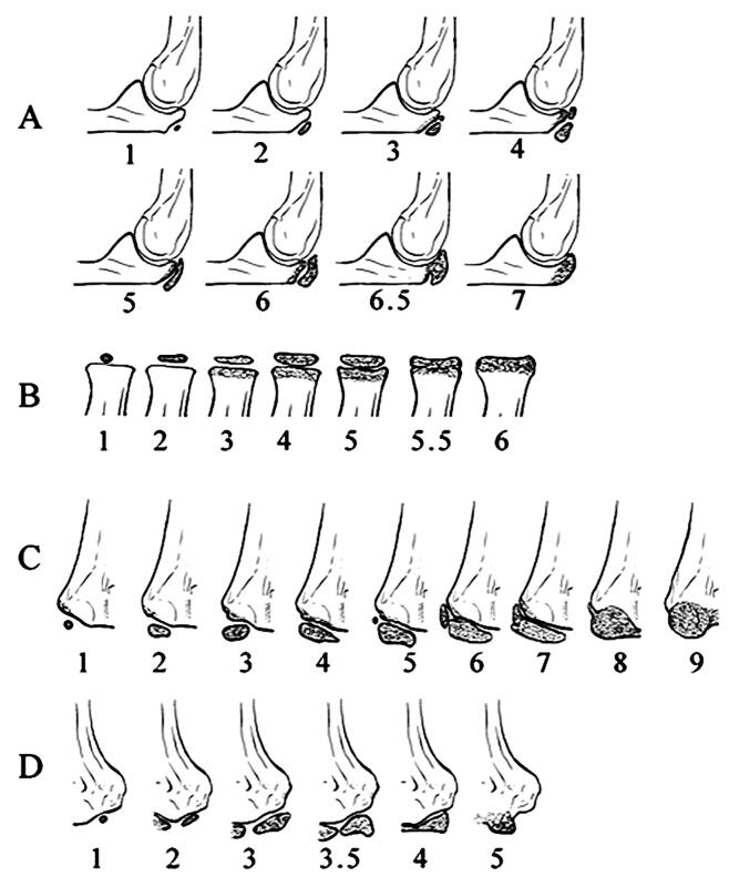
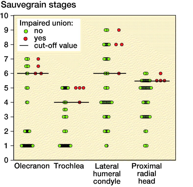
Background and purpose - Elastic stable intramedullary nailing (ESIN) is the preferred method of operative stabilization of unstable pediatric forearm shaft fractures. However, the decision whether to use ESIN or open reduction and internal fixation (ORIF) in older children or teenagers is not always straightforward. We hypothesized that the development stage of the elbow would aid in evaluating the eligibility of the patient for ESIN.Patients and methods - All eligible children, aged <16 years who were treated with ESIN in Oulu University Hospital, during 2010-2019 were included (N = 70). The development stages of 4 ossification centers were assessed according to the Sauvegrain and Diméglio scoring. The proportion of impaired union vs. union was analyzed according to bone maturity, by using the optimal cutoff-points determined with receiver operating characteristics (ROC).Results - Development stage ≥ 6 in the olecranon was associated with impaired union in 20% of patients, compared with none in stages 1-5 (95% CI of difference 8% to 24%). Trochlear ossification center ≥ 4 was associated with impaired union in 17% of patients (CI of difference 7% to 36%) and lateral condyle ≥ 6 in 13% of patients (CI of difference 3.4% to 30%). Proximal radial head ≥ 5.5 was associated with impaired union in 18% of patients (CI of difference 7% to 39%).Interpretation - Recognizing the rectangular or fused olecranon ossification center, referring to stage ≥ 6, was in particular associated with impaired fracture healing. This finding may aid clinicians to consider between ESIN and plating, when treating forearm shaft fracture of an older child or teenager.
Figures
Similar articles
-
Intramedullary nailing of forearm shaft fractures by biodegradable compared with titanium nails: Results of a prospective randomized trial in children with at least two years of follow-up.Biomaterials. 2018 Dec;185:383-392. doi: 10.1016/j.biomaterials.2018.09.011. Epub 2018 Sep 11. Biomaterials. 2018. PMID: 30292588 Clinical Trial.
-
Nonunion of forearm shaft fractures in children after intramedullary nailing.J Pediatr Orthop B. 2009 Nov;18(6):289-95. doi: 10.1097/BPB.0b013e32832f5b20. J Pediatr Orthop B. 2009. PMID: 19623087
-
Comparison of hybrid fixation versus dual intramedullary nailing fixation for forearm fractures in older children: Case-control study.Int J Surg. 2016 Jun;30:7-12. doi: 10.1016/j.ijsu.2016.03.070. Epub 2016 Apr 7. Int J Surg. 2016. PMID: 27063636
-
Treatment of pediatric femoral shaft fractures with elastic stable intramedullary nails versus external fixation: A meta-analysis.Orthop Traumatol Surg Res. 2020 Nov;106(7):1305-1311. doi: 10.1016/j.otsr.2020.06.012. Epub 2020 Oct 17. Orthop Traumatol Surg Res. 2020. PMID: 33082120
-
State-of-the-art treatment of forearm shaft fractures.Injury. 2005 Feb;36 Suppl 1:A25-34. doi: 10.1016/j.injury.2004.12.010. Injury. 2005. PMID: 15652933 Review.
Cited by
-
Child Fractures: Are We Getting More Surgical?Rev Bras Ortop (Sao Paulo). 2022 Jun 10;58(2):191-198. doi: 10.1055/s-0042-1748815. eCollection 2023 Apr. Rev Bras Ortop (Sao Paulo). 2022. PMID: 37252311 Free PMC article.
-
Forearm Fractures in Older Children and Adolescents: ORIF is Safer Than IMN With Equivalent Outcomes.J Pediatr Orthop. 2025 Mar 1;45(3):e218-e223. doi: 10.1097/BPO.0000000000002853. Epub 2024 Oct 24. J Pediatr Orthop. 2025. PMID: 39445700 Free PMC article.
References
-
- Abraham A, Kumar S, Chaudhry S, Ibrahim T.. Surgical interventions for diaphyseal fractures of the radius and ulna in children. Cochrane Database Syst Rev 2011; (11): CD007907. - PubMed
-
- Baldwin K, Morrison M J 3rd, Tomlinson L A, Ramirez R, Flynn J M.. Both bone forearm fractures in children and adolescents, which fixation strategy is superior—plates or nails? A systematic review and meta-analysis of observational studies. J Orthop Trauma 2014; 28(1): e8–e14. - PubMed
-
- Bhandari M, Guyatt G H, Swiontkowski M F, Tornetta 3rd P, Sprague S, Schemitsch E H.. A lack of consensus in the assessment of fracture healing among orthopaedic surgeons. J Orthop Trauma 2002; 16(8): 562–6. - PubMed
MeSH terms
LinkOut - more resources
Full Text Sources
Other Literature Sources
Medical