Review
. 2021 May 28;41(5):BSR20210427.
doi: 10.1042/BSR20210427.

Modulating skin colour: role of the thioredoxin and glutathione systems in regulating melanogenesis

Affiliations
Review

Modulating skin colour: role of the thioredoxin and glutathione systems in regulating melanogenesis

Yaoying Lu et al. Biosci Rep. .

Abstract

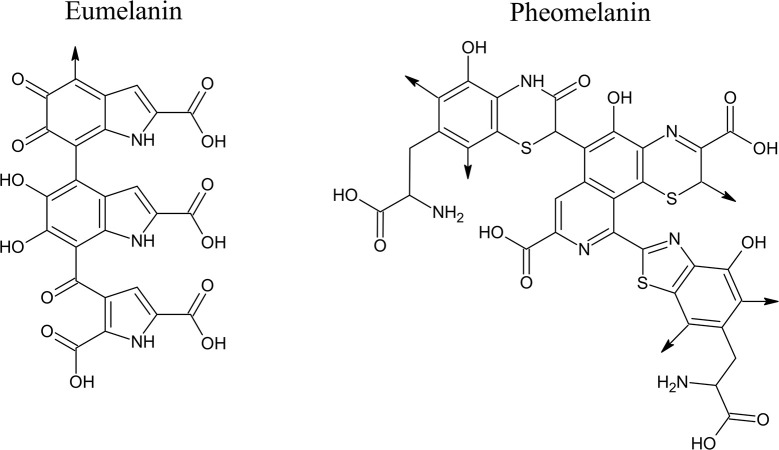
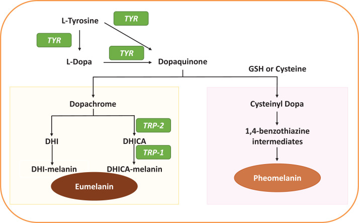
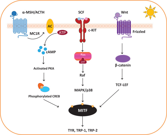
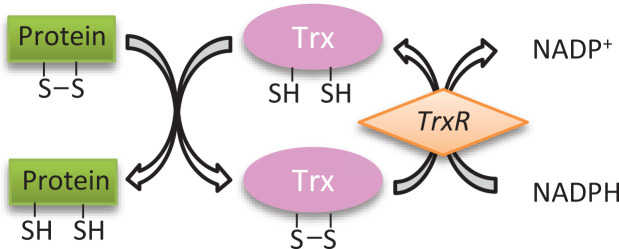
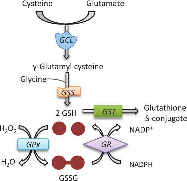
Different skin colour among individuals is determined by the varying amount and types of melanin pigment. Melanin is produced in melanocytes, a type of dendritic cell located in the basal layer of the epidermis, through the process of melanogenesis. Melanogenesis consists of a series of biochemical and enzymatic reactions catalysed by tyrosinase and other tyrosinase-related proteins, leading to the formation of two types of melanin, eumelanin and pheomelanin. Melanogenesis can be regulated intrinsically by several signalling pathways, including the cyclic adenosine monophosphate (cAMP)/protein kinase A (PKA), stem cell factor (SCF)/c-kit and wingless-related integration site (Wnt)/β-catenin signalling pathways. Ultraviolet radiation (UVR) is the major extrinsic factor in the regulation of melanogenesis, through the generation of reactive oxygen species (ROS). Antioxidants or antioxidant systems, with the ability to scavenge ROS, may decrease melanogenesis. This review focuses on the two main cellular antioxidant systems, the thioredoxin (Trx) and glutathione (GSH) systems, and discusses their roles in melanogenesis. In the Trx system, high levels/activities of thioredoxin reductase (TrxR) are correlated with melanin formation. The GSH system is linked with regulating pheomelanin formation. Exogenous addition of GSH has been shown to act as a depigmenting agent, suggesting that other antioxidants may also have the potential to act as depigmenting agents for the treatment of human hyperpigmentation disorders.

Keywords: antioxidants; glutathione; hyperpigmentation; melanogenesis; thioredoxin.


Conflict of interest statement

The authors declare that there are no competing interests associated with the manuscript.

Figures

Figure 1
Figure 1. Chemical structures of eumelanin and pheomelanin
The structure of eumelanin represents DHICA-melanin, which is a polymeric structure made of DHICA as the basic motif. The positions with –COOH group in the eumelanin structure can be substituted by –H to represent DHI-melanin. Both DHICA-melanin and DHI-melanin are regarded as eumelanin. The molecular weight of eumelanin shown above is 563.4 g/mol. The structure of pheomelanin consists of polymers composed of benzothiazine and benzothiazole units. The molecular weight of pheomelanin shown above is 806.8 g/mol. The arrows in the structures of eumelanin and pheomelanin indicate sites where polymerization can occur [15,16]. Abbreviations: DHI, 5,6-dihydroxyindole; DHICA, DHI-2-carboxylic acid. This figure is reproduced and used with permission from the publisher [15]. © 2007 The Authors. Journal Compilation. The American Society of Photobiology.
Figure 2
Figure 2. Melanogenesis formation pathway
Eumelanin and pheomelanin are produced through a multistep process of biochemical reactions, with the rate-limiting enzyme TYR using l-tyrosine and/or l-dopa as the initial substrates (Abbreviations: DHICA, DHI-2-carboxylic acid; TRP-2, dopachrome tautomerase).
Figure 3
Figure 3. Melanogenesis signalling pathways
The central melanogenesis regulator MITF can be regulated by different signalling pathways, including the cAMP/PKA signalling pathway, SCF/c-kit mediated signalling pathway and Wnt/β-catenin signalling pathway (Abbreviations: α-MSH, α-melanocyte-stimulating hormone; AC, adenylate cyclase; ACTH, adrenocorticotropic hormone; ATP, adenosine triphosphate; cKIT, tyrosine-protein kinase kit; CREB, cAMP response element-binding protein; GTP, guanosine-5′-triphosphate; MAPK, mitogen-activated protein kinase; MC1R, melanocortin 1 receptor; TCF-LEF, T-cell factor/lymphoid enhancer-binding factor; TRP-2, dopachrome tautomerase).
Figure 4
Figure 4. Mechanism of action of the Trx system
The active form of Trx has two free thiol groups to catalyse target protein substrate reduction during which a disulphide bond is formed between two cysteine residues. TrxR catalyses the reduction of oxidized Trx using NADPH.
Figure 5
Figure 5. The glutathione system
First, γ-glutamyl cysteine is synthesized from l-glutamate and cysteine using GCL. Then, GSS catalyses the condensation of γ-glutamyl cysteine and glycine to form GSH. GSH is used as a cofactor by GPx to reduce H2O2, resulting in the formation of GSSG. GR reduces GSSG to two GSH by NADPH (Abbreviation: GST, glutathione S-transferase).

Similar articles

Cited by

References

    1. Costin G.E. and Hearing V.J. (2007) Human skin pigmentation: melanocytes modulate skin color in response to stress. FASEB J. 21, 976–994 10.1096/fj.06-6649rev - DOI - PubMed
    1. Lin J.Y. and Fisher D.E. (2007) Melanocyte biology and skin pigmentation. Nature 445, 843–850 10.1038/nature05660 - DOI - PubMed
    1. Tapia C.V., Falconer M., Tempio F., Falcon F., Lopez M., Fuentes M.et al. . (2014) Melanocytes and melanin represent a first line of innate immunity against Candida albicans. Med. Mycol. 52, 445–454 10.1093/mmy/myu026 - DOI - PubMed
    1. Thody A.J., Higgins E.M., Wakamatsu K., Ito S., Burchill S.A. and Marks J.M. (1991) Pheomelanin as well as eumelanin is present in human epidermis. J. Invest. Dermatol. 97, 340–344 10.1111/1523-1747.ep12480680 - DOI - PubMed
    1. Hearing V.J. and Jimenez M. (1987) Mammalian tyrosinase–the critical regulatory control point in melanocyte pigmentation. Int. J. Biochem. 19, 1141–1147 10.1016/0020-711X(87)90095-4 - DOI - PubMed

Publication types

LinkOut - more resources