

Review
doi: 10.4269/ajtmh.20-1000.
Pathogenesis of Pulmonary Hemorrhagic Syndrome in Human Leptospirosis
Affiliations
- PMID: 33872210
- PMCID: PMC8176490
- DOI: 10.4269/ajtmh.20-1000
Item in Clipboard
Review
Pathogenesis of Pulmonary Hemorrhagic Syndrome in Human Leptospirosis
Am J Trop Med Hyg.
.
Abstract
Based on a previous study and by incorporating new knowledge, the goal of our study was to understand more fully the pathogenesis of hemorrhagic pneumonia of severe human leptospirosis, highlighting the onset of capillary lesions by Leptospira itself and/or its antigenic/toxic products acting on the endothelium and binding to cadherins. Both events lead to loss of endothelial integrity, alter permeability, cause rupture, and open intercellular junctions, contributing to the hemorrhagic phenomena associated with severe leptospirosis.
Figures
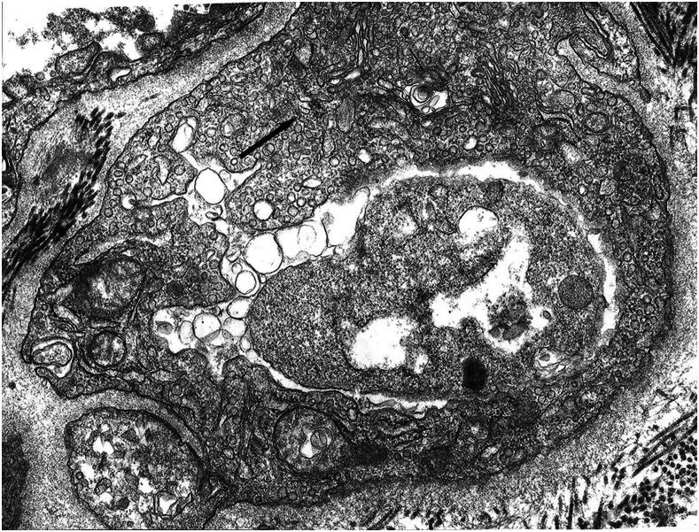
Septal capillary with edema and increased pinocytotic vesicles in endothelial cell, ×81.900 original magnification.
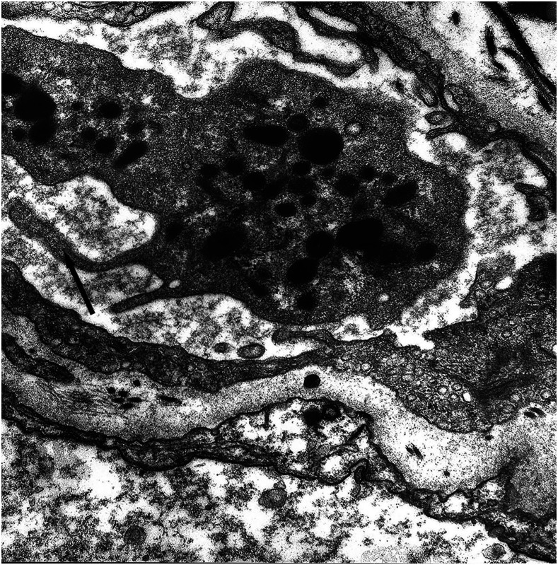
Septal capillary with pseudopod emission by the endothelial cell and platelet, ×42.560 original magnification.
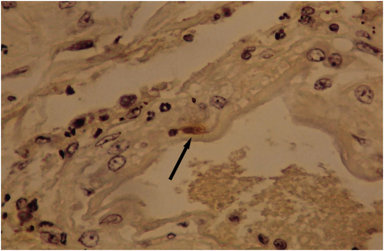
Granular leptospiral antigen (arrow) in an endothelial cell of pulmonary capillary; avidin–biotin stained, ×600 original magnification. This figure appears in color at www.ajtmh.org .
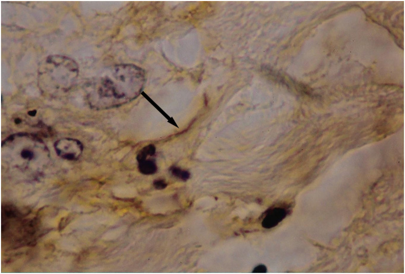
Filamentous leptospiral antigen (arrow) attached to the endothelium of a septal capillary; avidin–biotin stained, ×1,000 original magnification. This figure appears in color at www.ajtmh.org .
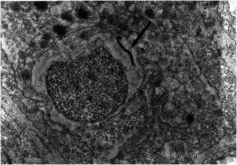
Leptospira (arrow) next to the mononuclear cell in septal capillary; conventional electron microscopy, 2.1 × 19.500.
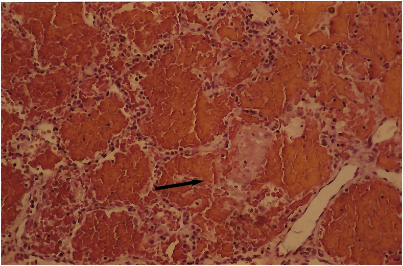
Alveoli filled with erythrocytes and ruptured alveolar septa; hematoxylin eosin stain, ×150 magnification. This figure appears in color at www.ajtmh.org .
References
-
- Ministério da Saúde , 2020. Available at: https://antigo.saude.gov.br/images/pdf/2020/fevereiro/07/obito-lepto-200....
Publication types
MeSH terms
LinkOut - more resources
Full Text Sources
Other Literature Sources
Medical
