. 2021 May 21;49(9):4954-4970.
doi: 10.1093/nar/gkab245.

Implication of repeat insertion domains in the trans-activity of the long non-coding RNA ANRIL

Affiliations

Implication of repeat insertion domains in the trans-activity of the long non-coding RNA ANRIL

Charbel Alfeghaly et al. Nucleic Acids Res. .

Abstract

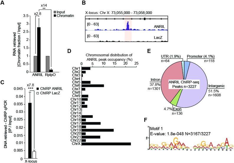
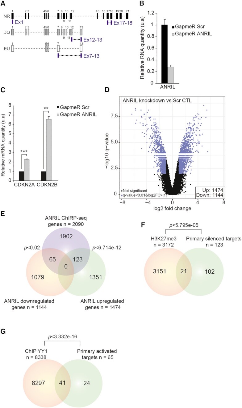
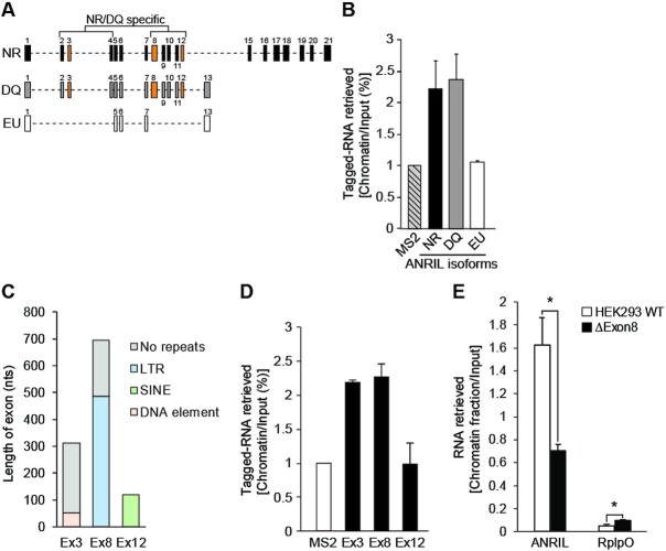
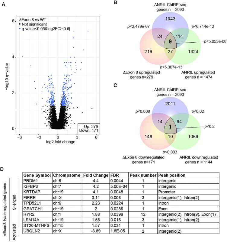
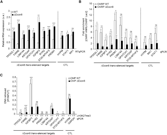
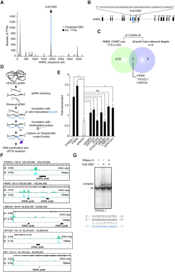
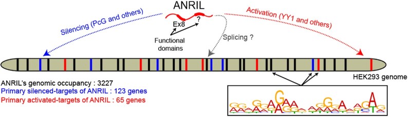
Long non-coding RNAs have emerged as critical regulators of cell homeostasis by modulating gene expression at chromatin level for instance. Here, we report that the lncRNA ANRIL, associated with several pathologies, binds to thousands of loci dispersed throughout the mammalian genome sharing a 21-bp motif enriched in G/A residues. By combining ANRIL genomic occupancy with transcriptomic analysis, we established a list of 65 and 123 genes potentially directly activated and silenced by ANRIL in trans, respectively. We also found that Exon8 of ANRIL, mainly made of transposable elements, contributes to ANRIL genomic association and consequently to its trans-activity. Furthermore, we showed that Exon8 favors ANRIL's association with the FIRRE, TPD52L1 and IGFBP3 loci to modulate their expression through H3K27me3 deposition. We also investigated the mechanisms engaged by Exon8 to favor ANRIL's association with the genome. Our data refine ANRIL's trans-activity and highlight the functional importance of TEs on ANRIL's activity.


Figures

Figure 1.
Figure 1.
ANRIL binds 3227 loci across the genome of HEK293 cells. (A) RNA extraction experiments showing ANRIL enrichment in the chromatin fraction (n = 3). Values are normalized to the Input. (B) Representative example of one of the ANRIL ChIRP-seq peaks located on the X chromosome. (C) ChIRP-qPCR validation of the peak localized on the X chromosome (n = 4). (D) ANRIL does not coat all the chromosomes to the same extent. Approximately 20% of the ANRIL peaks are localized on the X chromosome. (E) ANRIL ChIRP-seq peaks are mostly distributed in distal intergenic and intronic regions. The percentages of each class were calculated by normalizing the sum of nucleotides in ANRIL ChIRP-seq dataset relative to the human genome. The number of peaks within each DNA category is indicated. (F) MEME motif analysis identifying a G/A rich motif in ANRIL ChIRP-seq peaks (n = 3167/3227). Data are represented as mean ± SEM. P-values: moderated t-statistics, *P < 0.05, **P < 0.01, ***P < 0.001, ns: not significant.
Figure 2.
Figure 2.
ANRIL is likely to regulate the expression of 188 genes in a direct manner. (A) Schematic representation of the hybridization position of the four different LNA GapmeRs used to silence ANRIL. (B) RTqPCR analysis after LNA GapmeR transfection revealed up to 75% reduction in ANRIL’s expression (n = 3). Values are normalized to the GAPDH housekeeping gene. (C) RTqPCR analysis of CDKN2A and CDKN2B expression following ANRIL knockdown by LNA GapmeRs (n = 3). Values are normalized to the GAPDH housekeeping gene. (D) Volcano plot representing the significance versus fold change for all differentially expressed (n = 2618, FDR < 0.01, log2FC > |1|) genes upon ANRIL knockdown (n = 5). (E) Venn diagram showing the intersection between the ANRIL upregulated genes, downregulated genes, and the ChIRP-seq dataset. A list of 188 genes has been identified as potential direct trans-regulated targets. (F) Venn diagram showing the intersection between H3K27me3 ChIP-seq data in HEK293 cells and the 123 silenced primary targets of ANRIL. List of 21 genes identified to be potentially repressed through PcG dependent mechanism. (G) Venn diagram showing the intersection between YY1 ChIP-seq data in HEK293 cells and the 65 primary activated targets of ANRIL. Forty-one genes were identified to be potentially activated through YY1. The P-values of Venn diagrams were obtained by hypergeometric test using the whole set of microarray genes as a background. Data are represented as mean ± SEM. P-values: moderated t-statistics, *P < 0.05, **P < 0.01, ***P < 0.001.
Figure 3.
Figure 3.
Exon8 is involved in the association of ANRIL with the chromatin. (A) Schematic representation of the three major ANRIL isoforms NR, DQ, and EU. Exons and introns are represented by numbered rectangles and dashed lines, respectively. (B) RNA extraction experiments after transient overexpression of the MS2-tagged NR, DQ, and EU isoforms (n = 2). This identified the NR and DQ isoforms as DNA/chromatin binders but not the EU compared to the MS2-CTL. Values are normalized to Input. (C) RepeatMasker analysis showing the distribution of TEs in ANRIL’s exons 3, 8, and 12. (D) RNA extraction experiments after transient overexpression of the MS2-tagged exons 3, 8 and 12 (n = 2). This identified the exons 3 and 8 of ANRIL as DNA/chromatin binders but not the exon 12 compared to the MS2-CTL. Values are normalized to Input. (E) RNA extraction experiments from ΔExon8 HEK293 cell lines which revealed a reduction in chromatin association of ANRIL by 60% but not for RplpO compared to the HEK293 WT cell lines (n = 3). Data are represented as mean ± SEM. P-values: moderated t-statistics, *P < 0.05, **P < 0.01, ***P < 0.001.
Figure 4.
Figure 4.
The expression of 10 primary trans-targets depends on the presence of Exon8. (A) Volcano plot representing the significance versus fold change for all differentially expressed (n = 450, FDR < 0.05, log2FC > |0.6|) genes in ΔExon8 HEK293 cells (n = 5). (B) Venn diagram showing the intersection between the ANRIL ChIRP-seq genes and the upregulated genes in the ΔExon8 and ANRIL LNA knockdown datasets. A list of nine primary targets has been identified as being potentially silenced by the presence of Exon8 containing-ERVL in a direct manner. (C) Venn diagram showing the intersection between the ANRIL ChIRP-seq genes and the downregulated genes in the ΔExon8 and ANRIL LNA knockdown datasets. One target has been identified as being potentially activated by the Exon8 containing-ERVL in a direct manner. The P-values were obtained by hypergeometric test using the whole set of microarray genes as a background. (D) Table summarizing the 10 ΔExon8 trans-regulated genes. Gene symbol, chromosome, fold change, FDR, ANRIL ChIRP-seq peak number and peak position are represented.
Figure 5.
Figure 5.
ANRIL directly contacts specific genes throughout the genome using its Exon8 to regulate their expression through the deposition of H3K27me3. (A) RTqPCR validation of the differentially expressed ΔExon8 trans-silenced targets in ΔExon8 HEK293 cells (n = 5). (B) ChIRP-qPCR on the ΔExon8 trans-silenced targets shows that in the absence of Exon8, ANRIL significantly dissociates from some of these loci (n = 5). Values are normalized to the Input then fold enrichment is calculated by normalizing to LacZ. (C) ChIP-qPCR using H3K27me3 and control IgG antibodies on promoter regions of the ΔExon8 trans-silenced targets (n = 5). Values are normalized to the Input. Data are represented as mean ± SEM. P-values: moderated t-statistics, *P < 0.05, **P < 0.01, ***P < 0.001, ns: not significant.
Figure 6.
Figure 6.
Exon8 harbors a DBD potentially involved in triplex formation. (A) TDF prediction using ANRIL full-length against the ChIRP-seq dataset. This revealed the potential DBD on ANRIL’s sequence located in Exon8 (P-value = 0.0013) with its associated TTS (n = 422) hereafter called ChIRP-seq TTSs. (B) Schematic representation of the position and purine-rich sequence of Ex8-DBD. (C) Venn diagram showing the intersection between the predicted ChIRP-seq TTSs and the ΔExon8 trans-silenced targets. A list of three genes (FIRRE, TPD52L1 and LSM14A) was predicted to contain TTSs. The P-values were obtained by hypergeometric test using the whole set of microarray genes as a background. (D) Schematic representation of the RNA-based DNA capture assay adapted from (44) to validate triplex formation. An antisense biotinylated DNA oligo hybridizing to the Exon8 was used to capture triplex formed with full length in vitro transcribed Exon8 incubated with sheared genomic DNA. After recovery of triplex on streptavidin beads, associated DNA was eluted and analyzed by qPCR. (E) Enrichment of FIRRE, TPD52L1 and GPATCH1 peak sequences were obtained but not GAPDH nor the remaining ΔExon8 trans-silenced targets after capturing Exon8 using antisense biotinylated DNA oligo on streptavidin beads (n = 4). The enrichment is presented as the ratio between these loci and TERC which was used as a negative control. (F) ATAC-seq and DNase-seq data evaluating the chromatin accessibility in HEK293 cells at the TPD52L1, FIRRE, LSM14A, MX1 and KRTDAP loci. Signals observed represent open chromatin regions. (G) EMSA using 14 μM of synthetic Ex8-DBD (42 nts) with 100 fmol of double–stranded 32P-labeled double stranded oligonucleotides harboring a predicted TTS of TPD52L1 (Supplementary Table S7). Gel shift was resistant to RNaseH indicating a Hoogsteen base pairing. Potential Hoogsteen base pairing between Ex8-DBD represented in blue and TPD52L1 dsDNA sequences is shown; mismatches are marked *. Data are represented as mean ± SEM. P-values: moderated t-statistics, *P < 0.05, **P < 0.01, ***P < 0.001, ns: not significant.
Figure 7.
Figure 7.
Summary of ANRIL’s functions. ANRIL interacts with 3227 of loci dispersed throughout the HEK293 cell genome sharing a 21-bp motif enriched in G/A residues. Combining ANRIL genomic occupancy with transcriptomic analysis identified 65 and 123 genes potentially trans-activated and silenced by ANRIL in a direct manner, respectively. Exon8 of ANRIL, mainly made of TEs, contributes to ANRIL genomic association and consequently to its trans-activity. ANRIL might also act as splicing regulator.

References

    1. Zhao Y., Li H., Fang S., Kang Y., wu W., Hao Y., Li Z., Bu D., Sun N., Zhang M.Q.et al. .. NONCODE 2016: an informative and valuable data source of long non-coding RNAs. Nucleic Acids Res. 2016; 44:D203–D208. - PMC - PubMed
    1. Bao Z., Yang Z., Huang Z., Zhou Y., Cui Q., Dong D.. LncRNADisease 2.0: an updated database of long non-coding RNA-associated diseases. Nucleic Acids Res. 2019; 47:D1034–D1037. - PMC - PubMed
    1. Mercer T.R., Dinger M.E., Mattick J.S.. Long non-coding RNAs: insights into functions. Nat. Rev. Genet. 2009; 10:155–159. - PubMed
    1. Wapinski O., Chang H.Y.. Long noncoding RNAs and human disease. Trends Cell Biol. 2011; 21:354–361. - PubMed
    1. Yao R.-W., Wang Y., Chen L.-L.. Cellular functions of long noncoding RNAs. Nat. Cell Biol. 2019; 21:542–551. - PubMed

Publication types