. 2021 Jun:188:114564.
doi: 10.1016/j.bcp.2021.114564. Epub 2021 Apr 17.

Discovery of a AHR pelargonidin agonist that counter-regulates Ace2 expression and attenuates ACE2-SARS-CoV-2 interaction

Affiliations

Discovery of a AHR pelargonidin agonist that counter-regulates Ace2 expression and attenuates ACE2-SARS-CoV-2 interaction

Michele Biagioli et al. Biochem Pharmacol. 2021 Jun.

Abstract

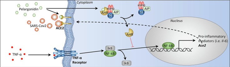
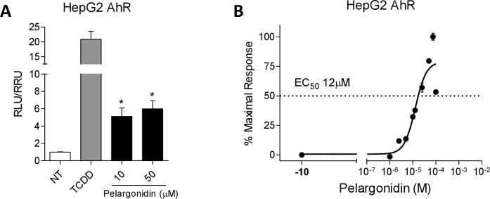
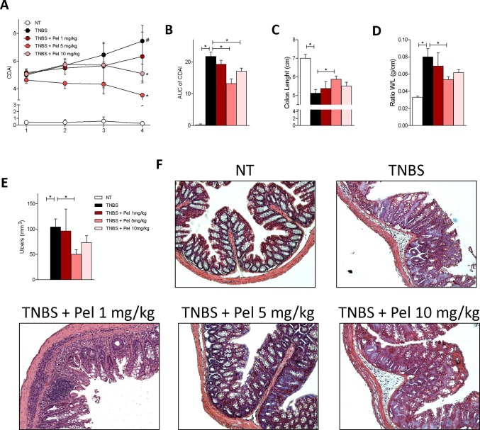
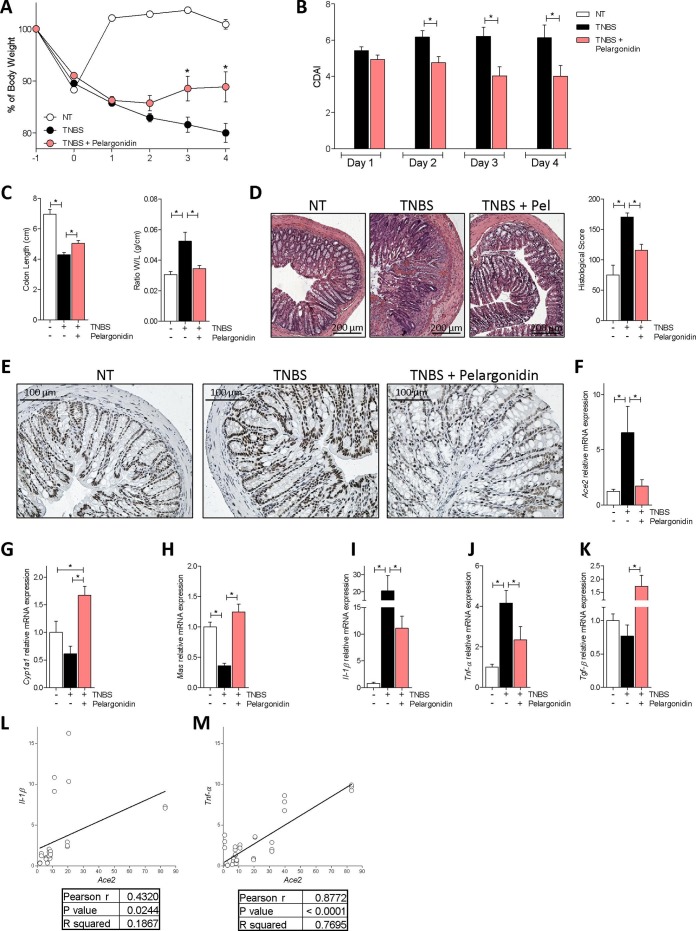
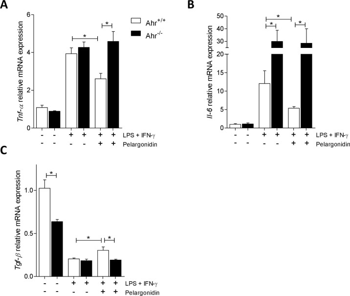
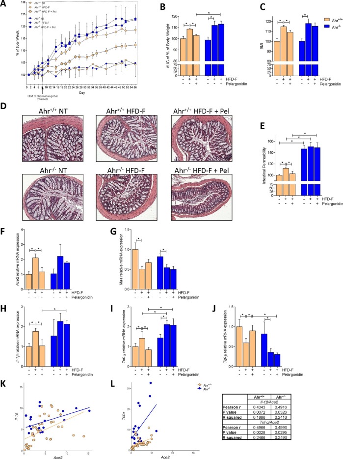
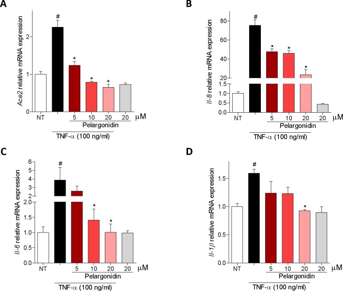
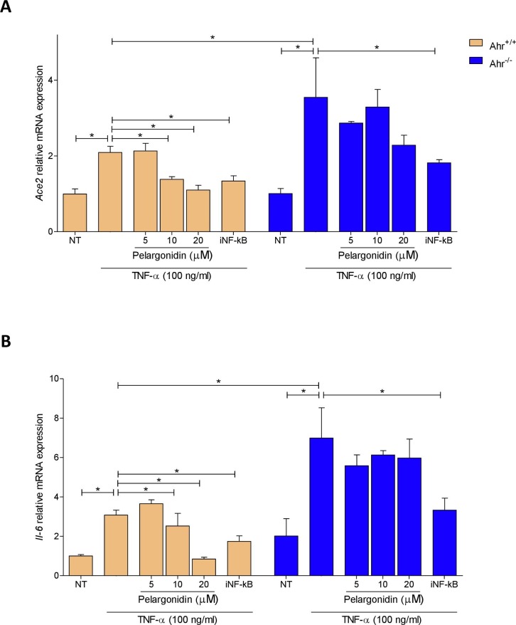
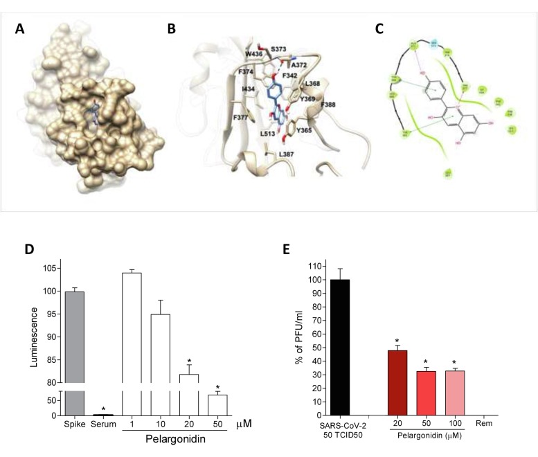
The severe acute respiratory syndrome (SARS)-CoV-2 is the pathogenetic agent of Corona Virus Induced Disease (COVID)19. The virus enters the human cells after binding to the angiotensin converting enzyme (ACE)2 receptor in target tissues. ACE2 expression is induced in response to inflammation. The colon expression of ACE2 is upregulated in patients with inflammatory bowel disease (IBD), highlighting a potential risk of intestinal inflammation in promoting viral entry in the human body. Because mechanisms that regulate ACE2 expression in the intestine are poorly understood and there is a need of anti-SARS-CoV-2 therapies, we have settled to investigate whether natural flavonoids might regulate the expression of Ace2 in intestinal models of inflammation. The results of these studies demonstrated that pelargonidin activates the Aryl hydrocarbon Receptor (AHR) in vitro and reverses intestinal inflammation caused by chronic exposure to high fat diet or to the intestinal braking-barrier agent TNBS in a AhR-dependent manner. In these two models, development of colon inflammation associated with upregulation of Ace2 mRNA expression. Colon levels of Ace2 mRNA were directly correlated with Tnf-α mRNA levels. Molecular docking studies suggested that pelargonidin binds a fatty acid binding pocket on the receptor binding domain of SARS-CoV-2 Spike protein. In vitro studies demonstrated that pelargonidin significantly reduces the binding of SARS-CoV-2 Spike protein to ACE2 and reduces the SARS-CoV-2 replication in a concentration-dependent manner. In summary, we have provided evidence that a natural flavonoid might hold potential in reducing intestinal inflammation and ACE2 induction in the inflamed colon in a AhR-dependent manner.

Keywords: ACE2; Ahr; Intestinal inflammation; NF-kB; Pelargonidin; SARS-CoV-2; TNF-α.


Conflict of interest statement

The authors declare that they have no known competing financial interests or personal relationships that could have appeared to influence the work reported in this paper.

Figures

None
Graphical abstract
Fig. 1
Fig. 1
Pelargonidin exerts its effects through the activation of the aryl hydrocarbon receptor (AHR). (A) Fold of induction of luciferase activity in cells transfected with AHR reporter gene and incubated with TCDD (5 nM) or Pelargonidin (10–50 µM). (B) Dose-response curve of pelargonidin to evaluate AHR activation; cells were stimulated with increasing concentrations of pelargonidin 1 μM to 100 μM. Results are expressed as mean ± standard error. ∗ p < 0.05 versus not treated cells (NT).
Fig. 2
Fig. 2
Pelargonidin reduces the severity of TNBS colitis in a dose-dependent manner. Colitis was induced by TNBS. After induction of colitis the mice were treated daily with Pelargonidin (1, 5 or 10 mg/Kg) or vehicle. Disease was monitored by daily evaluation of (A) changes in colitis disease activity index (CDAI) and by evaluation of the (B) Area Under the Curve (AUC). At the end of the experiment, we evaluated (C) colon length (cm) and (D) ratio of colon weight/colon length (g/cm). (E) Area of ulcers and (F) H&E staining of colon sections (10× magnification) and Histological Score from each experimental group. Results are expressed as mean ± SEM (n = 5–7); In graph A # TNBS Vs NT; * TNBS + Pel Vs TNBS; in all graphs # and * p < 0.05.
Fig. 3
Fig. 3
Pelargonidin effect on acute colitis. Colitis was induced by TNBS. After induction of colitis the mice were treated daily with Pelargonidin (5 mg/Kg) or vehicle. Disease was monitored by daily evaluation of (A) changes in body weight (%), (B) colitis disease activity index (CDAI), (C) colon length (cm) and ratio of colon weight/colon length (g/cm). (D) H&E staining of colon sections (10× magnification) and Histological Score from each experimental group. (E) The figure shows immunohistochemistry representative images of the colon of one mouse for each experimental group stained with anti-ACE2 Ab (20x magnification). RNA extracted from colon was used to evaluate, by quantitative real-time PCR, the relative mRNA expression of (F) Ace2, (G) Cyp1a1, (H) Mas, (I) Il-1β, (J) Tnf-α and (K) Tgf-β. Values are normalized relative to Gapdh mRNA. The values are expressed relative to those of control group (NT) which are arbitrarily set to one. Correlation graph of Ace2 mRNA expression and (L) Il-1β (M) Tnf-α. Results are expressed as mean ± SEM (n = 7–12); In graph A * TNBS + Pelargonidin Vs TNBS; in all graphs * p < 0.05.
Fig. 4
Fig. 4
Pelargonidin exert immunomodulatory effects through AHR. Spleen macrophages purified from AhR+/+ and AhR−/− mice were activated in vitro with LPS (5 ng/mL) in combination with IFN-γ (20 ng/mL) alone or plus Pelergonidin (20 µM) for 16 h. At the end of stimulation, the relative mRNA expression of pro-inflammatory cytokines (C) Tnf-α and (D) Il-6, and anti-inflammatory cytokines (E) Tgf-β, was evaluated by Real-Time PCR. Values are normalized relative to Gapdh mRNA and the values are expressed relative to those of control group which are arbitrarily set to one. Results are the mean ± SEM (n = 6); * p < 0.05.
Fig. 5
Fig. 5
Benefit of Pelargonidin administration in mouse model of NASH is lost in Ahr−/− strains. C57BL/6 male mice, (Ahr+/+) and their congenic littermates Ahr knock out (Ahr−/−) were fed a normal chow diet (NT) or a high fat diet with fructose in water (HFD-F) as described in Material and Methods section. (A) Changes in body weight (%) assessed for 56 days. (B) Areas under curve (AUC) of body weight expressed in arbitrary units. (C) Body Mass Index (BMI) is calculated at the end of the study as the ratio between body weight (g) and body length2 (cm2). (D) Histological sections, performed with H&E staining on colon (10x magnification) of Ahr+/+ and Ahr −/− mice for each experimental group. (E) Intestinal permeability was measured after 4 weeks of diet with FITC-dextran administration. At the end of experiment the total RNA extracted from colon was used to evaluate, by quantitative real-time PCR, the relative mRNA expression of (F) Ace2, (G) Mas, (H) Il-1β, (I) Tnf-α, (J) Tgf-β. Values are normalized relative to Gapdh mRNA. The values are expressed relative to those of control group (NT) which are arbitrarily set to one. Correlation graph of Ace2 mRNA expression and (K) Il-1β (L) Tnf-α. Results are expressed as mean ± SEM (n = 6–10); * p < 0.05.
Fig. 6
Fig. 6
Pelargonidin counteract TNF-α-inflammatory activation on Caco2-cells. Caco-2 cells, a human intestinal epithelial cell line, activated with TNF-α 100 ng/ml for 24 h alone or in combination with pelargonidin (5, 10, and 20 µM). At the end of stimulation, the relative mRNA expression of (A) ACE2, (B) IL-8, (C) IL-6 and (D) IL-1Β, was evaluated by Real-Time PCR. Values are normalized relative to Gapdh mRNA and the values are expressed relative to those of control group (NT) which are arbitrarily set to one. Results are the mean ± SEM (n = 5); # NT Vs TNF-α; * TNF-α Vs TNF-α + Pelargonidin; # and * p < 0.05.
Fig. 7
Fig. 7
TNF-α up-regulates Ace2 expression by NF-kB on intestinal epithelial cells and pelargonidin inhibits this pathway by activating AHR. Intestinal epithelial cells were purified from the colon of Ahr+/+ and Ahr−/− mice. Intestinal epithelial cells were cultured for 24 h with TNF-α 100 ng/ml and treated with Pelargonidin (5, 10, and 20 µM) or with the NF-κB inhibitor (iNF-kB 100 nM). At the end of stimulation, the relative mRNA expression of (A) Ace2 and (B) Il-6 was evaluated by Real-Time PCR. Values are normalized relative to Gapdh mRNA and the values are expressed relative to those of control group (Ahr+/+ NT) which are arbitrarily set to one. are expressed as mean ± SEM (n = 5); * p < 0.05.
Fig. 8
Fig. 8
Pelargonidin inhibits binding of the SARS-Cov-2 virus on the host cells. (A) Hydrophobic FA binding pocket in a surface representation; (B) Cartoon representation of binding mode of pelargonidin to SARS-CoV-2 receptor. The ligand is represented as blue sticks, whereas the interacting residues of the receptor are shown in tan and labelled. Oxygen atoms are depicted in red and nitrogens in blue. The receptors are represented as tan ribbons. Hydrogens are omitted for the sake of clarity; (C) Diagram of Pelargonidin interaction. (D) SARS-CoV-2 Spike binding to immobilized ACE2; Luminescence was measured using a Fluo-Star Omega fluorescent microplate reader. Pelargonidin was tested at different concentration (1, 10, 20 and 50 μM), to evaluate their ability to inhibit the binding of Spike protein (5 nM) to immobilized ACE2, by using the ACE2:SARS-CoV-2 Spike Inhibitor Screening assay Kit. Results are expressed as mean ± SEM (n = 5); * p < 0.05. To confirm the validity of the assay used in this study, we tested plasma samples of post COVID-19 patients as a control. (E) Virus growth in Vero 6E cells analyzed by plaque assay. Pelargonidin was tested at concentration of 20, 50 and 100 μM. To confirm the validity of the assay used in this study, we tested Remdesivir.

References

    1. Zhu N., Zhang D., Wang W., Li X., Yang B., Song J., Zhao X., Huang B., Shi W., Lu R., Niu P., Zhan F., Ma X., Wang D., Xu W., Wu G., Gao G.F., Tan W. A novel coronavirus from patients with pneumonia in China, 2019. N. Engl. J. Med. 2020;382(8):727–733. - PMC - PubMed
    1. Yan R., Zhang Y., Li Y., Xia L., Guo Y., Zhou Q. Structural basis for the recognition of SARS-CoV-2 by full-length human ACE2. Science. 2020;367(6485):1444–1448. - PMC - PubMed
    1. Gui M., Song W., Zhou H., Xu J., Chen S., Xiang Y., Wang X. Cryo-electron microscopy structures of the SARS-CoV spike glycoprotein reveal a prerequisite conformational state for receptor binding. Cell Res. 2017;27(1):119–129. - PMC - PubMed
    1. Wang Q., Zhang Y., Wu L., Niu S., Song C., Zhang Z., Lu G., Qiao C., Hu Y., Yuen K.Y., Zhou H., Yan J., Qi J. Structural and Functional Basis of SARS-CoV-2 Entry by Using Human ACE2. Cell. 2020;181(4):894–904.e9. - PMC - PubMed
    1. Yuan Y., Cao D., Zhang Y., Ma J., Qi J., Wang Q., Lu G., Wu Y., Yan J., Shi Y., Zhang X., Gao G.F. Cryo-EM structures of MERS-CoV and SARS-CoV spike glycoproteins reveal the dynamic receptor binding domains. Nat. Commun. 2017;8:15092. - PMC - PubMed

MeSH terms