. 2021 Jul:95:489-501.
doi: 10.1016/j.bbi.2021.04.010. Epub 2021 Apr 17.

Role of microglial and endothelial CD36 in post-ischemic inflammasome activation and interleukin-1β-induced endothelial activation

Affiliations

Role of microglial and endothelial CD36 in post-ischemic inflammasome activation and interleukin-1β-induced endothelial activation

Lidia Garcia-Bonilla et al. Brain Behav Immun. 2021 Jul.

Abstract

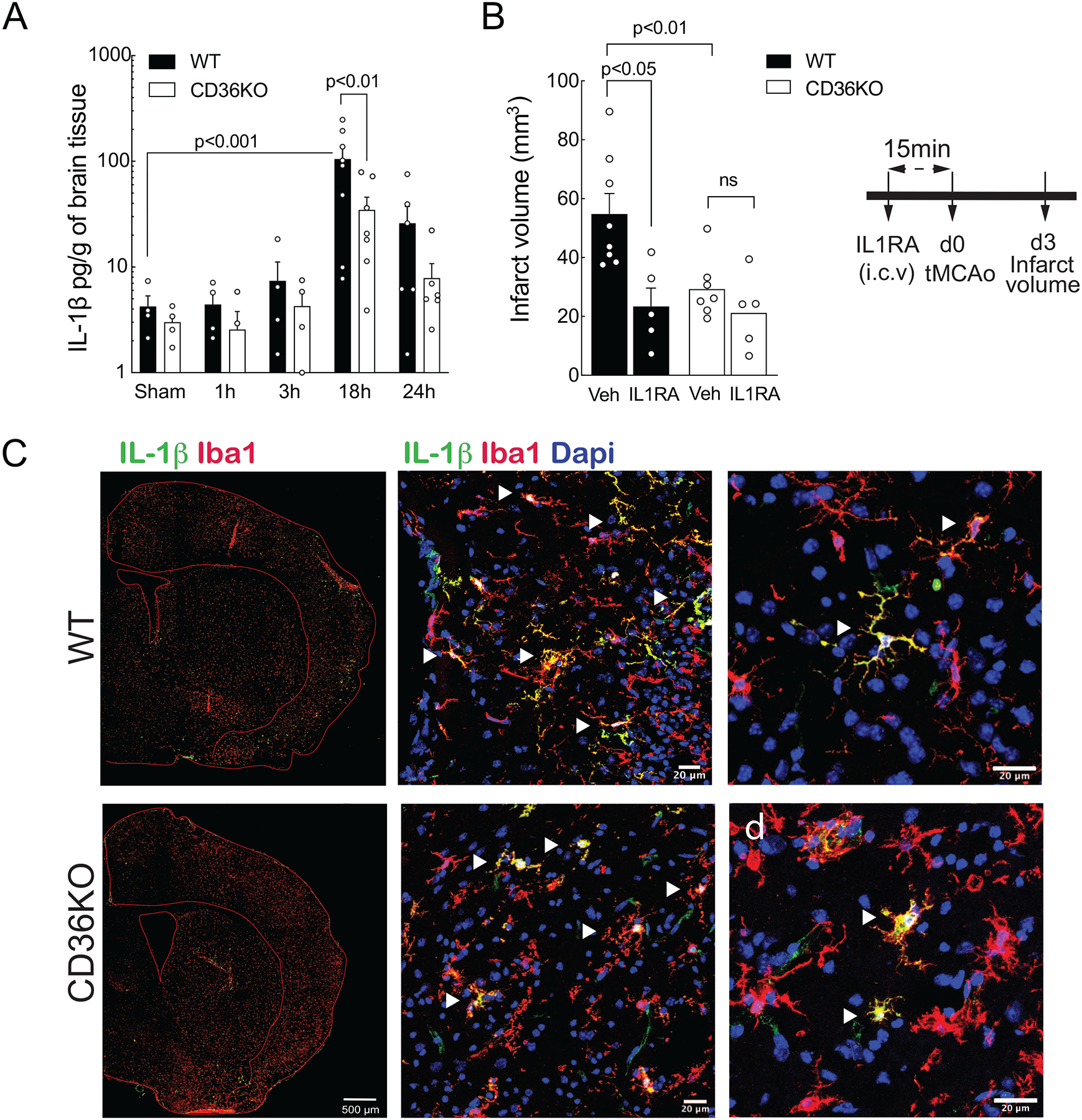
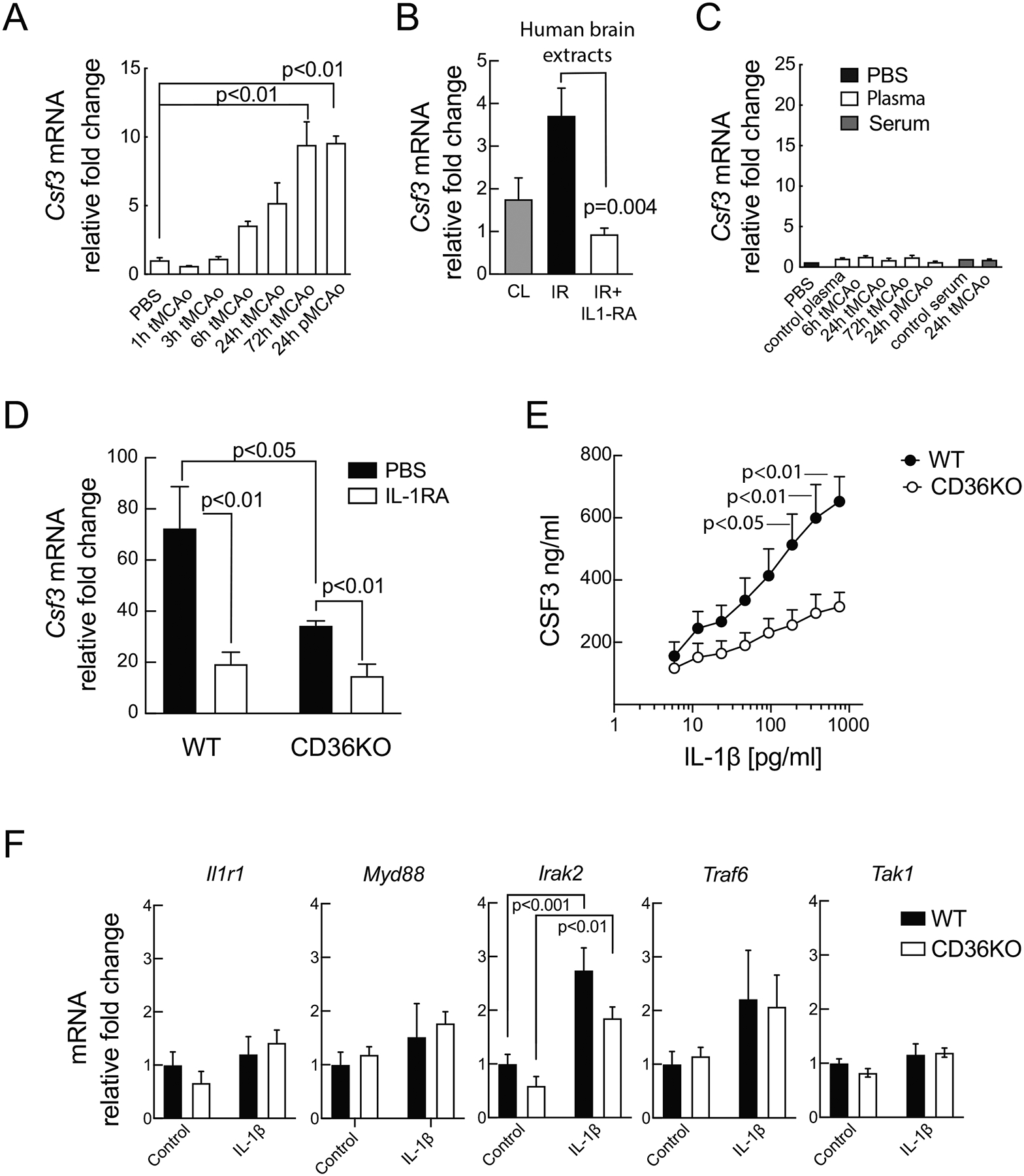
Cerebral ischemia is associated with an acute inflammatory response that contributes to the resulting injury. The innate immunity receptor CD36, expressed in microglia and endothelium, and the pro-inflammatory cytokine interleukin-1β (IL-1β) are involved in the mechanisms of ischemic injury. Since CD36 has been implicated in activation of the inflammasome, the main source of IL-1β, we investigated whether CD36 mediates brain injury through the inflammasome and IL-1β. We found that active caspase-1, a key inflammasome component, is decreased in microglia of CD36-deficient mice subjected to transient middle cerebral artery occlusion, an effect associated with a reduction in brain IL-1β. Conditional deletion of CD36 either in microglia or endothelium reduced ischemic injury in mice, attesting to the pathogenic involvement of CD36 in both cell types. Application of an ischemic brain extract to primary brain endothelial cell cultures from wild type (WT) mice induced IL-1β-dependent endothelial activation, reflected by increases in the cytokine colony stimulating factor-3, a response markedly attenuated in CD36-deficient endothelia. Similarly, the increase in colony stimulating factor-3 induced by recombinant IL-1β was attenuated in CD36-deficient compared to WT endothelia. We conclude that microglial CD36 is a key determinant of post-ischemic IL-1β production by regulating caspase-1 activity, whereas endothelial CD36 is required for the full expression of the endothelial activation induced by IL-1β. The data identify microglial and endothelial CD36 as critical upstream components of the acute inflammatory response to cerebral ischemia and viable putative therapeutic targets.

Keywords: Caspase-1; Csf3; IL-1β; Neuroinflammation; Pattern recognition receptors; Stroke.


Conflict of interest statement

Conflicts of interest to declare

CI serves on the Scientific Advisory Board of Broadview Ventures. The other authors have no conflict of interest to declare.

Figures

Figure 1.
Figure 1.. CD36 regulates IL-1β production after cerebral ischemia.
A. IL-1β protein was measured in the brain of wild type (WT) and CD36KO mice at 1, 3, 18 and 24 hours after tMCAo. IL-1β level peaked 18 hours after tMCAo. This increase was significantly reduced in CD36KO mice as compared to wild type WT mice (n=4–8 mice/group). Statistical analysis: two-way ANOVA (interaction, F (4, 41) = 1.833, P = 0.1410; time, F (4, 41) = 6.822, P = 0.0003; group, F (4, 41) = 2.841, P= 0.0995) followed by Sidak’s and Dunnett’s post-hoc test. B. Infarct volume in mice pretreated with recombinant IL-1 receptor antagonist (IL-1RA; i.c.v.) (timeline on the right). Administration of IL-1RA decreased infarct volume in WT but not in CD36KO mice (n=7–5 mice/group). Two-way ANOVA (interaction, F (1, 19) = 4.094, P = 0.056; treatment, F (1, 19) = 5.573, P = 0.0291; group, F (1, 19) = 11.68, P= 0.0029) followed by Sidak’s post-hoc test. C. Immunofluorescent images of the ischemic hemisphere 18 hours after tMCAo show specific cellular localization of IL-1β (green) with the microphage/microglia marker Iba-1 in both WT and CD36KO mice.
Figure 2.
Figure 2.. Microglial CD36 is involved in caspase-1 activation but may not contribute to inflammasome priming.
A. Gene expression of Il1b, Il1a and the inflammasome Nrlp3, Pycard, Casp1 genes in microglia sorted from WT and CD36KO mice 16 hours after tMCAo (n=5–6 pool/group; pool=2 mice). Both Il1b and Il1a mRNA levels were similarly increased in either WT or CD36KO microglia; Il1b: Brown-Forsythe ANOVA F=5.661 (2.00, 7.97), P=0.0295; Il1a: one-way ANOVA F (2, 14)=3.566, P=0.0560; No changes were observed in Nrlp3 mRNA levels (Brown-Forsythe ANOVA F=1.634 (2.00, 9.06), P=0.2478), whereas both Pycard and Casp1 genes were downregulated after tMCAo (Pycard: Kruskall-Wallis, H=7.906, P=0.0127; Casp1: one-way ANOVA F (2, 15)=63.85, P<0.0001). No differences were found between WT and CD36KO groups in gene expression levels. B. Active caspase-1 levels in microglia from WT and CD36KO mice undergoing tMCAO measured by FLICA assay and flow cytometry. In the graph (left), levels are expressed by the percentage of microglia with activate (cleaved) caspase-1 over total microglia. Only live microglia, propidium iodide (PI) negative cells, were considered. Active caspase-1 significantly increased at 18 hours post-tMCAo in microglia from WT mice but not CD36KO mice (n=3–7 mice/group). Right panels show the flow cytometry strategy used to identify microglial active caspase-1 in WT and CD36KO mice. Microglia were phenotyped as cells expressing intermediate levels of CD45 (CD45int) and high levels of CX3CR1 and CD11b. Active caspase-1 cells were identified as FLICA+ and PI-cells. Two-way ANOVA (interaction, F (4, 38) = 1.794, P = 0.1502; time, F (4, 38) = 10.70, P < 0.0001; group, F (1, 38) = 3.645, P= 0.0638) followed by Sidak’s post-hoc test. C. Mice were sacrificed after i.c.v. administration of the caspase-1 inhibitor Ac-YVAD-cmk and tMCAo, and neuronal degeneration was analyzed by Fluoro Jade (Fluoro J) staining (see top left panel for timeline). Lower left: quantification of Fluoro J positive cells in the ischemic hemisphere (lower left). Two-way ANOVA (interaction, F (1, 16) = 5.485, P = 0.0324; treatment, F (1, 16) = 6.013, P = 0.026; group, F (1, 16) = 18.96, P= 0.005) followed by Sidak’s post-hoc test. Lower right: representative images of Fluoro J staining of the ischemic hemisphere. AcY, Ac-YVAD-cmk.
Figure 3.
Figure 3.. Specific deletion of CD36 in either endothelium or microglia protects from ischemic brain injury.
A. Experimental timeline of tamoxifen treatment and tMCAo induction. P3: post-natal day 3; B. Specificity of endothelial and microglial Cre recombinase activity after tamoxifen injection was assessed in Ve-CreER (top panels) and CX3CR1-CreER mice (bottom panels) crossed with Td-Tomato reporter mice, respectively. Tomato fluorescence (red signal) examination and histological evaluation of EC on brain sections stained for the endothelial marker CD31 (green signal), showed complete colocalization of Ve-CreER recombinase activity with EC but not with Iba1+ microglia (cyan signal). Evaluation of tomato fluorescence signal (red) of CX3CR1-CreER recombinase activity showed complete colocalization of recombinase activity in microglia (Iba1+ cells, cyan) but not in EC (green). C. Relative presence of CD36 genomic DNA of deleted exon 5 over non-deleted exon 16 in flow-sorted isolated brain endothelial cells (EC) and microglia (MG) from EC-CD36KO mice and from MG-CD36KO tamoxifen treated mice. Infarct volume (D) and neurological deficit (E) in 8-week old CD36f/f, endothelial CD36KO (EC CD36KO) and microglial CD36KO (MG CD36KO) mice 3 days after tMCAo. D. Both EC-CD36KO and MG-CD36KO mice had smaller infarcts than CD36f/f control mice (n=13–24 mice/group). Statistical analysis: one-way ANOVA F (2, 47) =8.478, P=0.0007 followed by Bonferroni’s post-hoc test. E. Neurological deficits were lower in either EC CD36KO or MG CD36KO mice as compared to CD36f/f mice (n=21–12 mice/group). Wilcoxon signed rank test, theoretical media, 1; W=36, ap<0.0078; W=21, NS= 0.1094 and W=9, NS=0.5312, respectively. F. Survival rate did not differ among groups. Log-rank Mantel-Cox test χ2= 0.5224, p=0.7701. G. Infarct volume in 8-week old wild type (WT), constitutive CD36KO (CD36KO), non-treated CD36f/f (CD36f/f) and CD36f/f pan-Cre+ (Sox2Cre+) mice. Statistical analysis: one-way ANOVA F (3, 19) = 13.20, P=0.0001 followed by Sidak’s post-hoc test.
Figure 4.
Figure 4.. CD36 is required for IL-1β-induced upregulation of Csf3 in brain endothelia.
A. Mouse EC cultures (bEnd.3 cells) treated with brain extracts from WT mice undergoing 1 to 72 hours of transient cerebral ischemia (tMCAo) or 24 hours of permanent ischemia (pMCAo). One-way ANOVA F (6,15) = 10.52, P=0.0001 followed by Dunnett’s post-hoc test. B. WT mouse primary brain EC (MBEC) treated with post-mortem human brain extracts from the ischemic region (IR) or the contralateral side (CL), or co-treated with human brain extracts from the IR and IL-1RA. Kruskal-Wallis test (3, 15) =10.08, P<0.0009) followed by Dunn’s post-hoc test. C. Relative Csf3 mRNA fold change in mouse EC cultures (bEnd.3 cells) treated with plasma or serum from mice undergoing 6 to 72 hours of tMCAo or 24 hours of pMCAo. D. Relative Csf3 mRNA fold change in WT and CD36KO MBEC after stimulation with ischemic brain extracts and co-treatment with IL-1RA or PBS. Csf3 mRNA was suppressed in cells treated with IL-1RA. Two-way ANOVA (interaction, F (1, 14) = 2.91, P = 0.1102; IL-1RA, F (1, 14) = 13.77, P = 0.0023; group, F (1, 14) = 4.77, P = 0.0465) followed by Tukey’s post-hoc test. E. IL-1β log dose-response curves for secretion of CSF3 by WT or CD36KO MBEC for 24 hours. Two-way ANOVA (interaction, F (8, 188) = 2.193, P = 0.0297; IL-1RA, F (8, 188) = 11.03, P < 0.0001; group, F (1, 188) = 34.34, P < 0.0001) followed by Sidak’s post-hoc test. F. Relative mRNA levels of IL1r1, Myd88, Irak2, Traf6, and Tak1 in cultured WT or CD36KO MBEC. Irak2: Two-way ANOVA (interaction, F (1, 25) = 0.9587, P = 0.3369; IL-1β, F (1, 25) = 36.59, P < 0.0001; group, F (1, 25) = 6.779, P=0.0153) followed by Sidak’s post-hoc test.
Figure 5.
Figure 5.. Potential mechanisms by which CD36 contributes to neuroinflammation after cerebral ischemia.
After a brain ischemia, danger signals (DAMPs) released from dead and damaged neurons [1] are recognized by pattern-recognition receptor, such as CD36 and TLRs, expressed in microglia. In microglia, TLR signaling triggers the expression of pro-IL-1b and Nlrp3, providing the priming signal of the inflammasome [2]. A second signal modulated by CD36 triggers inflammasome activation [3] and caspase-1-dependent cleavage of pro-IL-1β into mature IL-1β [4]. Secreted IL-1β engages IL-1 receptors on endothelial cells, stimulating transcriptional NF-κb activity [5]. At the same time, DAMPs may bind to CD36 to enhance IL-1β signaling [5]. NF-κb transcriptional activity ultimately leads to increased production of CSF3 and other cytokines [6], which activate infiltrating neutrophils, aggravating inflammatory injury through free radical production and other pathogenic mechanisms [7].

References

    1. Abe T, Shimamura M, Jackman K, Kurinami H, Anrather J, Zhou P, Iadecola C, 2010. Key role of CD36 in Toll-like receptor 2 signaling in cerebral ischemia. Stroke 41, 898–904. - PMC - PubMed
    1. Abulafia DP, de Rivero Vaccari JP, Lozano JD, Lotocki G, Keane RW, Dietrich WD, 2009. Inhibition of the inflammasome complex reduces the inflammatory response after thromboembolic stroke in mice. J Cereb Blood Flow Metab 29, 534–544. - PubMed
    1. Allan SM, Tyrrell PJ, Rothwell NJ, 2005. Interleukin-1 and neuronal injury. Nat Rev Immunol 5, 629–640. - PubMed
    1. Amantea D, Nappi G, Bernardi G, Bagetta G, Corasaniti MT, 2009. Postischemic brain damage: pathophysiology and role of inflammatory mediators. FEBS J 276, 13–26. - PubMed
    1. Bao Y, Qin L, Kim E, Bhosle S, Guo H, Febbraio M, Haskew-Layton RE, Ratan R, Cho S, 2012. CD36 is involved in astrocyte activation and astroglial scar formation. J Cereb Blood Flow Metab 32, 1567–1577. - PMC - PubMed

Publication types

LinkOut - more resources