

Ultra-high field (7T) functional magnetic resonance imaging in amyotrophic lateral sclerosis: a pilot study
- PMID: 33872993
- PMCID: PMC8060594
- DOI: 10.1016/j.nicl.2021.102648
Ultra-high field (7T) functional magnetic resonance imaging in amyotrophic lateral sclerosis: a pilot study
Abstract
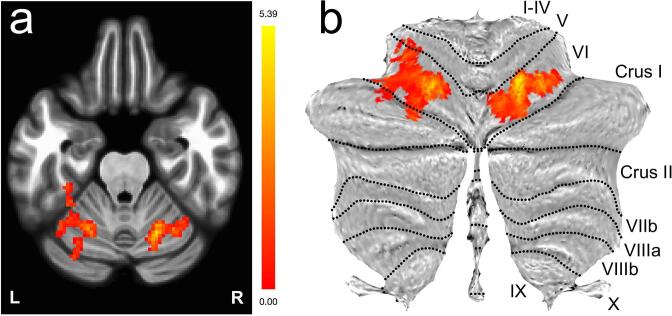
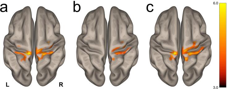
Amyotrophic lateral sclerosis (ALS) is a neurodegenerative disease of the central nervous system that results in a progressive loss of motor function and ultimately death. It is critical, yet also challenging, to develop non-invasive biomarkers to identify, localize, measure and/or track biological mechanisms implicated in ALS. Such biomarkers may also provide clues to identify potential molecular targets for future therapeutic trials. Herein we report on a pilot study involving twelve participants with ALS and nine age-matched healthy controls who underwent high-resolution resting state functional magnetic resonance imaging at an ultra-high field of 7 Tesla. A group-level whole-brain analysis revealed a disruption in long-range functional connectivity between the superior sensorimotor cortex (in the precentral gyrus) and bilateral cerebellar lobule VI. Post hoc analyses using atlas-derived left and right cerebellar lobule VI revealed decreased functional connectivity in ALS participants that predominantly mapped to bilateral postcentral and precentral gyri. Cerebellar lobule VI is a transition zone between anterior motor networks and posterior non-motor networks in the cerebellum, and is associated with a wide range of key functions including complex motor and cognitive processing tasks. Our observation of the involvement of cerebellar lobule VI adds to the growing number of studies implicating the cerebellum in ALS. Future avenues of scientific investigation should consider how high-resolution imaging at 7T may be leveraged to visualize differences in functional connectivity disturbances in various genotypes and phenotypes of ALS along the ALS-frontotemporal dementia spectrum.
Keywords: 7 Tesla; Amyotrophic lateral sclerosis; Cerebellum; Functional magnetic resonance imaging; Ultra-high field.
Copyright © 2021 The Author(s). Published by Elsevier Inc. All rights reserved.
Conflict of interest statement
Christina Triantafyllou, PhD, is currently employed by Siemens Healthineers. Nazem Atassi, MD, PhD, is currently employed by Sanofi Genzyme. The other authors declare that they have no known competing financial interests or personal relationships that could have appeared to influence the work reported in this paper.
Figures
References
-
- Abidi M., de Marco G., Couillandre A., Feron M., Mseddi E., Termoz N. Adaptive functional reorganization in amyotrophic lateral sclerosis: coexisting degenerative and compensatory changes. Eur. J. Neurol. 2020;27:121–128. - PubMed
-
- Abrahams S., Goldstein L.H., Kew J.J., Brooks D.J., Lloyd C.M., Frith C.D. Frontal lobe dysfunction in amyotrophic lateral sclerosis. A PET study. Brain. 1996;119(Pt 6):2105–2120. - PubMed
-
- Abrahams S., Leigh P.N., Goldstein L.H. Cognitive change in ALS: a prospective study. Neurology. 2005;64:1222–1226. - PubMed
-
- Agosta F., Pagani E., Petrolini M., Sormani M.P., Caputo D., Perini M. MRI predictors of long-term evolution in amyotrophic lateral sclerosis. Eur. J. Neurosci. 2010;32:1490–1496. - PubMed
Publication types
MeSH terms
Grants and funding
LinkOut - more resources
Full Text Sources
Other Literature Sources
Medical
Miscellaneous
