Review
. 2021 Jul 5:219:113446.
doi: 10.1016/j.ejmech.2021.113446. Epub 2021 Apr 10.

AAA ATPases as therapeutic targets: Structure, functions, and small-molecule inhibitors

Affiliations
Review

AAA ATPases as therapeutic targets: Structure, functions, and small-molecule inhibitors

Gang Zhang et al. Eur J Med Chem. .

Abstract

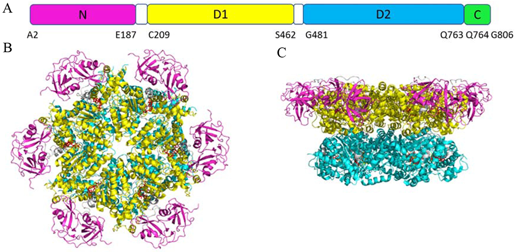
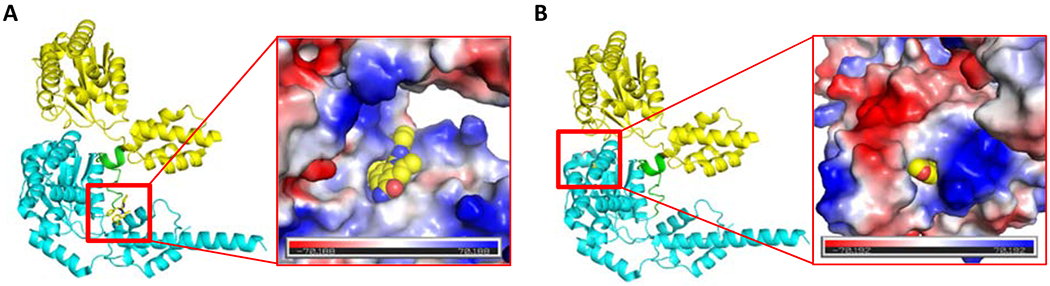
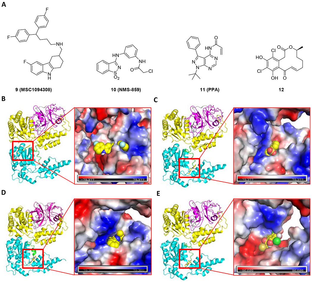
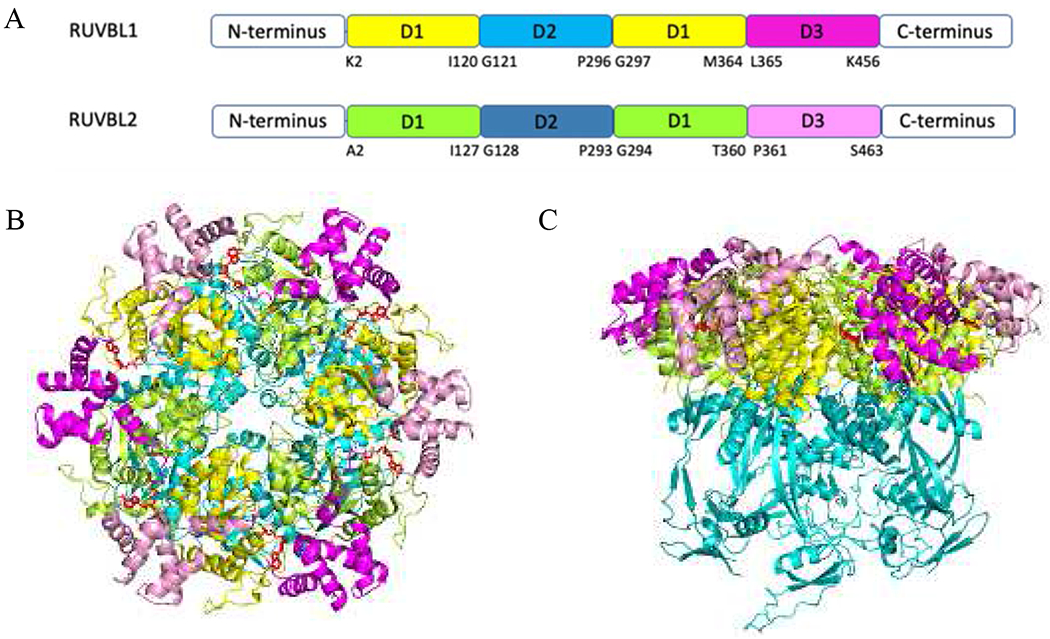
ATPases Associated with Diverse Cellular Activity (AAA ATPase) are essential enzymes found in all organisms. They are involved in various processes such as DNA replication, protein degradation, membrane fusion, microtubule serving, peroxisome biogenesis, signal transduction, and the regulation of gene expression. Due to the importance of AAA ATPases, several researchers identified and developed small-molecule inhibitors against these enzymes. We discuss six AAA ATPases that are potential drug targets and have well-developed inhibitors. We compare available structures that suggest significant differences of the ATP binding pockets among the AAA ATPases with or without ligand. The distances from ADP to the His20 in the His-Ser-His motif and the Arg finger (Arg353 or Arg378) in both RUVBL1/2 complex structures bound with or without ADP have significant differences, suggesting dramatically different interactions of the binding site with ADP. Taken together, the inhibitors of six well-studied AAA ATPases and their structural information suggest further development of specific AAA ATPase inhibitors due to difference in their structures. Future chemical biology coupled with proteomic approaches could be employed to develop variant specific, complex specific, and pathway specific inhibitors or activators for AAA ATPase proteins.

Keywords: AAA ATPases; ATAD2; RUVBL1/2; Small molecule inhibitors; p97.


Conflict of interest statement

Declaration of competing interest The authors declare that they have no known competing financial interests or personal relationships that could have appeared to influence the work reported in this paper.

Figures

Figure 1.
Figure 1.
(A) Schematic diagram of the p97 domain organization. (B) Top view of the crystal structure of p97 (PDB code 5FTK). (C) Side view of p97. N domain, D1, D2, and C-terminal are presented in magenta, yellow, cyan, and green, respectively.
Figure 2.
Figure 2.
Chemical structure of p97 inhibitors 1-8.
Figure 3.
Figure 3.
CB-5083 (PDB: 6MCK, A) and NMS-873 (proposed binding mode, PDB: 6MCK, B) in binding sites of p97 with a vacuum electrostatic surface. The D1, D1D2 linker, and D2 are presented as cartoons in yellow, green and cyan, respectively.
Figure 4.
Figure 4.
Chemical structure (A) and the proposed binding mode of MSC1094308 (9, PDB code 5FTJ, B), NMS-859 (10, PDB code 5FTJ, C), PPA (11, PDB code 5FTK, D) and curvularin (12, PDB code 5FTJ, E) with p97. The N, ND1 linker, D1, D1D2 linker, and D2 are presented as cartoons in magenta, gray, yellow, green, and cyan, respectively.
Figure 5.
Figure 5.
(A) Schematic diagram of the RUVBL1 and RUVBL2 domain organization. (B) Top view of the crystal structure of RUVBL1/2 complex (PDB code 5OAF). (C) Side view of RUVBL1/2 complex. For RUVBL1, D1, D2 and D3 are presented as a cartoon in yellow, cyan, and magenta; For RUVBL2, D1, D2 and D3 are presented as cartoons in limon, teal, and pink. ADP are presented as red sticks.
Figure 6.
Figure 6.
Overlay of RUVBL1/2 complex with (5OAF) or without (6IGM) ADP. (A) Overlay of full-length RUVBL1/2 complex, RUVBL1/2 complex with ADP (5OAF) is in green (ADP in red), RUVBL1/2 complex without ADP (6IGM) is in cyan. (B) Overlay of the binding site of ADP. ADP is in yellow. The interacted residues are presented as lines. The conserved HSH motif, walker A, walker B from RUVBL1 are presented as a cartoon.
Figure 7.
Figure 7.
Chemical structure of 13-19
Figure 8.
Figure 8.
Chemical structure of 20-27
Figure 9.
Figure 9.
Overlay of the binding site of phenyl sulfonamide (cyan, PDB code 6S55) and KAc (yellow, PDB code 5A5N). The four conserved water molecules (PDB code 5A5N) are presented as red spheres.
Figure 10.
Figure 10.
(A) Schematic diagram of the spastin domain organization. (B) The top view of the crystal structure of hexameric spastin (PDB code 6PEN). (C) The structure of the monomeric spastin.
Figure 11.
Figure 11.
(A) Chemical structure of 28-32. (B) The binding site of compound 29 on spastin with a vacuum electrostatic surface (PDB code 6NYV). (C) The superimposition of compounds 29 and 30 in the binding site (PDB code 6NYV). Compounds 29 and 30 are presented as yellow and cyan sticks, respectively. (D)The superimposition of compound 31 (PDB code 6P10) and compound 32 (PDB code 6P12) at the binding site of spastin. Compound 31 and 32 are presented as green sticks and cyan lines, respectively. The key residues are presented as sticks or lines in salmon.
Figure 12.
Figure 12.
(A) Schematic diagram of the Midasin and dynein. (B) Crystal structure of Midasin (PDB code 6YLH) and dynein (PDB code 4RH7). Midasin: N domain, AAA domains, Tail, and MIDAS are presented in blue, red, green, and pink, respectively. Dynein: Tail, Linker, AAA domains, Stalk, C domain, and MTBD are presented in gray, orange, red, teal, cyan, and violet, respectively.
Figure 13.
Figure 13.
Chemical structure of 33 - 37

References

    1. Ranson NA, White HE, Saibil HR, Chaperonins, Biochem. J, 333 (Pt 2) (1998) 233–242. - PMC - PubMed
    1. Hirokawa N, Noda Y, Okada Y, Kinesin and dynein superfamily proteins in organelle transport and cell division, Curr. Opin. Cell Biol, 10 (1998) 60–73. - PubMed
    1. Otsuji N, Iyehara H, Hideshima Y, Isolation and Characterization of an Escherichia coli ruv Mutant Which Forms Nonseptate Filaments After Low Doses of Ultraviolet Light Irradiation, J. Bacteriol, 117 (1974) 337–344. - PMC - PubMed
    1. Lee DG, Bell SP, ATPase switches controlling DNA replication initiation, Curr. Opin. Cell Biol, 12 (2000) 280–285. - PubMed
    1. Yang W, Structure and function of mismatch repair proteins, Mutat. Res, 460 (2000) 245–256. - PubMed

MeSH terms

LinkOut - more resources