. 2021;58(4):252-266.
doi: 10.1159/000514555. Epub 2021 Apr 19.

R-Ras Deficiency in Pericytes Causes Frequent Microphthalmia and Perturbs Retinal Vascular Development

Affiliations

R-Ras Deficiency in Pericytes Causes Frequent Microphthalmia and Perturbs Retinal Vascular Development

Jose Luis Herrera et al. J Vasc Res. 2021.

Abstract

Purpose: The retinal vasculature is heavily invested by pericytes. Small GTPase R-Ras is highly expressed in endothelial cells and pericytes, suggesting importance of this Ras homolog for the regulation of the blood vessel wall. We investigated the specific contribution of pericyte-expressed R-Ras to the development of the retinal vasculature.

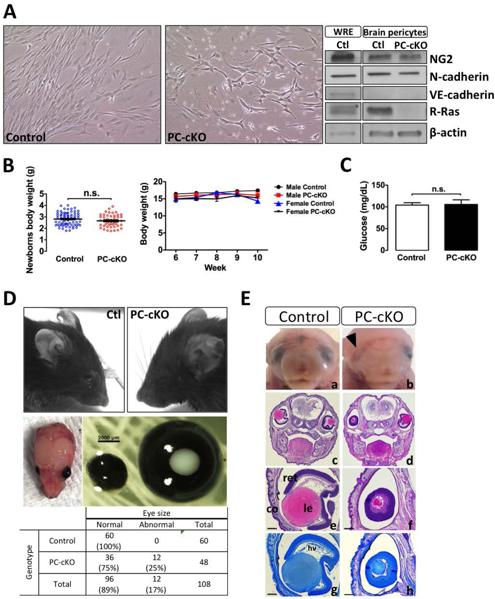
Methods: The effect of R-Ras deficiency in pericytes was analyzed in pericyte-targeted conditional Rras knockout mice at birth and during the capillary plexus formation in the neonatal retina.

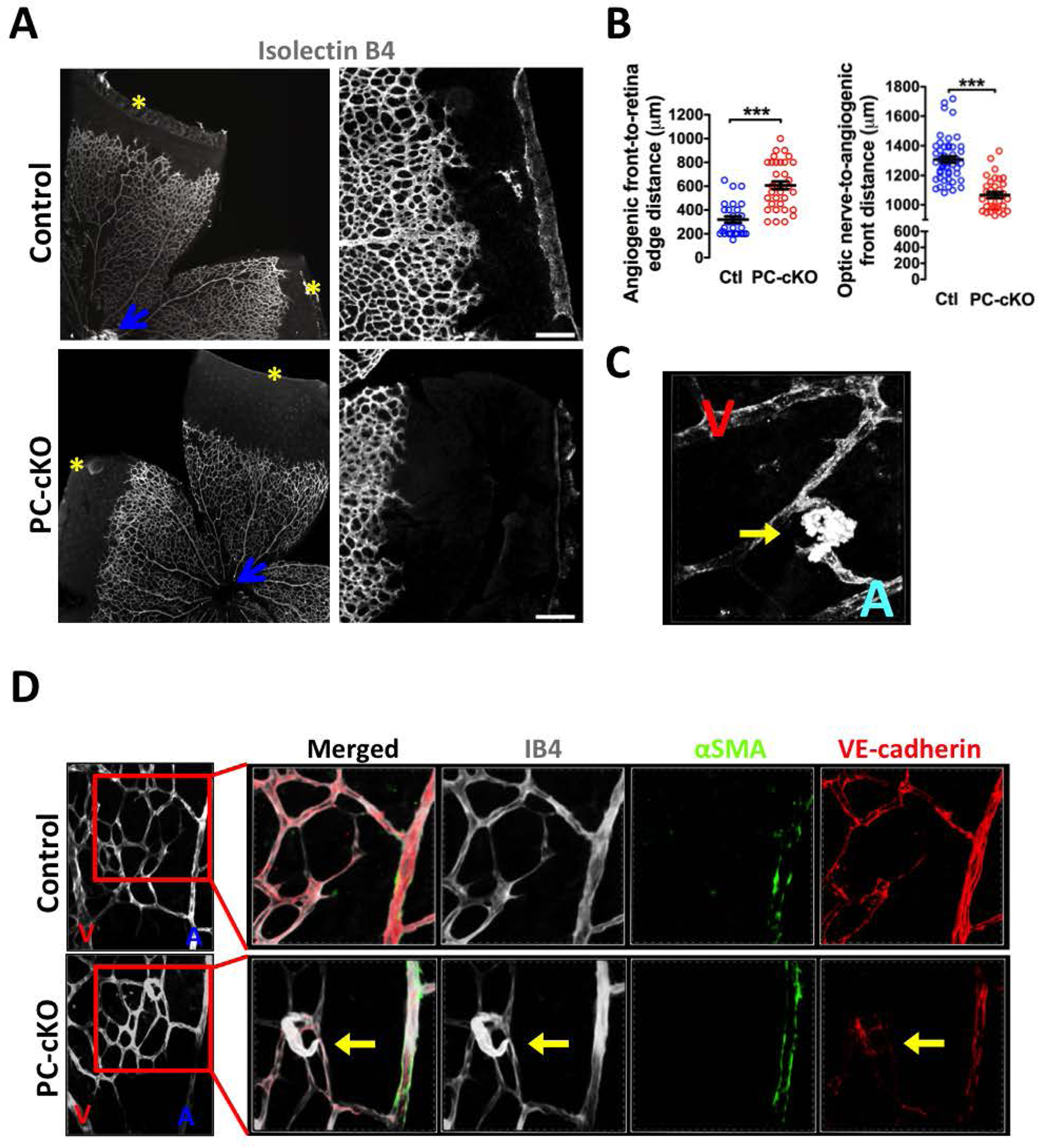
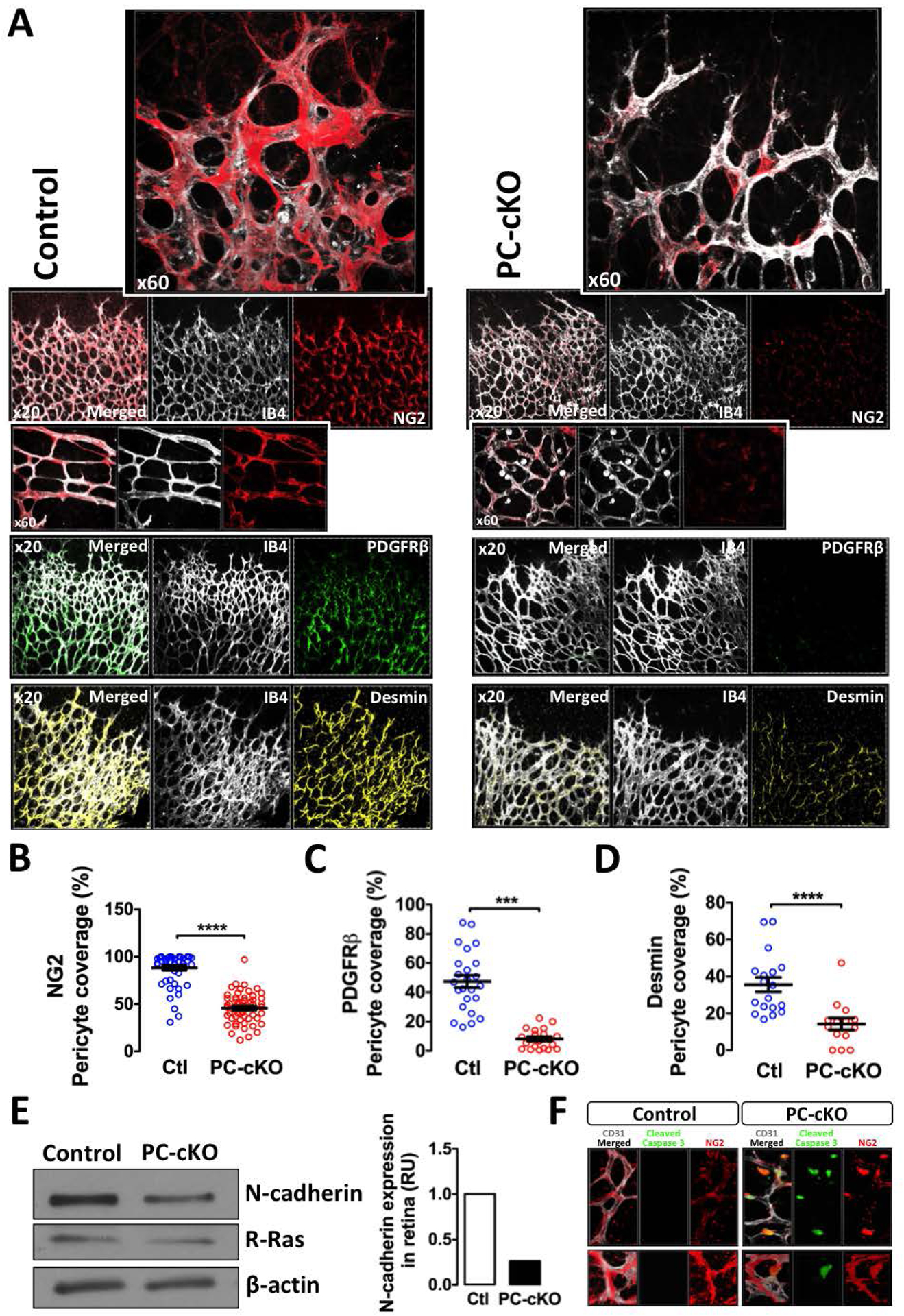
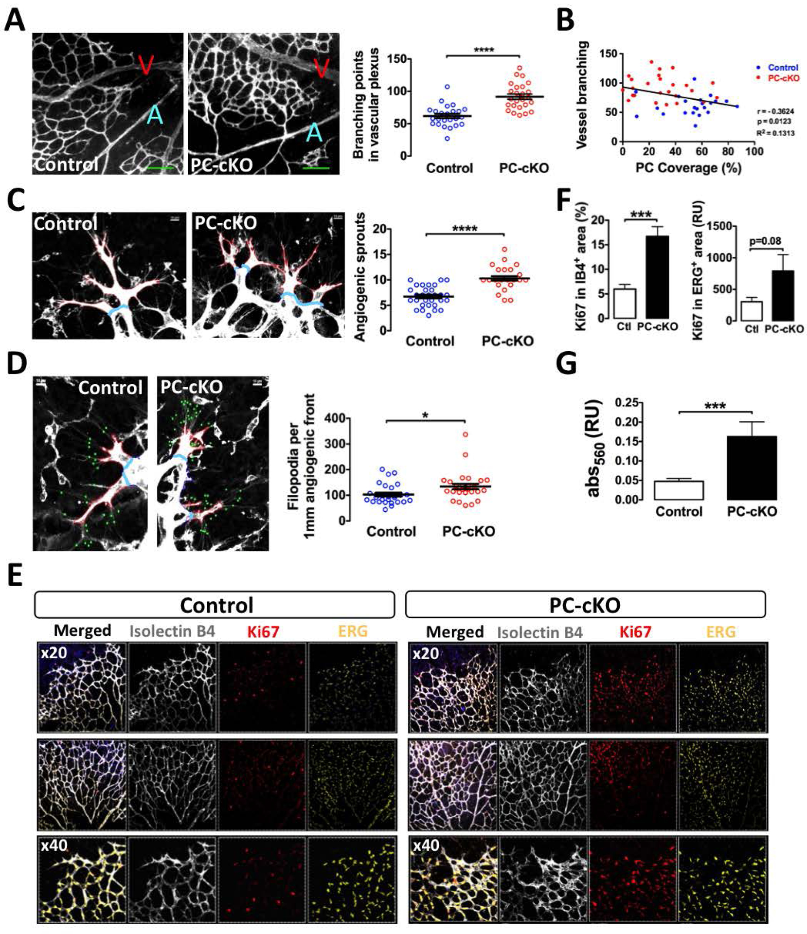
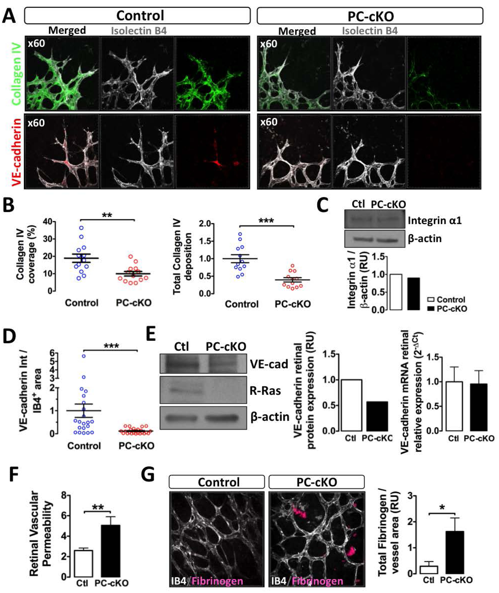
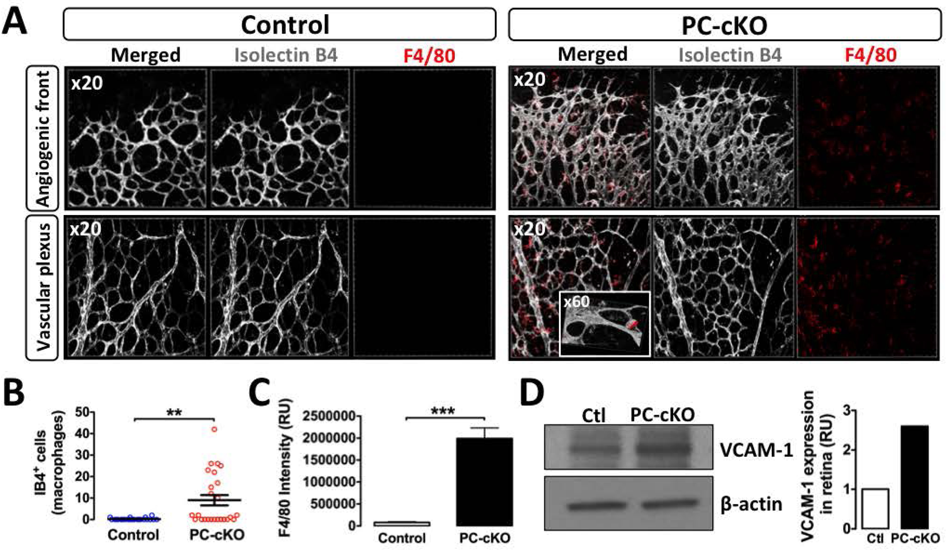
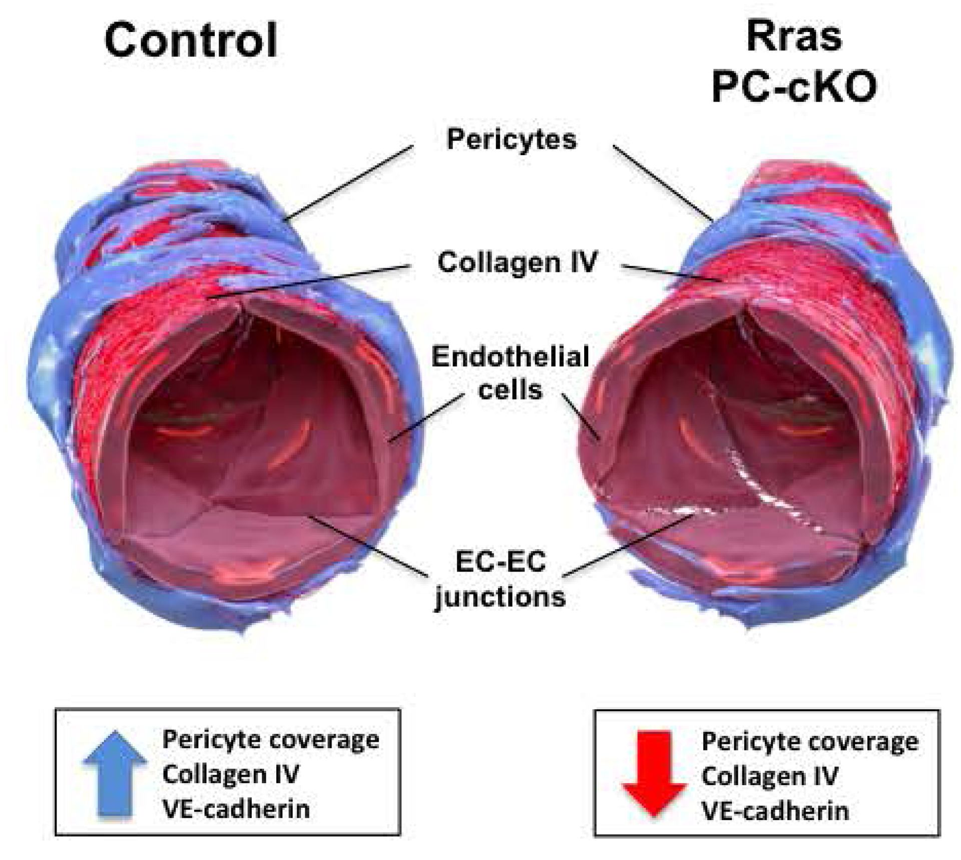
Results: The offspring of these mice frequently exhibited unilateral microphthalmia. Analyses of the developing retinal vasculature in the eyes without microphthalmia revealed excessive endothelial cell proliferation, sprouting, and branching of the capillary plexus in these animals. These vessels were structurally defective with diminished pericyte coverage and basement membrane formation. Furthermore, these vessels showed reduced VE-cadherin staining and significantly elevated plasma leakage indicating the breakdown of the blood-retinal barrier. This defect was associated with considerable macrophage infiltration in the retina.

Conclusions: The normal retinal vascular development is dependent on R-Ras expression in pericytes, and the absence of it leads to unattenuated angiogenesis and significantly weakens the blood-retinal barrier. Our findings underscore the importance of R-Ras for pericyte function during the normal eye development.

Keywords: Angiogenesis; Pericyte; R-Ras; Retina; Vascular permeability.


Conflict of interest statement

Conflict of interest statement

The authors have no conflicts of interest to declare.

Figures

Fig. 1.
Fig. 1.. Rras PC-cKO mice are frequently born with microphthalmia
(A) Cultured brain microvascular pericytes isolated from Rras PC-cKO mice exhibit altered morphology. Lysate of these cells was analyzed by western blot for the expression of NG2 (pericyte marker), VE-cadherin (EC marker), R-Ras, N-cadherin, and β-actin (loading control) to demonstrate the lack of R-Ras in pericytes. Pooled whole retina extract (WRE) from control mice was used as a control for antibodies. Eight retinas were pooled. (B) Bodyweight at P5 and 6–10 weeks old was compared between Rras PC-cKO and control mice. n=45 or 73 P5 mice were examined for Rras PC-cKO or control group. n=5 or 6 mice for 6–10 weeks old bodyweight. Results are shown as Mean ± S.E.M. No significant statistical difference was found (n.s.) (C) Blood glucose level analyzed at P5 (D) Twenty five percent of Rras PC-cKO mice exhibits microphthalmia in one eye at birth. Pictures of microphthalmia and contralateral eyes of adult Rras PC-cKO mice are shown. (E) Gross anatomical features of PC-cKO mice. Microphthalmia is present at birth (a, b; arrowhead). H&E (c-f) and toluidine blue (g, h) staining of histological sections of a P0 PC-cKO mouse displays a microphthalmic eye with a small lens surrounded by retina (f, h). The contralateral eyes of the mice displaying microphthalmia did not show any sign of abnormal gross anatomy (d). Ret, retina; le, lens; co, cornea; hv, hyaloid vessels. Scale bar, 100 μm.
Fig. 2.
Fig. 2.. Abnormal vascular development in Rras PC-cKO neonatal retina
(A) PC-cKO and control retinas at P5 were cut into four segments, flat-mounted, and stained with isolectin B4 (IB4) to visualize the endothelium of the retinal vasculature. * edge of the retina; blue arrow: central retina. Scale bar, 0.5 μm (B) Distance from the angiogenic front to the edge of the retina in all four segments of each retina (Control: 7 retinas, PC-cKO: 8 retinas; left graph) and distance from the optic nerve to the angiogenic front (Control: 14 retinas, PC-cKO: 9 retinas, right graph). Each dot represents the distance in one segment of retina. P<0.0001 (C) IB4 staining of capillary plexus shows abnormal endothelial growth near capillary branching in the PC-cKO retina (arrow). V, vein; A, artery (D) Abnormal endothelial growth in the PC-cKO capillary plexus co-immunostained with IB4 and VE-cadherin antibody (arrows).
Fig. 3.
Fig. 3.. Absence of R-Ras in pericytes impairs pericyte coverage of capillary plexus
(A) Retinas were harvested at P5, whole-mounted and double-stained with Alexa Fluor-conjugated IB4 (white) for EC staining and an antibody for pericyte marker NG2 (red), PDGFRβ (green) or desmin (yellow). 3-D images of the stained vascular plexus are shown (B-D) Intersecting areas between ECs (IB4+) and NG2+ (B), PDGFRβ+ (C) or desmin+ (D) pericytes were quantified in 3-D images and presented as % pericyte coverage. For NG2, at least 52 pictures from 5 retinas were examined for each group. For PDGFRβ, at least 21 pictures from 3 retinas were examined. For desmin, at least 14 pictures from 3 retinas were examined. Results are shown as Mean values ± S.E.M. P<0.0001 (E) Western blot analysis of N-cadherin in whole retina extract. Twelve retinas were pooled for each group (F) Retinas (n=3) were triple-stained with Isolectin B4 (white), a pericyte marker NG2 (red) and an antibody for apoptosis Cleaved Caspase-3 (green). Confocal 3-D images show the presence of apoptotic pericytes in the PC-cKO retinal vessels. A: Artery.
Fig. 4.
Fig. 4.. R-Ras deficiency in pericytes leads to excessive angiogenic activities of ECs
(A) Analysis of vessel branching in the capillary plexus. Branching points were defined as junctions of capillary segments and counted in the observation field. Total 25 retina pictures from 4 mice were examined for each group. P<0.0001, V=vein, A=artery. Scale bar, 100 μm (B) Correlation analysis between pericyte coverage and vessel branching in the capillary plexus. An inverse correlation exists between the two parameters (Pearson’s r = −0.3624, P= 0.0123, R2= 0.1313) (C) Endothelial sprouts (red lines) were counted in the angiogenic front of the growing capillary plexus. Blue lines indicate the base of the sprouts. Total 30 retina pictures of 4 mice were examined for each group. P<0.0001, Scale bar, 10 μm (D) The number of filopodia (green dots) was counted per endothelial vessel length (red lines). Total 28 retina pictures of 4 mice were examined per group. P=0.019, Scale bar, 10 μm (E-F) EC proliferation was assessed by anti-Ki-67 (red) and IB4 (white; left graph) or anti-ERG (yellow; right graph) double staining of P5 retina to identify Ki-67+ cycling cells in the endothelium. Proliferation of ECs was assessed by the % fraction of Ki-67+IB4+ area in total IB4+ area (left) or by the Ki-67 intensity within ERG+ nuclei normalized for the total ERG+ area in the image (right). Six mice were examined for each group. P<0.0001 (G) MTT assay to assess proliferation of cultured brain microvascular pericytes isolated from R-Ras PC-cKO or control mice. Mean ± S.D. P<0.0001
Fig. 5.
Fig. 5.. Impaired basement membrane formation and disruption of endothelial barrier in Rras PC-cKO retinal vasculature
(A) Representative images of collagen IV (green) and VE-cadherin (red) immunofluorescence of the retina at the angiogenic front. IB4 staining (white) highlights the endothelium. 3-D images were reconstituted by confocal microscopy. (B) Quantification of immunostaining in the 3-D images. The vessel surface area covered by collagen IV matrix (coverage) and the amount of collagen IV deposition deduced from the staining intensity per vessel area (relative value compared with the control) are shown. Three to four 3-D pictures were analyzed for each retina. Four or five mice were examined for each group. **P=0.0031, ***P<0.0001 (C) Integrin α1 protein expression level in brain-isolated pericytes analyzed by western blot (D) A similar 3-D analysis for VE-cadherin. **P=0.0067 (E) VE-cadherin protein and mRNA expression levels in pooled whole retina extract were analyzed by western blot and RT-qPCR. Eight to twelve retinas were pooled for each group. (F) Permeability of the retinal vasculature was quantified by Miles assay and presented with vascular permeability factor. Five to seven retinas were examined. P=0.0089 (G) Vascular permeability was also assessed by the plasma leakage determined by fibrinogen immunostaining of the whole-mounted retina. Data are presented as the total fluorescence intensity of fibrinogen (RU) normalized by the vessel area. Three retinas were examined for each group. P=0.0247
Fig. 6.
Fig. 6.. Infiltrating macrophages accumulate in Rras PC-cKO retina
(A) Whole-mounted P5 retinas were immunostained for a macrophage marker F4/80, co-stained with IB4, and analyzed by confocal 3-D imaging. Inset, a macrophage doubly stained for F4/80+ and IB4+ shown in higher magnification (×60) (B, C) Quantification of IB4+ cell count outside the vessels (macrophages, B) and F4/80 immunofluorescence intensity (C) in the observation. Total of 25 retina pictures of 3 animals were examined for each group. **P=0.0013, ***P<0.0001 (D) Western blot analysis of VCAM-1 in pooled whole retina protein extract. Eight or twelve retinas are pooled for the control or PC-cKO group, respectively.
Fig. 7.
Fig. 7.. Schematic diagram depicting the role of pericyte-expressed R-Ras in retinal capillary vessel stability.
R-Ras in pericytes controls endothelial sprouting, branching, and pericyte coverage, and stabilizes the integrity of endothelial lining by VE-cadherin clustering and collagen IV basement membrane formation, leading to the stabilization of the blood-retinal-barrier. EC, endothelial cell; BM, basement membrane; AJ, adherens junctions.

Similar articles

Cited by

References

    1. Chatterjee S, Naik UP. Pericyte-endothelial cell interaction. Cell Adh Migr. 2012;6(3);159–9. - PMC - PubMed
    1. Jain RK. Molecular regulation of vessel maturation. Nat Med. 2003;9;685–693. - PubMed
    1. Armulik A, Abramsson A, Betsholtz C. Endothelial/Pericyte Interactions. Circ Res. 2005;97:512–523. - PubMed
    1. Vähätupa M, Prince S, Vataja S, Mertimo T, Kataja M, Kinnunen K, et al. Lack of R-Ras leads to increased vascular permeability in ischemic retinopathy. Invest Ophthalmol Vis Sci. 2016;57:4898–4909. - PMC - PubMed
    1. Gaengel K, Genové G, Armulik A, Betsholtz C. Endothelial-mural cell signaling in vascular development and angiogenesis. Arterioscler Thromb Vasc Biol. 2009;29:630–638. - PubMed

Publication types

MeSH terms