Review
. 2021 Nov;29(11):983-992.
doi: 10.1016/j.tim.2021.03.014. Epub 2021 Apr 16.

Respiratory mucus as a virus-host range determinant

Affiliations
Review

Respiratory mucus as a virus-host range determinant

Louisa E Wallace et al. Trends Microbiol. 2021 Nov.

Abstract

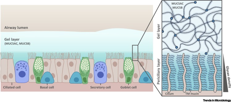
Efficient penetration of the mucus layer is needed for respiratory viruses to avoid mucociliary clearance prior to infection. Many respiratory viruses bind to glycans on the heavily glycosylated mucins that give mucus its gel-like characteristics. Influenza viruses, some paramyxoviruses, and coronaviruses avoid becoming trapped in the mucus by releasing themselves by means of their envelope-embedded enzymes that destroy glycan receptors. For efficient infection, receptor binding and destruction need to be in balance with the host receptor repertoire. Establishment in a novel host species requires resetting of the balance to adapt to the different glycan repertoire encountered. Growing understanding of species-specific mucosal glycosylation patterns and the dynamic interaction with respiratory viruses identifies the mucus layer as a major host-range determinant and barrier for zoonotic transfer.


Conflict of interest statement

There are no interests to declare.

Figures

Figure 1
Figure 1
Schematic diagram of the respiratory mucus layer. The airway surface liquid (ASL) overlays the respiratory epithelia and consists of two layers: the gel layer and the periciliary layer (PCL). The gel layer contains soluble mucins MUC5AC and MUC5B, secreted primarily from goblet cells and mucous cells within submucosal glands (not shown), respectively. The soluble mucins are major contributors to the viscosity and gel-like properties of this layer which enables the impediment of airway pollutants to be cleared by mucociliary clearance. Compared to the gel layer, the PCL – the height of which is approximately that of outstretched cilia – is free of soluble mucins and is therefore less viscous, which provides favorable conditions for ciliary beating. Cilia present on the epithelial surface are rich in transmembrane (TM) mucins (MUC1, MUC4, MUC16, and MUC20) which create a glycan meshwork that increases in density closer to the cell surface, aiding the exclusion of molecules and invading pathogens. This figure represents a general schematic representation of the ciliated respiratory epithelium, thus the term 'secretory cell' may refer to different cell types including club or dense-core granulated cells of the airway epithelium [76].
Figure 2
Figure 2
Mucins and their glycosylation. (A) The domain structure of the soluble mucins MUC5B (5762 amino acids) and MUC5AC (5654 amino acids). The N- and C-terminal von Willebrand factor (vWF)-like regions and cysteine-rich domains are highly conserved between MUC5B and MUC5AC as well as between species. The four central proline/threonine/serine-rich (PTS) regions consist of imperfect repeats (aa; amino acids). PTS repeats are densely decorated with O-linked glycans and their low sequence conservation between species will result in spatial differences in glycan presentation that could potentially affect the binding of a specific virus. (B) Diversity of sialylated O-linked glycan structures present on high-molecular-weight human mucins. Structures were interpreted from the glycan compositions reported in the most extensive analysis of glycans on mucus to date [39]. Note that di-sialylated bi-antennary structures with multiple LacNAc repeats are present. Such structures on mucins are likely to have differential effects on the binding of viruses as has been reported for N-linked glycans [77]. Sulfation of the sialoglycans shown here was also abundant [39] but is not indicated. Abbreviations: Gal, galactose; GalNAc, N-acetylgalactosamine; GlcNAc, N-acetylglucosamine; Neu5Ac, N-acetyl-neuraminic acid.

References

    1. Lillehoj E.P., et al. Cellular and molecular biology of airway mucins. Int. Rev. Cell Mol. Biol. 2013;303:139–202. - PMC - PubMed
    1. Zanin M., et al. The interaction between respiratory pathogens and mucus. Cell Host Microbe. 2016;19:159–168. - PMC - PubMed
    1. Matrosovich M., et al. In: SialoGlyco Chemistry and Biology II: Tools and Techniques to Identify and Capture Sialoglycans. Gerardy-Schahn R., et al., editors. Springer International Publishing; 2015. Sialic acid receptors of viruses; pp. 1–28.
    1. Barnard K.N., et al. Modified sialic acids on mucus and erythrocytes inhibit influenza A virus hemagglutinin and neuraminidase functions. J. Virol. 2020;94 - PMC - PubMed
    1. Varki A., et al. In: Essentials of Glycobiology. Varki A., et al., editors. Cold Spring Harbor Laboratory Press; 2017. Sialic acids and other nonulosonic acids.

LinkOut - more resources