Case of right ventricular and aortic thrombi in a patient with severe COVID-19
- PMID: 33875503
- PMCID: PMC8057564
- DOI: 10.1136/bcr-2020-240745
Case of right ventricular and aortic thrombi in a patient with severe COVID-19
Abstract
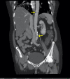
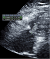
Emerging evidence suggests that novel COVID-19 is associated with increased prothrombotic state and risk of thromboembolic complications, particularly in severe disease. COVID-19 is known to predispose to both venous and arterial thrombotic disease. We describe a case of a 61-year-old woman with history of type II diabetes, hypertension and hyperlipidaemia who presented with dry cough and acute abdominal pain. She was found to have a significantly elevated D-dimer, prompting imaging that showed thrombi in her right ventricle and aorta. She had rapid clinical deterioration and eventually required tissue plasminogen activator with subsequent durable clinical improvement. This case highlights a rare co-occurrence of venous and arterial thrombi in a patient with severe COVID-19. Further studies are needed to clarify the molecular mechanism of COVID-19 coagulopathy, the utility of D-dimer to predict and stratify risk of thrombosis in COVID-19, and the use of fibrinolytic therapy in patients with COVID-19.
Keywords: COVID-19; haematology (incl blood transfusion); venous thromboembolism.
© BMJ Publishing Group Limited 2021. No commercial re-use. See rights and permissions. Published by BMJ.
Conflict of interest statement
Competing interests: None declared.
Figures


Similar articles
-
Aortic Arch Thrombus and Pulmonary Embolism in a COVID-19 Patient.J Emerg Med. 2021 Feb;60(2):223-225. doi: 10.1016/j.jemermed.2020.08.009. Epub 2020 Aug 4. J Emerg Med. 2021. PMID: 32917441 Free PMC article.
-
Aortic thrombus in patients with severe COVID-19: review of three cases.J Thromb Thrombolysis. 2021 Jan;51(1):237-242. doi: 10.1007/s11239-020-02219-z. J Thromb Thrombolysis. 2021. PMID: 32648092 Free PMC article.
-
Pulmonary thromboembolism and right heart thromboemboli--an experience with tenecteplase.J Assoc Physicians India. 2012 Oct;60:52-4. J Assoc Physicians India. 2012. PMID: 23777027
-
The Impact of COVID-19 Disease on Platelets and Coagulation.Pathobiology. 2021;88(1):15-27. doi: 10.1159/000512007. Epub 2020 Oct 13. Pathobiology. 2021. PMID: 33049751 Free PMC article. Review.
-
[COVID-19 and stroke].Rinsho Shinkeigaku. 2020 Dec 26;60(12):822-839. doi: 10.5692/clinicalneurol.cn-001529. Epub 2020 Nov 20. Rinsho Shinkeigaku. 2020. PMID: 33229838 Review. Japanese.
Cited by
-
COVID-19 and Peripheral Artery Thrombosis: A Mini Review.Curr Probl Cardiol. 2022 Oct;47(10):100992. doi: 10.1016/j.cpcardiol.2021.100992. Epub 2021 Sep 24. Curr Probl Cardiol. 2022. PMID: 34571103 Free PMC article. Review.
-
Arterial Thrombosis in Acute Respiratory Infections: An Underestimated but Clinically Relevant Problem.J Clin Med. 2024 Oct 9;13(19):6007. doi: 10.3390/jcm13196007. J Clin Med. 2024. PMID: 39408067 Free PMC article. Review.
-
Spectrum of Thrombotic Complications in Fatal Cases of COVID-19: Focus on Pulmonary Artery Thrombosis In Situ.Viruses. 2023 Aug 2;15(8):1681. doi: 10.3390/v15081681. Viruses. 2023. PMID: 37632023 Free PMC article.
References
-
- COVID-19 Dashboard by the center for systems science and engineering (CSSE) at Johns Hopkins University (JHU). Available: https://coronavirus.jhu.edu/map.html [Accessed 10 Aug 2020].
Publication types
MeSH terms
Substances
LinkOut - more resources
Full Text Sources
Other Literature Sources
Medical
