

Selenomethionine protects hematopoietic stem/progenitor cells against cobalt nanoparticles by stimulating antioxidant actions and DNA repair functions
- PMID: 33875618
- PMCID: PMC8109066
- DOI: 10.18632/aging.202865
Selenomethionine protects hematopoietic stem/progenitor cells against cobalt nanoparticles by stimulating antioxidant actions and DNA repair functions
Abstract
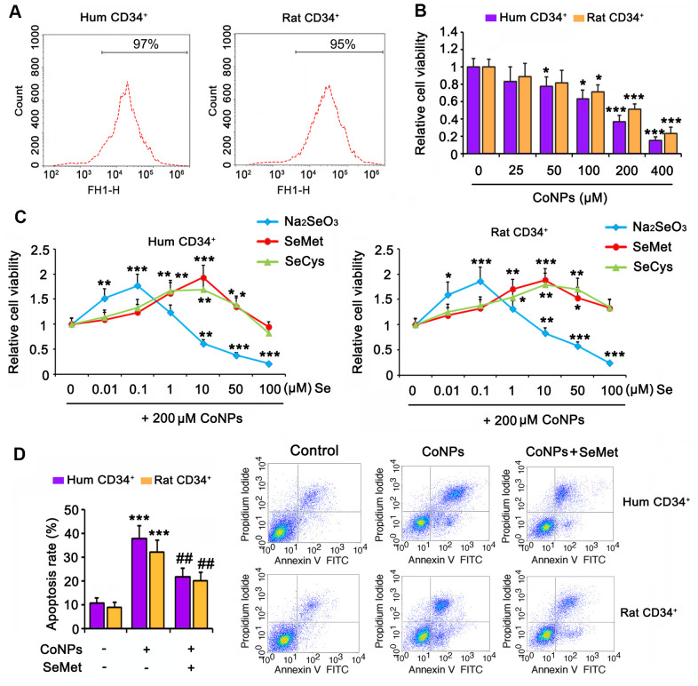
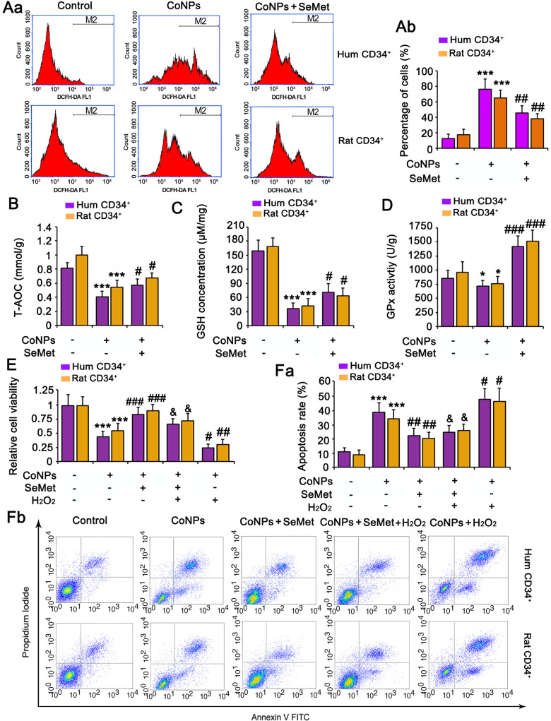
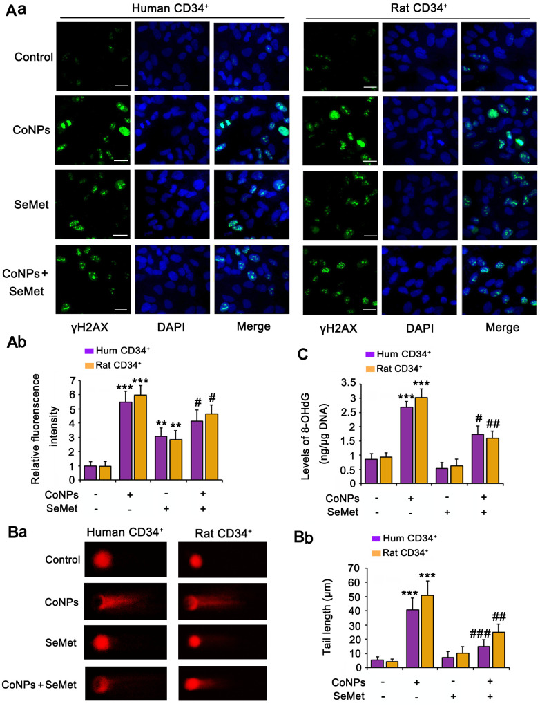
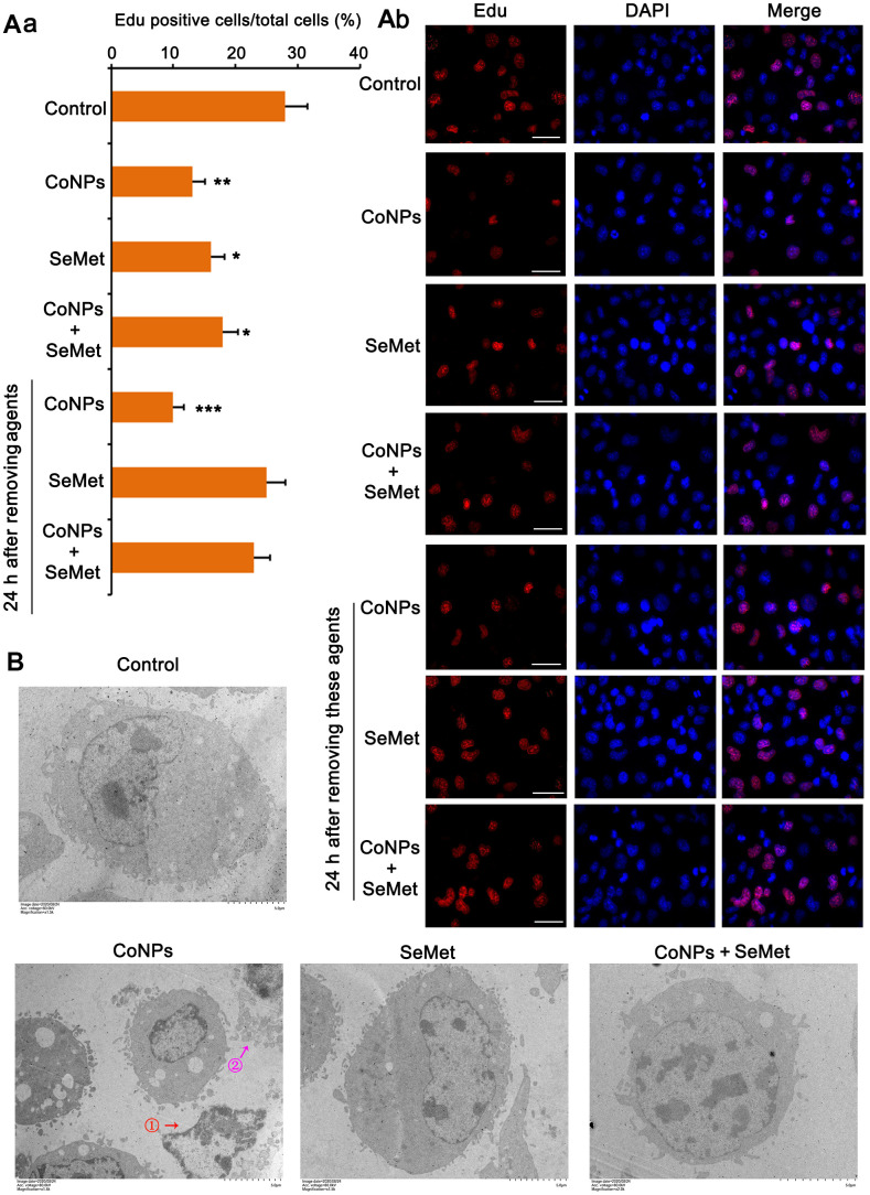
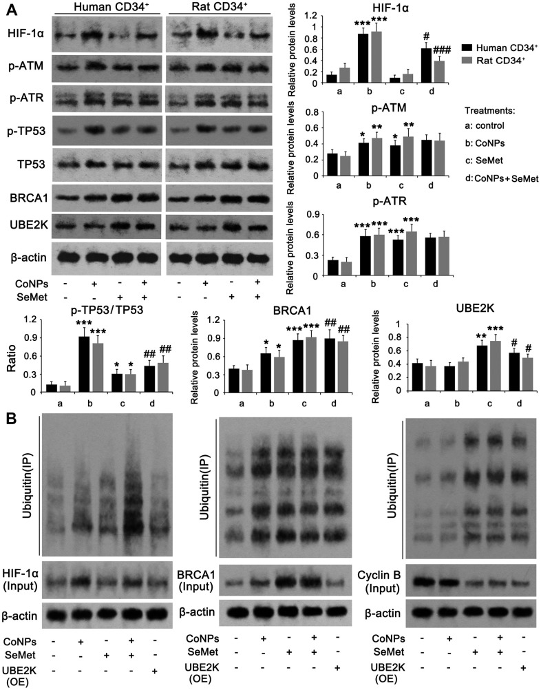
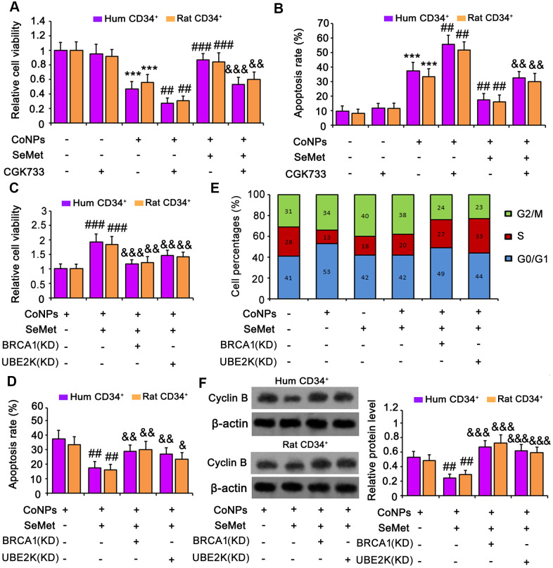
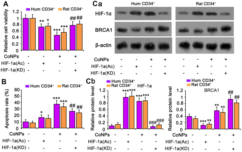
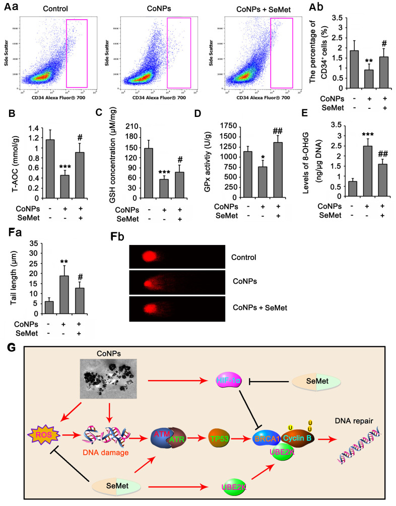
Hematopoietic stem cells (HSCs) and hematopoietic progenitor cells (HPCs) can differentiate into all blood lineages to maintain hematopoiesis, wound healing, and immune functions. Recently, cobalt-chromium alloy casting implants have been used extensively in total hip replacements; however, cobalt nanoparticles (CoNPs) released from the alloy were toxic to HSCs and HPCs. We aimed to investigate the mechanism underlying the toxic effect of CoNPs on HSCs/HPCs and to determine the protective effect of selenomethionine (SeMet) against CoNPs in vitro and in vivo. Human and rat CD34+ HSCs/HPCs were isolated from cord blood and bone marrow, respectively. CoNPs decreased the viability of CD34+ HSCs/HPCs and increased apoptosis. SeMet attenuated the toxicity of CoNPs by enhancing the antioxidant ability of cells. The protective effect of SeMet was not completely abolished after adding H2O2 to abrogate the improvement of the antioxidant capacity by SeMet. SeMet and CoNPs stimulated ATM/ATR DNA damage response signals and inhibited cell proliferation. Unlike CoNPs, SeMet did not damage the DNA, and cell proliferation recovered after removing SeMet. SeMet inhibited the CoNP-induced upregulation of hypoxia inducible factor (HIF)-1α, thereby disrupting the inhibitory effect of HIF-1α on breast cancer type 1 susceptibility protein (BRCA1). Moreover, SeMet promoted BRCA1-mediated ubiquitination of cyclin B by upregulating UBE2K. Thus, SeMet enhanced cell cycle arrest and DNA repair post-CoNP exposure. Overall, SeMet protected CD34+ HSCs/HPCs against CoNPs by stimulating antioxidant activity and DNA repair.
Keywords: DNA repair function; antioxidant action; cobalt nanoparticles; hematopoietic stem/progenitor cells; selenium.
Conflict of interest statement
Figures
Similar articles
-
A paradox: Fe2+-containing agents decreased ROS and apoptosis induced by CoNPs in vascular endothelial cells by inhibiting HIF-1α.Biosci Rep. 2021 Jan 29;41(1):BSR20203456. doi: 10.1042/BSR20203456. Biosci Rep. 2021. PMID: 33345265 Free PMC article.
-
Long-term exposures to low doses of cobalt nanoparticles induce cell transformation enhanced by oxidative damage.Nanotoxicology. 2015 Mar;9(2):138-47. doi: 10.3109/17435390.2014.900582. Epub 2014 Apr 9. Nanotoxicology. 2015. PMID: 24713074
-
L-Ascorbic Acid Protected Against Extrinsic and Intrinsic Apoptosis Induced by Cobalt Nanoparticles Through ROS Attenuation.Biol Trace Elem Res. 2017 Feb;175(2):428-439. doi: 10.1007/s12011-016-0789-x. Epub 2016 Jul 5. Biol Trace Elem Res. 2017. PMID: 27377067
-
Radioprotective agents against the ionizing radiation-induced hematopoietic stem and progenitor cell injury; Foundation review.Crit Rev Oncol Hematol. 2025 Jul;211:104713. doi: 10.1016/j.critrevonc.2025.104713. Epub 2025 Apr 3. Crit Rev Oncol Hematol. 2025. PMID: 40187710 Review.
-
The importance of hypoxia and extra physiologic oxygen shock/stress for collection and processing of stem and progenitor cells to understand true physiology/pathology of these cells ex vivo.Curr Opin Hematol. 2015 Jul;22(4):273-8. doi: 10.1097/MOH.0000000000000144. Curr Opin Hematol. 2015. PMID: 26049746 Free PMC article. Review.
Cited by
-
Research progress on nanotoxicity and detoxification of cobalt in metal-based implants.Ann Med. 2025 Dec;57(1):2532120. doi: 10.1080/07853890.2025.2532120. Epub 2025 Jul 11. Ann Med. 2025. PMID: 40644368 Free PMC article. Review.
-
Targeting SLFN11-regulated pathways restores chemotherapy sensitivity in AML.Blood Neoplasia. 2024 Aug 28;1(4):100037. doi: 10.1016/j.bneo.2024.100037. eCollection 2024 Dec. Blood Neoplasia. 2024. PMID: 40552140 Free PMC article.
-
Proteinase 3 depletion attenuates leukemia by promoting myeloid differentiation.Cell Death Differ. 2024 Jun;31(6):697-710. doi: 10.1038/s41418-024-01288-4. Epub 2024 Apr 8. Cell Death Differ. 2024. PMID: 38589495 Free PMC article.
-
Evaluation of the Neuroprotective Effect of Organic Selenium Compounds: An in Vitro Model of Alzheimer's Disease.Biol Trace Elem Res. 2024 Jul;202(7):2954-2965. doi: 10.1007/s12011-023-03893-9. Epub 2023 Oct 7. Biol Trace Elem Res. 2024. PMID: 37803188
-
Cobalt exposure and pulmonary function reduction in chronic obstructive pulmonary disease patients: the mediating role of club cell secretory protein.Respir Res. 2024 Aug 24;25(1):324. doi: 10.1186/s12931-024-02950-8. Respir Res. 2024. PMID: 39182083 Free PMC article.
References
-
- Sabbioni E, Fortaner S, Farina M, Del Torchio R, Olivato I, Petrarca C, Bernardini G, Mariani-Costantini R, Perconti S, Di Giampaolo L, Gornati R, Di Gioacchino M. Cytotoxicity and morphological transforming potential of cobalt nanoparticles, microparticles and ions in Balb/3T3 mouse fibroblasts: an in vitro model. Nanotoxicology. 2014; 8:455–64. 10.3109/17435390.2013.796538 - DOI - PubMed
-
- Laovitthayanggoon S, Henderson CJ, McCluskey C, MacDonald M, Tate RJ, Grant MH, Currie S. Cobalt administration causes reduced contractility with parallel increases in TRPC6 and TRPM7 transporter protein expression in adult rat hearts. Cardiovasc Toxicol. 2019; 19:276–86. 10.1007/s12012-018-9498-3 - DOI - PMC - PubMed
Publication types
MeSH terms
Substances
LinkOut - more resources
Full Text Sources
Other Literature Sources
Medical
Research Materials
Miscellaneous
