. 2021 Apr 20;13(8):10920-10933.
doi: 10.18632/aging.202992. Epub 2021 Apr 20.

Pentraxin-3-mediated complement activation in a swine model of renal ischemia/reperfusion injury

Affiliations

Pentraxin-3-mediated complement activation in a swine model of renal ischemia/reperfusion injury

Chiara Divella et al. Aging (Albany NY). .

Abstract

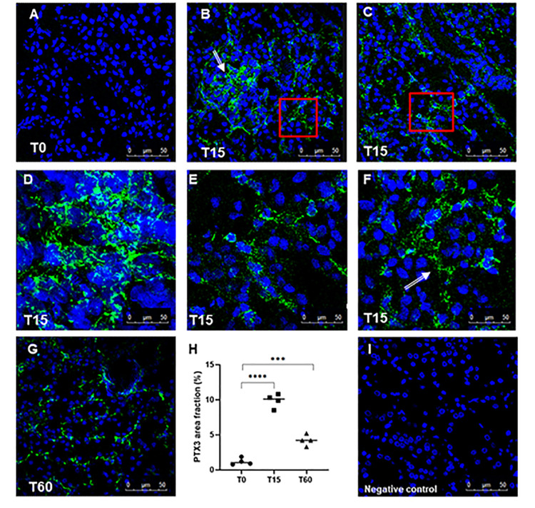
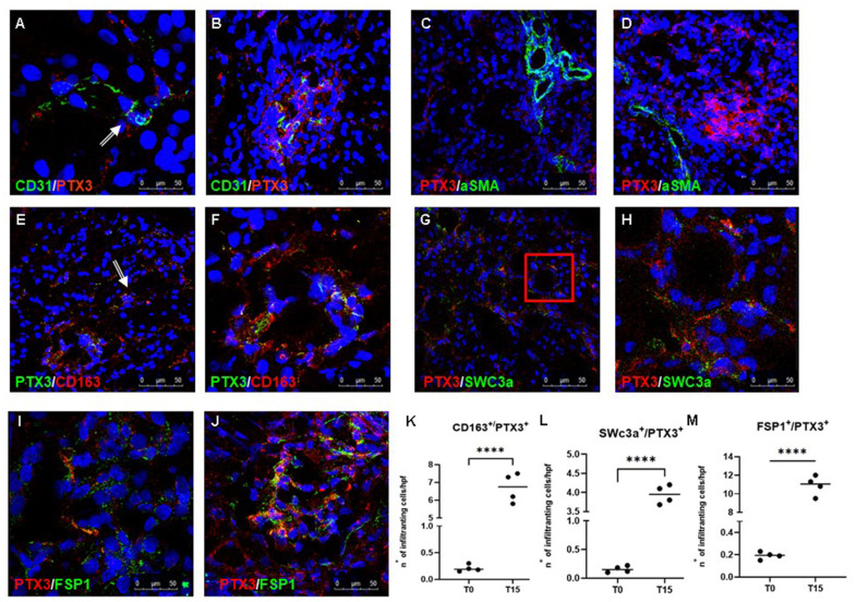
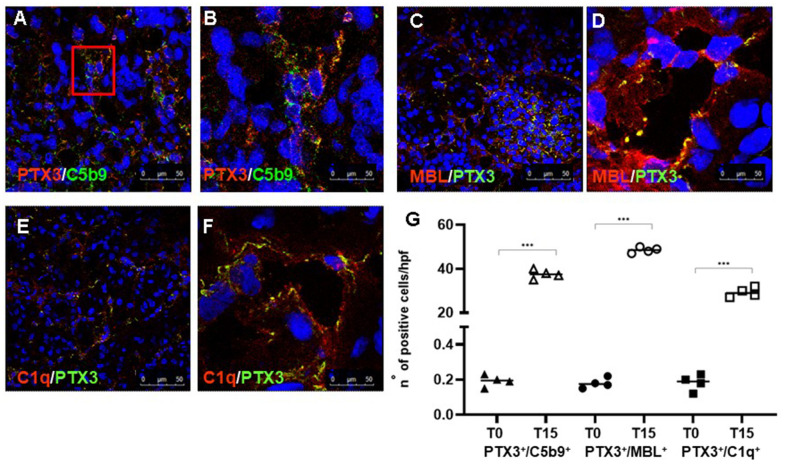
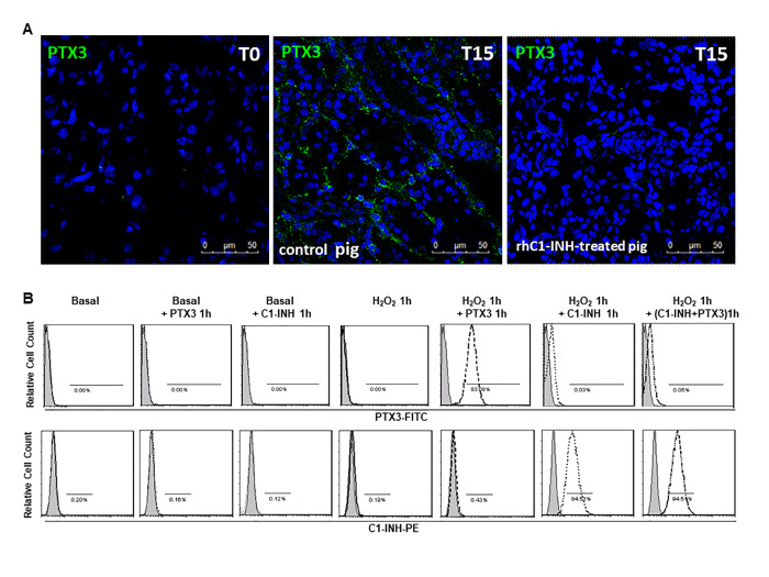
Pentraxins are a family of evolutionarily conserved pattern recognition molecules with pivotal roles in innate immunity and inflammation, such as opsonization of pathogens during bacterial and viral infections. In particular, the long Pentraxin 3 (PTX3) has been shown to regulate several aspects of vascular and tissue inflammation during solid organ transplantation. Our study investigated the role of PTX3 as possible modulator of Complement activation in a swine model of renal ischemia/reperfusion (I/R) injury. We demonstrated that I/R injury induced early PTX3 deposits at peritubular and glomerular capillary levels. Confocal laser scanning microscopy revealed PTX3 deposits co-localizing with CD31+ endothelial cells. In addition, PTX3 was associated with infiltrating macrophages (CD163), dendritic cells (SWC3a) and myofibroblasts (FSP1). In particular, we demonstrated a significant PTX3-mediated activation of classical (C1q-mediated) and lectin (MBL-mediated) pathways of Complement. Interestingly, PTX3 deposits co-localized with activation of the terminal Complement complex (C5b-9) on endothelial cells, indicating that PTX3-mediated Complement activation occurred mainly at the renal vascular level. In conclusion, these data indicate that PTX3 might be a potential therapeutic target to prevent Complement-induced I/R injury.

Keywords: classical pathway; complement system; ischemia/reperfusion injury; kidney; pentraxin 3.


Conflict of interest statement

CONFLICTS OF INTEREST: The authors declare that they have no conflicts of interest.

Figures

Figure 1
Figure 1
Analysis of PTX3 deposits in a swine model of I/R injury. Indirect immunofluorescence for PTX3 was performed on frozen pig kidney sections. A limited presence of PTX3 was observed in the biopsies at T0 (A). PTX3 deposits were observed after 15 min of reperfusion (B, C) at interstitial (E, zoomed image), peri-tubular (F, zoomed image) and glomerular (D) capillary levels. After 60 min the PTX3 deposits were still described at the level of peritubular capillaries (G). (I) Negative staining control for immunofluorescence was performed on cryosections with irrelevant primary antibodies for experimental conditions. Nuclei were highlighted with TO-PRO 3 in blue. Magnification 630X. (H) Quantification of PTX3 demonstrated a statistically significant increase after 15 min of reperfusion compared to basal biopsies. Results were expressed as % ± s.d. of positive area /high power field (hpf). *p<0.05 versus T0.
Figure 2
Figure 2
Characterization of the PTX3-associated cellular pattern in I/R injury. Frozen pig kidney sections were analyzed by indirect immunofluorescence to characterize the PTX3 source after 15 min of reperfusion. Co-localization between CD31 and PTX3 on renal EC was evident (A, B yellow staining). Activated myofibroblasts identified by alpha-smooth muscle actin (green) were negative for PTX3 (red; α-SMA+/PTX3-, C, D). Monocytes/macrophages identified by CD163 (red) co-localized with PTX3 (green; CD163+/PTX3+ yellow, (E) particular of E, F). Dendritic cells identified by SWC3a (green) were intensively positive for PTX3 (red; SWC3a+/PTX3+ yellow, (G) particular of G, H). Myofibroblasts identified by fibroblast-specific protein 1 (FSP1, red) co-localized with PTX3 (green; FSP1+/PTX3+ yellow, I, J). Nuclei were highlighted with TO-PRO 3 in blue. Original magnifications were x630. Quantification of CD163+/PTX3+ (K), SWC3a+/PTX3+ (L) and FSP1+/PTX3+ (M) cells demonstrated a statistically significant increase after 15 min of reperfusion compared to basal biopsies. Results were expressed as mean ± s.d. of infiltrating cells/high power field (hpf). *p<0.05 versus T0.
Figure 3
Figure 3
PTX3-mediated Complement activation in a pig model of I/R injury. Frozen pig kidney sections were examined by indirect immunofluorescence to investigate the co-localization (yellow staining) of C5b9 (green) and PTX3 (red) deposits (A, B). The co-localization between PTX3 (green) with MBL (red, C, D) and C1q (E, F) was investigated by immunofluorescence/confocal microscopy. PTX3 co-localized with MBL (C, D, yellow staining) and C1q (E, F, merge) at peri-glomerular (E) and peri-tubular (D, F) capillary sites. In confocal microscopy images nuclei were stained with TO-PRO 3 (blue). (G) Quantification of C5b9+/PTX3+, MBL+/PTX3+and C1q+/PTX3+cells compared to basal biopsies. Results were expressed as % ± s.d. of positive area /high power field (hpf). *p<0.05 versus T0.
Figure 4
Figure 4
C1-inhibitor prevents PTX-3 binding on endothelial cells. (A) Frozen pig kidney sections were analyzed by indirect immunofluorescence to characterize the PTX3 source after 15 min of reperfusion in control and rhC1-INH treated pigs. PTX3 deposits were observed at the level of peritubular capillaries in control pigs. rhC1-INH infusion prevented PTX3 deposits on ECs. (B) FACS showed that ECs in basal condition did not bind both rhC1-INH and PTX3. Both PTX3 and rhC1-INH presented a significant binding on H2O2-activated ECs. When H2O2-activated EC were co-stimulated with PTX3 and rhC1-INH, rhC1INH prevented PTX3 binding. Results are representative of three independent experiments.

References

    1. Bonventre JV, Yang L. Cellular pathophysiology of ischemic acute kidney injury. J Clin Invest. 2011; 121:4210–21. 10.1172/JCI45161 - DOI - PMC - PubMed
    1. Jang HR, Rabb H. The innate immune response in ischemic acute kidney injury. Clin Immunol. 2009; 130:41–50. 10.1016/j.clim.2008.08.016 - DOI - PMC - PubMed
    1. Ricklin D, Hajishengallis G, Yang K, Lambris JD. Complement: a key system for immune surveillance and homeostasis. Nat Immunol. 2010; 11:785–97. 10.1038/ni.1923 - DOI - PMC - PubMed
    1. Thurman JM, Holers VM. The central role of the alternative complement pathway in human disease. J Immunol. 2006; 176:1305–10. 10.4049/jimmunol.176.3.1305 - DOI - PubMed
    1. Franzin R, Stasi A, Fiorentino M, Stallone G, Cantaluppi V, Gesualdo L, Castellano G. Inflammaging and Complement System: A Link Between Acute Kidney Injury and Chronic Graft Damage. Front Immunol. 2020; 11:734. 10.3389/fimmu.2020.00734 - DOI - PMC - PubMed

Publication types

MeSH terms