Review
. 2022 Jan;27(1):445-465.
doi: 10.1038/s41380-021-01092-3. Epub 2021 Apr 19.

Mechanisms of synaptic transmission dysregulation in the prefrontal cortex: pathophysiological implications

Affiliations
Review

Mechanisms of synaptic transmission dysregulation in the prefrontal cortex: pathophysiological implications

Zhen Yan et al. Mol Psychiatry. 2022 Jan.

Abstract

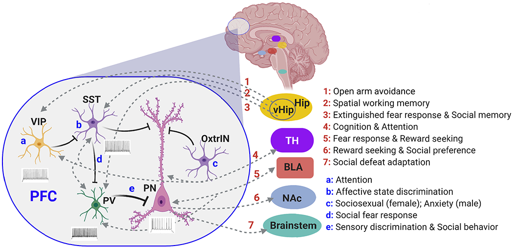
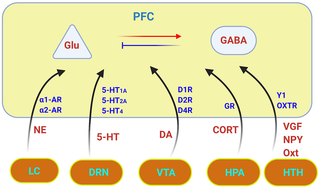
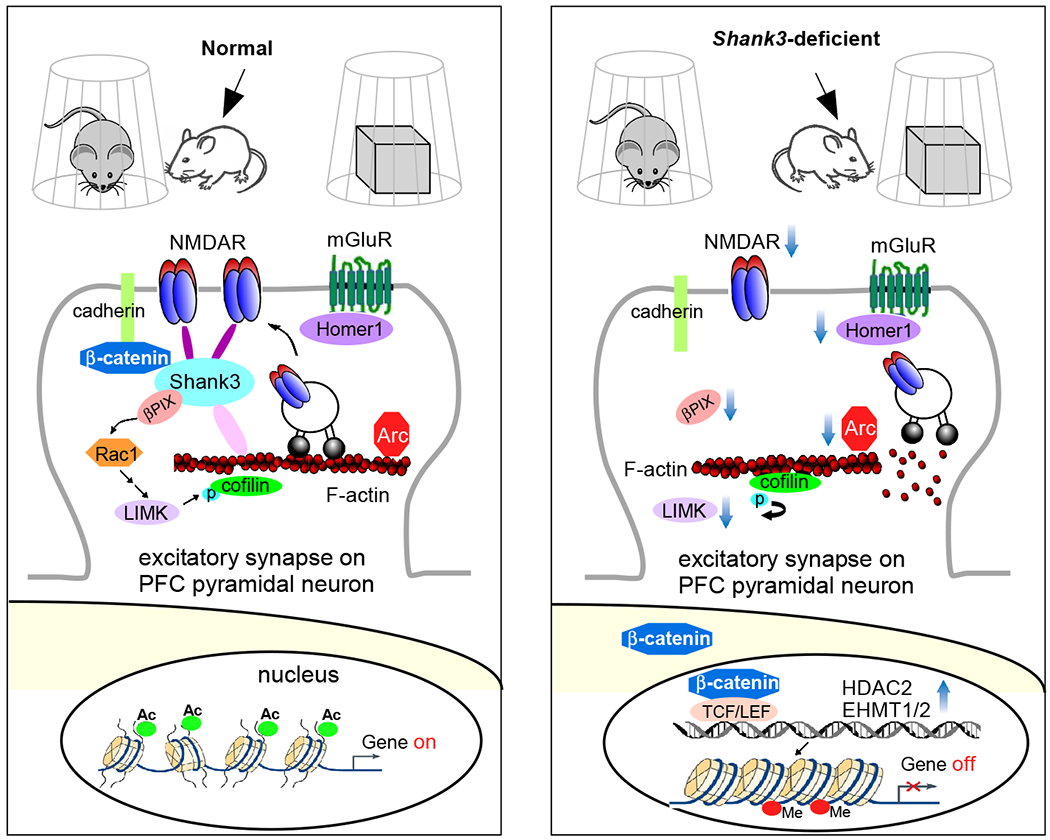
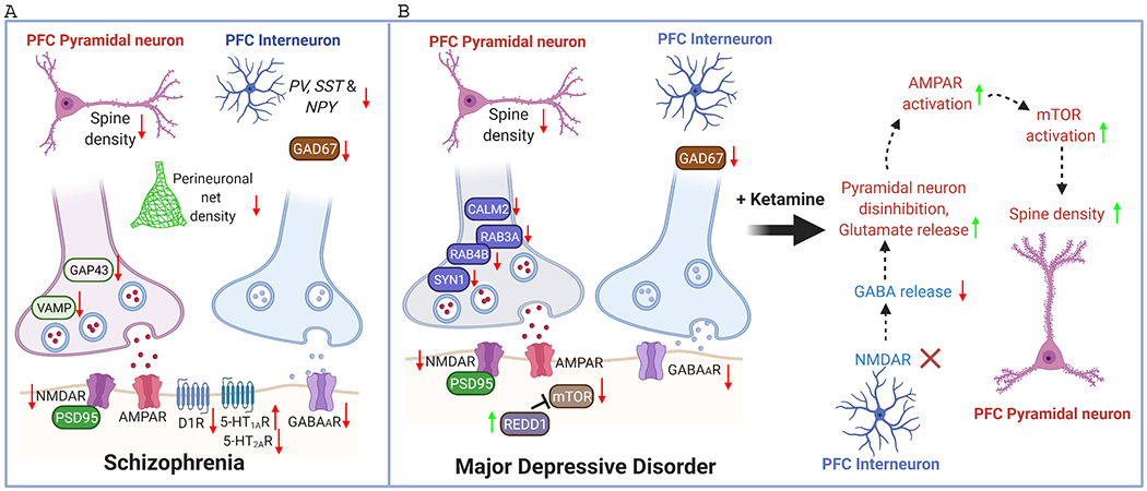
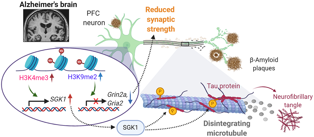
The prefrontal cortex (PFC) serves as the chief executive officer of the brain, controlling the highest level cognitive and emotional processes. Its local circuits among glutamatergic principal neurons and GABAergic interneurons, as well as its long-range connections with other brain regions, have been functionally linked to specific behaviors, ranging from working memory to reward seeking. The efficacy of synaptic signaling in the PFC network is profundedly influenced by monoaminergic inputs via the activation of dopamine, adrenergic, or serotonin receptors. Stress hormones and neuropeptides also exert complex effects on the synaptic structure and function of PFC neurons. Dysregulation of PFC synaptic transmission is strongly linked to social deficits, affective disturbance, and memory loss in brain disorders, including autism, schizophrenia, depression, and Alzheimer's disease. Critical neural circuits, biological pathways, and molecular players that go awry in these mental illnesses have been revealed by integrated electrophysiological, optogenetic, biochemical, and transcriptomic studies of PFC. Novel epigenetic mechanism-based strategies are proposed as potential avenues of therapeutic intervention for PFC-involved diseases. This review provides an overview of PFC network organization and synaptic modulation, as well as the mechanisms linking PFC dysfunction to the pathophysiology of neurodevelopmental, neuropsychiatric, and neurodegenerative diseases. Insights from the preclinical studies offer the potential for discovering new medical treatments for human patients with these brain disorders.


Conflict of interest statement

Conflict of Interests:

The authors declare no conflict of interest.

Figures

Figure 1.
Figure 1.
Plot of PFC organization highlighting the functional mapping of some long-range and local circuits of PFC. VIP+ interneurons (VIP) in PFC sends inhibitory inputs to SST+ interneurons (SST), which causes disinbition of PV+ interneurons (PV), resulting in the strong somatic inhibition and weak dendritic inhibition of pyramidal neurons (PN). PFC interneurons and PN are innervated by hippocampus (HIP) or ventral HIP (vHIP), thalamus (TH) and basolateral amygdala (BLA), while PN send output to TH, BLA, nucleus accumbens (NAc) and brainstem. Some behavioral correlates of these neuronal circuits are listed, along with the functional role of VIP+ or SST+ interneurons, as well as a subtype of SST+ interneurons expressing oxytocin receptor (OxtrIN).
Figure 2.
Figure 2.
Plot of PFC regulation illustrating some neurochemical influences on glutamatergic (Glu) excitation and GABAergic (GABA) inhibition in PFC neurons. The major modulators include dopamine (DA) from ventral tegmental area (VTA), serotonin (5-HT) from dorsal raphe (DRN), norepinephrine (NE) from locus ceruleus (LC), corticosterone (CORT) stress hormone from hypothalamic–pituitary–adrenal (HPA) axis, and neuropeptides, such as VGF nerve growth factor (VGF), Neuropeptide Y (NPY), and oxytocin (OT), from hypothalamus (HTH). These monoaminergic and homornal modulations are achieved by activing corresponding receptors. The complex intracellular signaling pathways initiated by each neuromodulator can be found in relevant papers or previous reviews.
Figure 3.
Figure 3.
Schematic diagram illustrating a potential mechanism underlying the social deficits in Shank3 autism models. Normally, Shank3 crosslinks NMDARs to actin cytoskeleton, and binds to the adhesive junction-associated protein β-catenin. Loss of Shank3 leads to the translocation of β-catenin from synapses to nucleus, inducing the upregulation of histone modifiers HDAC2 and EHMT1/2. The ensuing transcriptional suppression of actin regulators, such as βPIX (Rac1 activator) and LIMK, and synaptic plasticity genes, such as Arc and Homer1 (mGluR anchor), results in the disruption of actin filaments (via cofilin-mediated depolymerization) and the diminished actin-based synaptic delivery of NMDARs in PFC pyramidal neurons. Consequently, the autism-like social preference deficits are manifested. Treatment with HDAC or EHMT inhibitors restores or elevates many target genes, which collectively leads to the normalization of NMDAR synaptic function in PFC and the rescue of social deficits,,.
Figure 4.
Figure 4.
Schematic diagram illustrating the synaptic changes in PFC of schizophrenia (SZ) and major depressive disorder (MDD). NMDAR hypofunction, GABA deficiency, dopamine hypoactivity, serotonin dysregulation, and dendritic spine loss are major aberrations in SZ PFC. The reduced density of perineuronal nets (PNN) may contriubute to the destabilization of synapses. Similar reduction on NMDAR, GABA system and spine density is found in MDD PFC. Some presynaptic molecules involved in transmitter release, including CALM2, SYN1, RAB3A, RAB4B, are also diminished in MDD PFC. In addition, mTOR signaling is decreased in MDD PFC because of the elevated expression of the endogenous inhibitor REDD1. The fast-acting antidepressant ketamine primarily blocks NMDARs on PFC interneurons, leading to the disinhibition of PFC pyramidal neurons and the increase of mTORC signaling, resulting in the restoration of dendritic spines in MDD PFC.
Figure 5.
Figure 5.
Schematic diagram illustrating the potential epigenetic mechanisms underlying gene dysregulation and synaptic deficits in PFC of AD. The elevation of repressive histone mark H3K9me2 in AD leads to the downregulation of genes involved in synaptic transmission, such as AMPAR subunit Gria2 and NMDAR subunit Grin2a, resulting in the reduction of synaptic strength. On the other hand, the elevation of permissive histone mark H3K4me3 in AD leads to the upregulation of genes involved in cell stress, such as serum- and glucocorticoid-inducible kinase 1 (SGK1), resulting in hyperphosphorylation of tau, disintegration of microtubules and disruption of vital protein transport. Targeting histone methytransferases to normalize histone modification or key target genes is the potential new therapeutic strategy for treating synaptic and cognitive deficits in AD.

Similar articles

Cited by

References

    1. Bourgeois JP, Goldman-Rakic PS & Rakic P Synaptogenesis in the prefrontal cortex of rhesus monkeys. Cerebral cortex (New York, N.Y. : 1991) 4, 78–96 (1994). - PubMed
    1. Diamond A Executive functions. Annu Rev Psychol 64, 135–168 (2013). - PMC - PubMed
    1. Stokes MG ‘Activity-silent’ working memory in prefrontal cortex: a dynamic coding framework. Trends Cogn Sci 19, 394–405 (2015). - PMC - PubMed
    1. Fuster JM & Alexander GE Neuron activity related to short-term memory. Science 173, 652–654 (1971). - PubMed
    1. Goldman-Rakic PS Cellular basis of working memory. Neuron 14, 477–485 (1995). - PubMed

Publication types

LinkOut - more resources