

Cardioprotection by post-conditioning with exogenous triiodothyronine in isolated perfused rat hearts and isolated adult rat cardiomyocytes
- PMID: 33876304
- PMCID: PMC8055637
- DOI: 10.1007/s00395-021-00868-6
Cardioprotection by post-conditioning with exogenous triiodothyronine in isolated perfused rat hearts and isolated adult rat cardiomyocytes
Abstract
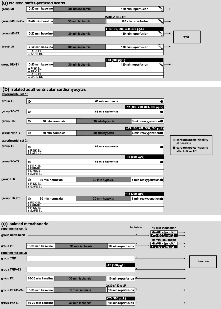
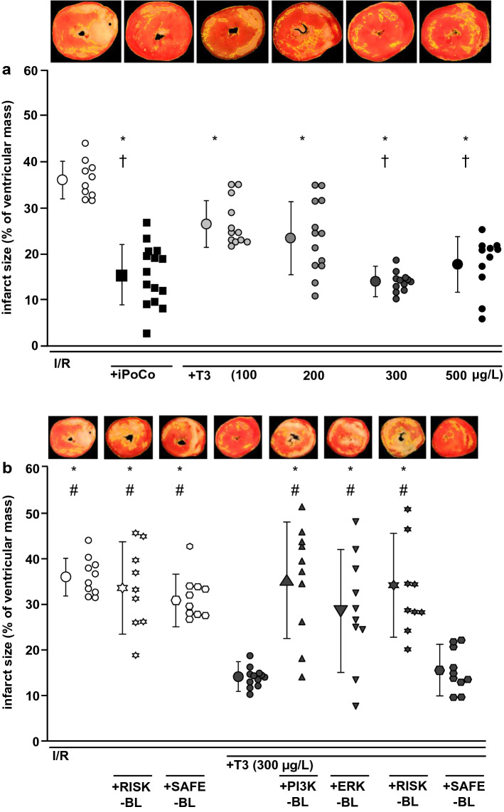
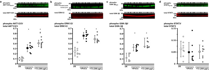
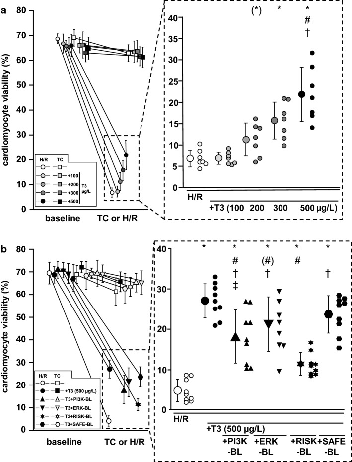
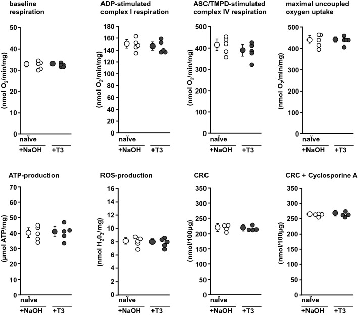
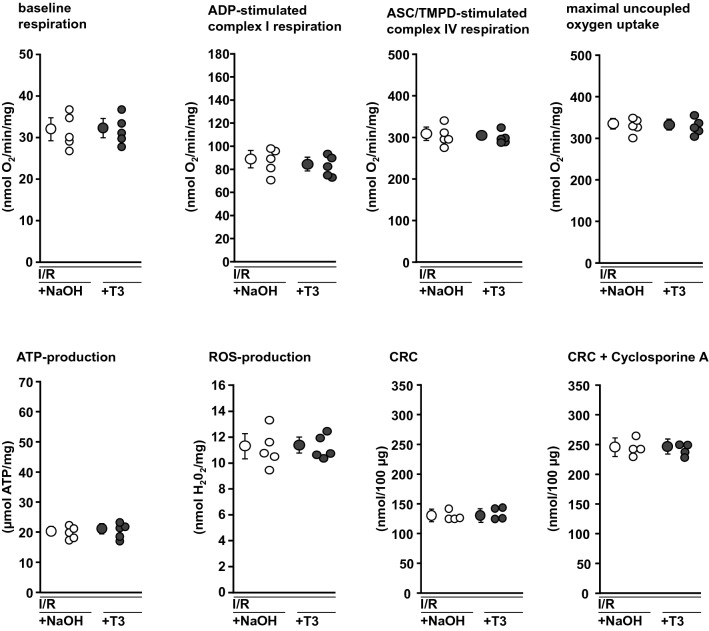
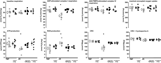
Ischemic post-conditioning (iPoCo) by coronary re-occlusion/reperfusion during immediate reperfusion after prolonged myocardial ischemia reduces infarct size. Mechanical manipulation of culprit lesions, however, carries the risk of coronary microembolization which may obscure iPoCo's cardioprotection. Pharmacological post-conditioning with exogenous triiodothyronine (T3) could serve as an alternative conditioning strategy. Similar to iPoCo, T3 may activate cardioprotective prosurvival pathways. We aimed to study T3's impact on infarct size and its underlying signal transduction. Hearts were isolated from male Lewis rats (200-380 g), buffer-perfused and subjected to 30 min/120 min global zero-flow ischemia/reperfusion (I/R). In additional hearts, either iPoCo (2 × 30 s/30 s I/R) was performed or T3 (100-500 µg/L) infused at reperfusion. Infarct size was demarcated with triphenyl tetrazolium chloride staining and calculated as percent of ventricular mass. Infarct size was reduced with iPoCo to 16 ± 7% vs. 36 ± 4% with I/R only. The maximum infarct size reduction was observed with 300 µg/L T3 (14 ± 2%). T3 increased the phosphorylation of protein kinase B and mitogen extracellular-regulated-kinase 1/2, both key enzymes of the reperfusion injury salvage kinase (RISK) pathway. Pharmacological RISK blockade (RISK-BL) during reperfusion abrogated T3's cardioprotection (35 ± 10%). Adult ventricular cardiomyocytes were isolated from buffer-perfused rat hearts and exposed to 30 min/5 min hypoxia/reoxygenation (H/R); reoxygenation was initiated without or with T3, respectively, and without or with RISK-BL, respectively. Maximal preservation of viability was observed with 500 µg/L T3 after H/R (27 ± 4% of all cells vs. 5 ± 3% in time-matched controls). Again, RISK-BL abrogated protection (11 ± 3%). Mitochondria were isolated at early reperfusion from buffer-perfused rat hearts without or with iPoCo or 300 µg/L T3, respectively, at reperfusion. T3 improved mitochondrial function (i.e.: increased respiration, adenosine triphosphate production, calcium retention capacity, and decreased reactive oxygen species formation) to a similar extent as iPoCo. T3 at reperfusion reduces infarct size by activation of the RISK pathway. T3's protection is a cardiomyocyte phenomenon and targets mitochondria.
Keywords: Cardioprotection; Ischemia/reperfusion; Ischemic conditioning; Post-conditioning; Triiodothyronine.
Conflict of interest statement
The authors declare that they have no competing interests.
Figures
Similar articles
-
Irisin plays a pivotal role to protect the heart against ischemia and reperfusion injury.J Cell Physiol. 2017 Dec;232(12):3775-3785. doi: 10.1002/jcp.25857. Epub 2017 May 3. J Cell Physiol. 2017. PMID: 28181692 Free PMC article.
-
Humoral transfer and intramyocardial signal transduction of protection by remote ischemic perconditioning in pigs, rats, and mice.Am J Physiol Heart Circ Physiol. 2018 Jul 1;315(1):H159-H172. doi: 10.1152/ajpheart.00152.2018. Epub 2018 Mar 23. Am J Physiol Heart Circ Physiol. 2018. PMID: 29569956
-
Sevoflurane post-conditioning protects isolated rat hearts against ischemia-reperfusion injury via activation of the ERK1/2 pathway.Acta Pharmacol Sin. 2014 Dec;35(12):1504-13. doi: 10.1038/aps.2014.78. Epub 2014 Oct 27. Acta Pharmacol Sin. 2014. PMID: 25345742 Free PMC article.
-
The coronary circulation in acute myocardial ischaemia/reperfusion injury: a target for cardioprotection.Cardiovasc Res. 2019 Jun 1;115(7):1143-1155. doi: 10.1093/cvr/cvy286. Cardiovasc Res. 2019. PMID: 30428011 Free PMC article. Review.
-
Myocardial reperfusion injury: looking beyond primary PCI.Eur Heart J. 2013 Jun;34(23):1714-22. doi: 10.1093/eurheartj/eht090. Epub 2013 Mar 27. Eur Heart J. 2013. PMID: 23536610 Review.
Cited by
-
Coronary microvascular obstruction and dysfunction in patients with acute myocardial infarction.Nat Rev Cardiol. 2024 May;21(5):283-298. doi: 10.1038/s41569-023-00953-4. Epub 2023 Nov 24. Nat Rev Cardiol. 2024. PMID: 38001231 Review.
-
The Potential of Thyroid Hormone Therapy in Severe COVID-19: Rationale and Preliminary Evidence.Int J Environ Res Public Health. 2022 Jun 30;19(13):8063. doi: 10.3390/ijerph19138063. Int J Environ Res Public Health. 2022. PMID: 35805716 Free PMC article. Review.
-
Cardioprotection in cardiovascular surgery.Basic Res Cardiol. 2024 Aug;119(4):545-568. doi: 10.1007/s00395-024-01062-0. Epub 2024 Jun 10. Basic Res Cardiol. 2024. PMID: 38856733 Review.
-
Higher Peripheral Thyroid Sensitivity Is Linked to a Lower Risk of Heart Failure After Acute Myocardial Infarction.J Clin Endocrinol Metab. 2023 Oct 18;108(11):2950-2960. doi: 10.1210/clinem/dgad240. J Clin Endocrinol Metab. 2023. PMID: 37104944 Free PMC article.
-
Effect of sleeve gastrectomy, Roux-en-Y gastric bypass, and ileal transposition on myocardial ischaemia-reperfusion injury in non-obese non-diabetic rats.Sci Rep. 2021 Dec 13;11(1):23888. doi: 10.1038/s41598-021-03283-y. Sci Rep. 2021. PMID: 34903800 Free PMC article.
References
-
- Bergh JJ, Lin HY, Lansing L, Mohamed SN, Davis FB, Mousa S, Davis PJ. Integrin alphavbeta3 contains a cell surface receptor site for thyroid hormone that is linked to activation of mitogen-activated protein kinase and induction of angiogenesis. Endocrinology. 2005;146:2864–2871. doi: 10.1210/en.2005-0102. - DOI - PubMed
-
- Bøtker HE, Hausenloy D, Andreadou I, Antonucci S, Boengler K, Davidson SM, Deshwal S, Devaux Y, Di Lisa F, Di Sante M, Efentakis P, Femmino S, Garcia-Dorado D, Giricz Z, Ibanez B, Iliodromitis E, Kaludercic N, Kleinbongard P, Neuhauser M, Ovize M, Pagliaro P, Rahbek-Schmidt M, Ruiz-Meana M, Schluter KD, Schulz R, Skyschally A, Wilder C, Yellon DM, Ferdinandy P, Heusch G. Practical guidelines for rigor and reproducibility in preclinical and clinical studies on cardioprotection. Basic Res Cardiol. 2018;113:39. doi: 10.1007/s00395-018-0696-8. - DOI - PMC - PubMed
Publication types
MeSH terms
Substances
LinkOut - more resources
Full Text Sources
Other Literature Sources
Medical
