. 2021 Dec;13(6):1644-1657.
doi: 10.1007/s12602-021-09789-1. Epub 2021 Apr 20.

Assessment of the Safety of Lactobacillus casei IMV B-7280 Probiotic Strain on a Mouse Model

Affiliations

Assessment of the Safety of Lactobacillus casei IMV B-7280 Probiotic Strain on a Mouse Model

Lazarenko L M et al. Probiotics Antimicrob Proteins. 2021 Dec.

Abstract

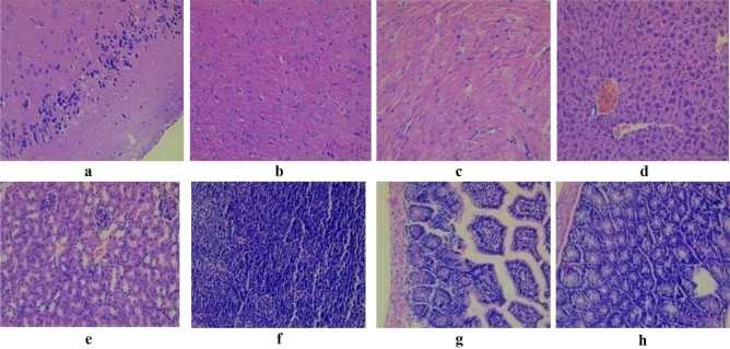
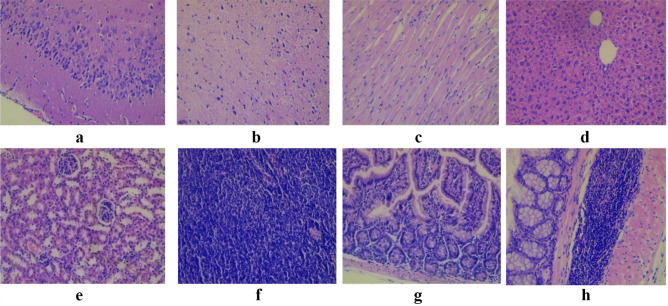
Probiotics, in particular Lactobacillus (lactic acid bacteria, LAB) strains, are widely used in clinical practice. Despite that these probiotics have GRAS (generally regarded as safe) and qualified presumption of safety (QPS) statuses, the safety of particular strains still needs to be thoroughly studied. The aim of the study was to evaluate the safety of Lact. casei IMV B-7280 strain by investigating toxicity and the effects on gut microbiota in experimental animal model. Male BALB/c mice (7-8 weeks, weight 20-24 g) were treated with amounts of Lact. casei IMV B-7280 strain: 5 × 10⁶, 5 × 10⁸, or 5 × 10⁹ CFU/animal once per day during 7 days, or in the amount of 1 × 10¹⁰ CFU/animal once per day during 3 days (most of the proposed probiotic doses for humans-from 108 to 109 CFU) and monitored during 14 days. Blood tests and serum biochemistry were conducted; the cecal content from mice of the experimental and control groups were freshly collected and analyzed. At the end of the experiments (15th day), the presence of LAB in the heart, liver, kidney, and mesenteric lymph nodes and peripheral blood was determined; histology of the brain, liver, heart, fragments of the small and large intestine, and mesenteric lymph nodes was conducted. Survival rate of BALB/c mice treated with Lact. casei IMV B-7280 strain in different concentrations in toxicity experiments during 14 days was 100%. We observed no signs of toxicity as changes in gait, lethargy, sleep, somatomotor activity as well as changes in fur, eyes, skin and mucous membranes, tremors, behavior pattern, convulsions, salivation, diarrhea, and local injuries in mice from all experimental groups. After administration of probiotic strain, the number of opportunistic bacteria in cecal contents, such as Staphylococcus spp., Candida spp., Pseudomonas spp., and total aerobic and optionally anaerobic bacteria decreased compared to controls; the population of beneficial bacteria such as lactobacilli increased in cecal contents of these mice. LAB were not detected in the peripheral blood, heart, liver, kidneys, and mesenteric lymph nodes after administration of this strain to intact mice. Lact. casei IMV B-7280 strain is safe at dose up to 1010 CFU/animal during 3- and 7-day oral administration to mice and has a positive effect on the gut microbiota composition; it could be potentially considered as safe probiotic for humans.

Keywords: In vivo; Lactobacillus; Lactobacillus casei IMV B-7280; Mouse model; Probiotic strain; Safety.


Conflict of interest statement

The authors declare no competing interests.

Figures

Fig. 1
Fig. 1
The scheme of oral toxicity study of Lact. casei IMV B-7280 strain
Fig. 2
Fig. 2
Light micrograph of the cortex a and white matter b of the brain cerebrum; cardiac muscle fibers c; lever d; cortical substance of the kidney e; mesenteric lymph node f; fragments of the small intestine g; fragments of the colon h intact mice that did not receive probiotic bacteria. Staining with hematoxylin and eosin. × 200. No cellular and inflammatory changes as well as hemorrhage were observed in cortex and white matter of the brain (cerebrum); cardiac muscle fibers; lever; cortical substance of the kidney; mesenteric lymph node; fragments of the small intestine, and colon
Fig. 3
Fig. 3
Light micrograph of the cortex a and white matter b of the brain (cerebrum); cardiac muscle fibers c; liver d; cortical substance of the kidney e; mesenteric lymph node f; fragments of the small intestine g; fragments of the colon h of mice that administrated with Lact. casei IMV B-7280 strain in the dose of 5 × 1010 CFU/animal (experimental group 1). Staining with hematoxylin and eosin. × 200. No cellular and inflammatory changes as well as hemorrhage were observed in cortex and white matter of the brain (cerebrum); cardiac muscle fibers; liver; cortical substance of the kidney; mesenteric lymph node; fragments of the small intestine, and colon. There was a slight perivascular edema in the cerebral cortex and white matter of the brain. In the cerebral cortex, there were no signs of pericellular edema. There were no perivascular or parenchymal hemorrhages in the brain. Inflammatory infiltration of the cerebral cortex and white matter is absent. There was mild interstitial edema in the myocardium of the heart, but inflammatory stroma infiltration was absent. Myocardial vessels are moderately full-blooded; no evidence of vascular wall edema, or perivascular hemorrhage in the myocardium of the heart was detected. There was no hyperplasia of lymphoid tissue in mesenteric lymph nodes, small and large intestine
Fig. 4
Fig. 4
Light micrograph of the cortex a and white matter b of the brain (cerebrum); cardiac muscle fibers c; liver d; cortical substance of the kidney e; mesenteric lymph node f; fragments of the small intestine g; fragments of the colon h of mice that administrated with Lact. casei IMV B-7280 strain in the dose of 5 × 106 CFU/animal (experimental group 2). Staining with hematoxylin and eosin. × 200. No cellular and inflammatory changes as well as hemorrhage were observed in cortex and white matter of the brain (cerebrum); cardiac muscle fibers; liver; cortical substance of the kidney; mesenteric lymph node; fragments of the small intestine, and colon. A slight perivascular edema was observed focally in the brain. Inflammatory infiltration of the cerebral cortex, meninges, and white matter was not detected. Perivascular or parenchymal hemorrhage in the brain was absent. In the liver, a small fraction of hepatocytes had more hyperchromic nucleus with multiple nucleoli d. The cell cytoplasm had fine-grained evenly eosinophilic staining; however, histological signs of hepatocyte damage, inflammatory infiltration or fibrotic changes, signs of bile stasis were not detected. Mild interstitial edema was observed in the myocardium of the heart, kidneys, and muscle layer, the walls of the small and large intestine. There was no hyperplasia of lymphoid tissue in mesenteric lymph nodes, small and large intestine
Fig. 5
Fig. 5
Light micrograph of the cortex a and white matter b of the brain (cerebrum); cardiac muscle fibers c; liver d; cortical substance of the kidney e; mesenteric lymph node f; fragments of the small intestine g; fragments of the colon h of mice that administrated with Lact. casei IMV B-7280 strain in the dose of 5 × 108 CFU/animal (experimental group 3). Staining with hematoxylin and eosin. × 200. No cellular and inflammatory changes as well as hemorrhage were observed in cortex and white matter of the brain (cerebrum); cardiac muscle fibers; liver; cortical substance of the kidney; mesenteric lymph node; fragments of the small intestine, and colon. Slightly expressed perivascular edema was observed in the cortex and white matter of the brain. Inflammatory infiltration of the cerebral cortex, meninges, and white matter was not detected. Perivascular or parenchymal hemorrhage in the brain was absent. There was mild interstitial edema in the heart myocardium, but inflammatory infiltration, vascular wall edema, or perivascular hemorrhages were absent c. Liver hepatocytes were polymorphic, of different size, had rounded, clearly delimited, moderately basophilic nucleus with nucleolus. No histological signs of hepatocyte damage, bile stasis, inflammatory infiltration, or fibrotic changes were detected d. A slight interstitial edema was found in the kidneys and the muscular layer of the wall of the small and large intestine. There was no hyperplasia of lymphoid tissue in mesenteric lymph nodes, fragments of the small and large intestine
Fig. 6
Fig. 6
Light micrograph of the cortex a and white matter b of the brain (cerebrum); cardiac muscle fibers c; liver d; cortical substance of the kidney e; mesenteric lymph node f; fragments of the small intestine g; fragments of the colon h of mice that administrated with Lact. casei IMV B-7280 strain in the dose of 5 × 108 CFU/animal (experimental group 4). Staining with hematoxylin and eosin. × 200. No cellular and inflammatory changes were observed in cortex and white matter of the brain (cerebrum); cardiac muscle fibers; liver; cortical substance of the kidney; mesenteric lymph node; fragments of the small intestine, and colon. There were no signs of pericellular edema in the cerebral cortex and white matter of the brain. Perivascular edema and perivascular hemorrhage in the major cortex were also absent. Mild interstitial edema was detected in the myocardium of the heart, but there was no inflammatory stroma infiltration. Myocardial vessels were moderately full-blooded, no signs of vascular wall swelling or perivascular hemorrhage were observed in the mice of this group. Hepatocytes had different sizes and rounded, clearly delineated, moderately basophilic nucleus with nucleolus. The cytoplasm of cells had fine-grained eosinophilic staining. Histological signs of hepatocyte damage and signs of bile stasis were not detected. Mild interstitial edema was detected in the kidneys and the muscular layer, the walls of the small and large intestine. There was no hyperplasia of lymphoid tissue in mesenteric lymph nodes, small and large intestine

References

    1. Williams NT (2010) Probiotics. Am J Health Syst Pharm 67:449-458. 10.2146/ajhp090168 - PubMed
    1. Falagas ME, Betsi GI, Tokas T, Athanasiou S (2006) Probiotics for prevention of recurrent urinary tract infections in women: a review of the evidence from microbiological and clinical studies. Drugs 66:1253-1261. 10.2165/00003495-200666090-00007 - PubMed
    1. Di Cerbo A, Palmieri B, Aponte M, Morales-Medina JC, Iannitti T (2016) Mechanisms and therapeutic effectiveness of lactobacilli. J Clin Pathol 69:187-203. 10.1136/jclinpath-2015-202976 - PMC - PubMed
    1. Kasińska MA, Drzewoski J (2015) Effectiveness of probiotics in type 2 diabetes: a meta-analysis. Pol Arch Med Wewn 125:803-813. 10.20452/pamw.3156 - PubMed
    1. Hendijani F, Akbari V (2018) Probiotic supplementation for management of cardiovascular risk factors in adults with type II diabetes: A systematic review and meta-analysis. Clin Nutr 37:532-541. 10.1016/j.clnu.2017.02.015 - PubMed

Publication types

LinkOut - more resources