Review
. 2021 Mar;78(3):77-96.
doi: 10.1002/cm.21662. Epub 2021 Apr 28.

Composition and function of ciliary inner-dynein-arm subunits studied in Chlamydomonas reinhardtii

Affiliations
Review

Composition and function of ciliary inner-dynein-arm subunits studied in Chlamydomonas reinhardtii

Ryosuke Yamamoto et al. Cytoskeleton (Hoboken). 2021 Mar.

Abstract

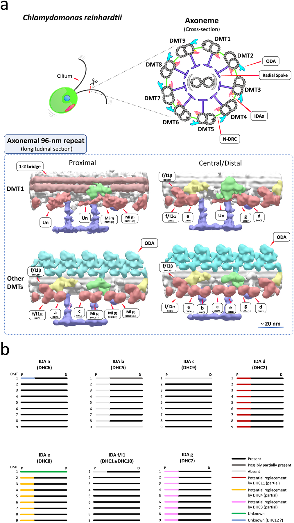
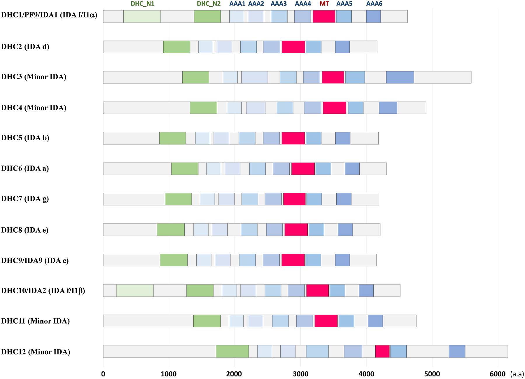
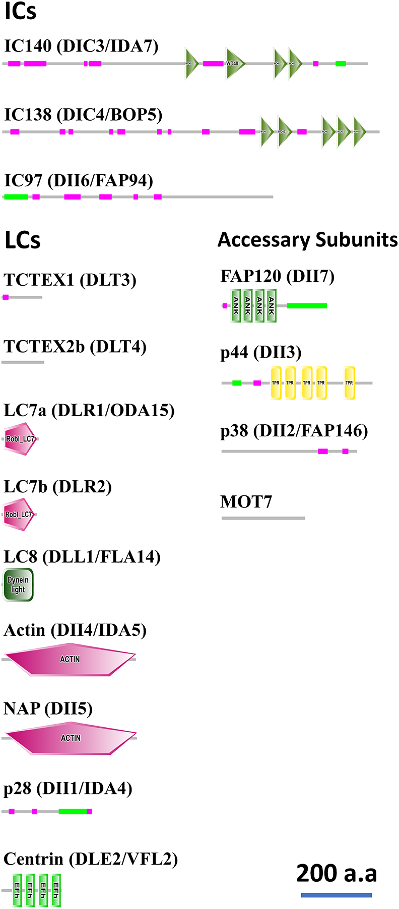
Motile cilia (also interchangeably called "flagella") are conserved organelles extending from the surface of many animal cells and play essential functions in eukaryotes, including cell motility and environmental sensing. Large motor complexes, the ciliary dyneins, are present on ciliary outer-doublet microtubules and drive movement of cilia. Ciliary dyneins are classified into two general types: the outer dynein arms (ODAs) and the inner dynein arms (IDAs). While ODAs are important for generation of force and regulation of ciliary beat frequency, IDAs are essential for control of the size and shape of the bend, features collectively referred to as waveform. Also, recent studies have revealed unexpected links between IDA components and human diseases. In spite of their importance, studies on IDAs have been difficult since they are very complex and composed for several types of IDA motors, each unique in composition and location in the axoneme. Thanks in part to genetic, biochemical, and structural analysis of Chlamydomonas reinhardtii, we are beginning to understand the organization and function of the ciliary IDAs. In this review, we summarize the composition of Chlamydomonas IDAs particularly focusing on each subunit, and discuss the assembly, conservation, and functional role(s) of these IDA subunits. Furthermore, we raise several additional questions/challenges regarding IDAs, and discuss future perspectives of IDA studies.

Keywords: Chlamydomonas; IDA; cilia; flagella; inner-arm dynein; motility; subunit.


Conflict of interest statement

Conflict of Interest

The authors declare there is no conflict of interest in this work.

Figures

Figure 1.
Figure 1.. Potential arrangements of IDAs in Chlamydomonas cilia
(a) (top) Drawings of a Chlamydomonas cell and ciliary axonemal cross-section. (bottom) Hypothesized models of IDA arrangements on surface-rendering images of the 96-nm repeats of Chlamydomonas ciliary doublet microtubules (DMTs). The images are the results of sub-tomogram averages from reconstructed cryo-electron tomograms. Surface-rendering images of the proximal and central/distal regions of DMT1 and other averaged DMTs (DMTs 2–8 or 2–9) are shown. In Chlamydomonas, DMT1 totally lacks ODAs, and also has particularly interesting features including the arrangement of IDAs (Bui et al., 2012; Hoops & Witman, 1983). In the proximal region of ciliary axonemes, the arrangement of IDAs differs from that in the central/distal region, lacking IDA b (DHC5) and possibly minor IDAs replacing some major IDAs (Bui et al., 2012). Approximate locations of ODAs, IDAs, the IC/LC complex of IDA f/I1, N-DRC, radial spokes and the 1–2 bridges are shown in water blue, old-rose red, yellow, green, purple, and light brown, respectively. A light pink arrowhead indicates the missing IDA b location in the proximal portion of the DMT2-9 average. Mi, minor IDA; Un, unknown density likely an IDA species (Bui et al., 2012). The tomograms were reconstructed and published in the previous publication (Bui et al., 2012), and refined for this review. The accession IDs of the density maps (Bui et al., 2012) in EMDataBank (http://www.emdatabank.org/) used to make this figure are as follows: DMT1 proximal region, EMD-2119; DMT1 central region, EMD-2113; DMT2-9 proximal region, EMD-2131; DMT2-8 central/distal region, EMD-2132. (b) A summary of proposed arrangements of IDAs in Chlamydomonas cilia. The figure is adapted/refined from (Bui et al., 2012). Chlamydomonas cilia have heterogeneity in the arrangement of IDAs among DMTs, and this heterogeneity could contribute to generation of proper ciliary waveform. IDA d and e may be partially replaced by minor IDAs DHC11 and DHC4 in the proximal portion of the axonemes (Bui et al., 2012). Also, replacement of IDA g by DHC3 was proposed in (Bustamante-Marin et al., 2020). DMT1 has unique organization of IDAs throughout the axonemes (Bui et al., 2012; Hoops & Witman, 1983). P, Proximal; D, Distal.
Figure 2.
Figure 2.. Domain structure of IDA HCs in Chlamydomonas
Domains/motifs in DHCs of Chlamydomonas ciliary IDAs. “Dynein heavy chain, N-terminal region 1 (DHC_N1)”, “dynein heavy chain, N-terminal region 2 (DHC_N2)”, and “microtubule-binding stalk of dynein motor (MT)” domains were predicted using the pfam analyses (https://pfam.xfam.org/)(Punta et al., 2012). Appropriate locations of 6 “ATPases-associated-with-diverse-cellular-activities (AAA)” domains/rings in the IDA DHCs were predicted by aligning the IDA sequences with the Tripneustes gratilla ODAβ DHC [X59603.1 (NCBI)](Mocz & Gibbons, 2001). Note that two minor IDAs, DHC3 and DHC12, have longer molecular length than other IDAs. The accession Nos/IDs in the Phytozome Chlamydomonas v5.5 (https://phytozome.jgi.doe.gov/pz/portal.html#!info?alias=Org_Creinhardtii) used for these domain predictions were as follows: DHC1/PF9/IDA1, Cre12.g484250.t1.1; DHC2, Cre09.g392282.t1.1; DHC3, Cre06.g265950.t1.1; DHC4, Cre02.g107350.t1.1; DHC5, Cre02.g107050.t1.1; DHC6, Cre05.g244250.t1.2; DHC7, Cre14.g627576.t1.1; DHC8, Cre16.g685450.t1.1; DHC9/IDA9, Cre02.g141606.t1.1; DHC10/IDA2, Cre14.g624950.t1.1; DHC11, Cre12.g555950.t1.2; DHC12, Cre06.g297850.t1.1.
Figure 3.
Figure 3.. Domain structure of IDA ICs/LCs/accessary subunits in Chlamydomonas
Domains/motifs in ICs/LCs/accessary subunits of Chlamydomonas ciliary IDAs were predicted using the SMART analyses (http://smart.embl-heidelberg.de/)(Letunic et al., 2021; Schultz et al., 1998) with the default setting and outputs with scale modification were shown. The green thin lines indicate the coiled-coil regions, and the pink thin lines indicate the low-complexity regions. While seven WD repeats were identified in the original descriptions both for the IC140 and IC138 molecules (Hendrickson et al., 2004; Yang & Sale, 1998), fewer repeats were predicted by SMART analyses in this figure due to the threshold. The domain structures of FAP120 and p44 were re-analyzed from (Ikeda et al., 2009; Yamamoto et al., 2008). The accession Nos/IDs in NCBI (https://www.ncbi.nlm.nih.gov/) or Phytozome Chlamydomonas v5.5 (https://phytozome.jgi.doe.gov/pz/portal.html#!info?alias=Org_Creinhardtii) used for these domain predictions were as follows: IC140/DIC3/IDA7, XP_001695786.1 (NCBI); IC138/DIC4/BOP5, XP_001696921.1 (NCBI); IC97/DII6/FAP94, ACN22075 (NCBI); TCTEX1/DLT3, Cre01.g004250.t1.2 (Phytozome); TCTEX2b/DLT4, DAA05278 (NCBI); LC7a/DLR1/ODA15, Cre08.g376550.t1.2 (Phytozome); LC7b/DLR2, Cre12.g546400.t1.2 (Phytozome); LC8/DLL1/FLA14, Cre03.g181150.t1.1 (Phytozome); Actin/DII4/IDA5, Cre13.g603700.t1.2 (Phytozome); NAP/DII5, Cre03.g176833.t1.1 (Phytozome); p28/DII1/IDA4, CAA88139 (NCBI); Centrin/DLE2/VFL2, Cre11.g468450.t1.2 (Phytozome); FAP120/DII3, BAN15819 (NCBI); p44/DII3, AB353122.2 (NCBI); p38/DII2/FAP146, BAG07147 (NCBI); MOT7, Cre01.g038750.t1.2 (Phytozome).

Similar articles

Cited by

References

    1. Afzelius BA (1976). A human syndrome caused by immotile cilia. Science, 193(4250), 317–319. - PubMed
    1. Alford LM, Wirschell M, Yamamoto R, & Sale WS (2012). 11 - Control of Axonemal Inner Dynein Arms A2 - King, Stephen M. In Dyneins (pp. 312–335). Academic Press.
    1. Aravind Kumar M, Singh V, Naushad SM, Shanker U, & Lakshmi Narasu M (2018). Microarray-based SNP genotyping to identify genetic risk factors of triple-negative breast cancer (TNBC) in South Indian population. Mol Cell Biochem, 442(1–2), 1–10. - PubMed
    1. Awata J, Song K, Lin J, King SM, Sanderson MJ, Nicastro D, & Witman GB (2015). DRC3 connects the N-DRC to dynein g to regulate flagellar waveform. Mol Biol Cell, 26(15), 2788–2800. - PMC - PubMed
    1. Barbar E (2008). Dynein light chain LC8 is a dimerization hub essential in diverse protein networks. Biochemistry, 47(2), 503–508. - PubMed

Publication types

LinkOut - more resources