Review
. 2021 Jun;10(11):e2001986.
doi: 10.1002/adhm.202001986. Epub 2021 Apr 19.

Multi-Dimensional Printing for Bone Tissue Engineering

Affiliations
Review

Multi-Dimensional Printing for Bone Tissue Engineering

Moyuan Qu et al. Adv Healthc Mater. 2021 Jun.

Abstract

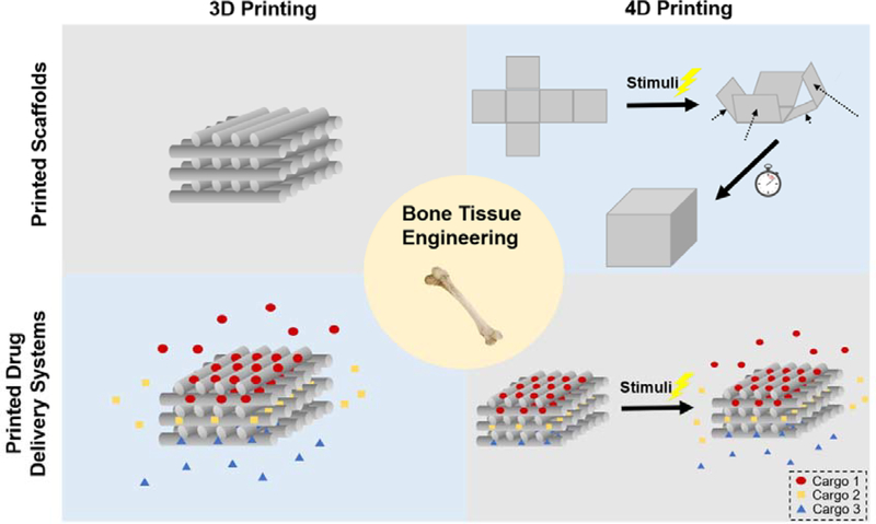
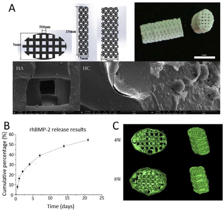
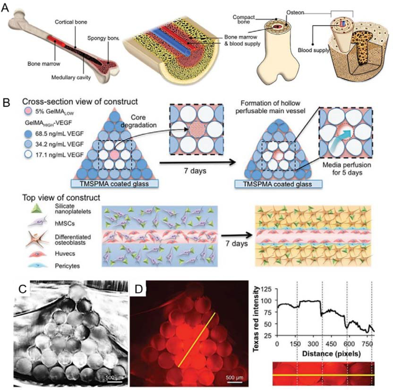
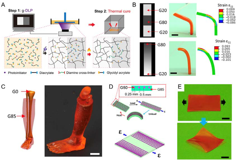
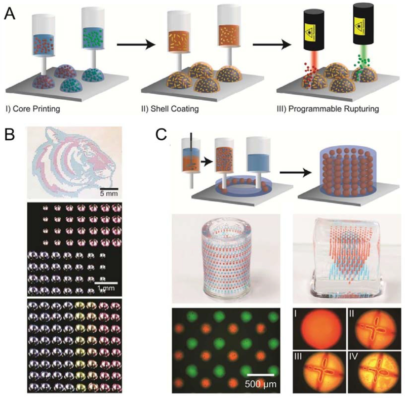
The development of 3D printing has significantly advanced the field of bone tissue engineering by enabling the fabrication of scaffolds that faithfully recapitulate desired mechanical properties and architectures. In addition, computer-based manufacturing relying on patient-derived medical images permits the fabrication of customized modules in a patient-specific manner. In addition to conventional 3D fabrication, progress in materials engineering has led to the development of 4D printing, allowing time-sensitive interventions such as programed therapeutics delivery and modulable mechanical features. Therapeutic interventions established via multi-dimensional engineering are expected to enhance the development of personalized treatment in various fields, including bone tissue regeneration. Here, recent studies utilizing 3D printed systems for bone tissue regeneration are summarized and advances in 4D printed systems are highlighted. Challenges and perspectives for the future development of multi-dimensional printed systems toward personalized bone regeneration are also discussed.

Keywords: 3D printing; 4D printing; bone; drug delivery; tissue engineering.


Conflict of interest statement

Conflict of Interest

The authors declare no conflict of interest.

Figures

Figure 1.
Figure 1.
Schematic illustration of multi-dimensional printed scaffolds and DDS for bone tissue engineering. 3D printing techniques enable control of scaffold microstructures as well as composition. With 3D printing, it is possible to fabricate personalized DDS with multiple components and achieve personalized and programed drug delivery. In addition to customized structures and compositions, 4D printed scaffolds and DDS are expected to sense signals within the physiological environment and change functionalities correspondingly, resulting in auto-adjusted structures or drug release profiles.
Figure 2.
Figure 2.
A. Schematic illustration and SEM images of 3D printed Haversian bone–mimicking scaffolds with Ca2MgSi2O7 bioceramic, 45S5 bioactive glass and photosensitive resin. Thsese scaffolds were integrated with Haversian canals, Volkmann canals and cancellous bone structures. hBMSCs were seeded in cancellous bone mimic structures and HUVECs were seeded on Haversian canals. B-F. Optical microscope images of 3D printed Haversian bone–mimicking scaffolds with various diameters and numbers of Haversian canals (red arrows). Scale bars: 1 mm. a-e. Micro-CT images exhibited the connection between Volkmann canals (blue arrows) and Haversian canals in the interior of scaffolds. Scale bars, 1 mm. G-J. SEM images displayed the surface microstructure of the scaffolds. Scale bar: 400 μm. K. Well-sintered surface of 3D printed Haversian bone–mimicking scaffolds. Scale bar: 6 μm. Reproduced with permission.[74] Copyright 2020, AAAS.
Figure 3.
Figure 3.
3D printed scaffolds consisting of tri-block poly (lactide-co-propylene glycol-co-lactide) dimethacrylate (PmLnDMA) and hydroxyethyl methacrylate (HEMA)-functionalized hydroxyapatite nanoparticles (nHAMA). A. Schematic illustration of material synthesis. B. Schematic illustration of 3D printing process. The PmLnDMA-nHAMA co-crosslinked network improved the interfacial interaction and further enhanced the mechanical strength of scaffolds. C. Images of printed scaffolds with multiple 3D shapes printed with P7L2DMA/50%nHAMA composited ink. Reproduced with permission.[88] Copyright 2020, Elsevier.
Figure 4.
Figure 4.
3D printed tissue engineering scaffold with hyaluronic acid-PLGA encapsulating BMP-2/PEG complex. A. Schematic illustrations and photographs of the preparation of feeding solution and instruments for 3D printing of scaffolds using a multihead deposition system. B. Photographs and SEM images of 3D printed hyaluronic acid-PLGA/PEG/BMP-2 scaffold and BMP-2/PEG complexes within the fiber. C. μCT images of regenerated bones in calvarial bone defect model. (From top to bottom: control, hyaluronic acid-PLGA scaffold, and hyaluronic acid-PLGA/PEG/BMP-2 scaffold respectively). Reproduced with permission.[132] Copyright 2011, WILEY-VCH Verlag GmbH & Co. KGaA, Weinheim.
Figure 5.
Figure 5.
The 3D printed porous HA scaffold coated with BMP-2 loaded chitosan microspheres used for bone regeneration. A. Design, photograph and SEM images of 3D printed porous HA scaffold, collagen coated scaffold (HC) and collagen coated scaffold with BMP-2 loaded chitosan microspheres (HCC); B. in vitro release profile of BMP-2 from HCC; C. μCT images of ectopic bone formation with HCC after implantations for 4 week and 8 weeks. Reproduced with permission.[141] Copyright 2016, Elsevier.
Figure 6.
Figure 6.
3D bioprinting of bone mimetic 3D architecture with osteogenic and vasculogenic gradients. A. Schematic illustration of natural bone structure; B. Schematic illustration of 3D architecture with concentration gradients of VEGF. Central channel able to quickly degrade and favor the growth of HUVECs. C. Cross-section image of the printed hydrogel. D. Cross-section image of the printed gradual hydrogel with gradual concentrations of fluorochrome (Texas Red). Reproduced with permission.[64] Copyright 2017, WILEY-VCH Verlag GmbH & Co. KGaA, Weinheim.
Figure 7.
Figure 7.
A. Schematics of two-stage light and heating curing process of the g-DLP printing via graded material using hybrid ink. B. g-DLP printing via a discrete gradient and continuous gradient greyscale patterns and corresponding strain simulations. Scale bars, 5 mm. C. g-DLP printing of an artificial limb structure with stiff bone (G0) surrounded by soft muscle (G85). Scale bars, 1 cm. D. Design of a composite shape-shifting film by distributing fibers (G50) within the film (G85). E. Pictures of the printed shape-shifting film before and after the strain applications at room temperature. Scale bars, 1 cm. Reproduced with permission.[172] Copyright 2019, AAAS.
Figure 8.
Figure 8.
3D printed responsive capsule with core/shell structures which can achieve programmable release of multiple drugs within a hydrogel matrix. A. Schematic illustration of the fabrication and rupturing of the responsive capsules, using laser. B. Optical micrographs of arrays with 3D printed capsules with different loading volume, distribution or multiple drug compositions; C. 3D printed hydrogel matrix with responsive capsules composed of multiple drugs allowing controlled capsule distributions. I-IV: Programed rupturing of the PLGA shell with plasmonic gold nanorods (AuNRs). (I: before laser rupture; II, III, IV: 15 min, 1 h, and 2 h after laser rupture) Reproduced with permission.[176] Copyright 2015, American Chemical Society.

References

    1. Bose S, Vahabzadeh S, Bandyopadhyay A, Materials today 2013, 16, 496.
    1. Jones AC, Arns CH, Sheppard AP, Hutmacher DW, Milthorpe BK, Knackstedt MA, Biomaterials 2007, 28, 2491. - PubMed
    1. Seol Y-J, Kang T-Y, Cho D-W, Soft Matter 2012, 8, 1730.
    1. Khademhosseini A, Langer R, Nature protocols 2016, 11, 1775. - PubMed
    1. Stevens MM, Materials today 2008, 11, 18.

Publication types

LinkOut - more resources