Impact of prenatal maternal cytokine exposure on sex differences in brain circuitry regulating stress in offspring 45 years later
- PMID: 33876747
- PMCID: PMC8054010
- DOI: 10.1073/pnas.2014464118
Impact of prenatal maternal cytokine exposure on sex differences in brain circuitry regulating stress in offspring 45 years later
Abstract
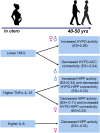
Stress is associated with numerous chronic diseases, beginning in fetal development with in utero exposures (prenatal stress) impacting offspring's risk for disorders later in life. In previous studies, we demonstrated adverse maternal in utero immune activity on sex differences in offspring neurodevelopment at age seven and adult risk for major depression and psychoses. Here, we hypothesized that in utero exposure to maternal proinflammatory cytokines has sex-dependent effects on specific brain circuitry regulating stress and immune function in the offspring that are retained across the lifespan. Using a unique prenatal cohort, we tested this hypothesis in 80 adult offspring, equally divided by sex, followed from in utero development to midlife. Functional MRI results showed that exposure to proinflammatory cytokines in utero was significantly associated with sex differences in brain activity and connectivity during response to negative stressful stimuli 45 y later. Lower maternal TNF-α levels were significantly associated with higher hypothalamic activity in both sexes and higher functional connectivity between hypothalamus and anterior cingulate only in men. Higher prenatal levels of IL-6 were significantly associated with higher hippocampal activity in women alone. When examined in relation to the anti-inflammatory effects of IL-10, the ratio TNF-α:IL-10 was associated with sex-dependent effects on hippocampal activity and functional connectivity with the hypothalamus. Collectively, results suggested that adverse levels of maternal in utero proinflammatory cytokines and the balance of pro- to anti-inflammatory cytokines impact brain development of offspring in a sexually dimorphic manner that persists across the lifespan.
Keywords: functional brain imaging; prenatal immune programming; prenatal stress; sex; stress circuitry.
Conflict of interest statement
Competing interest statement: J.M.G. is on the scientific advisory board for, and has equity interest in, Cala Health (a neuromodulation company). J.M.G.’s interests are managed by Massachusetts General Hospital and Mass General Brigham HealthCare in accordance with their conflict of interest policies. However, the work in the study presented here was conducted prior to that relationship, so there is no conflict of interest.
Figures


Similar articles
-
Prenatal immune origins of brain aging differ by sex.Mol Psychiatry. 2025 May;30(5):1887-1896. doi: 10.1038/s41380-024-02798-w. Epub 2024 Nov 21. Mol Psychiatry. 2025. PMID: 39567743 Free PMC article.
-
Prenatal immune programming of the sex-dependent risk for major depression.Transl Psychiatry. 2016 May 31;6(5):e822. doi: 10.1038/tp.2016.91. Transl Psychiatry. 2016. PMID: 27244231 Free PMC article.
-
Functional Connectivity to the Amygdala in the Neonate Is Impacted by the Maternal Anxiety Level During Pregnancy.J Neuroimaging. 2025 Jan-Feb;35(1):e70004. doi: 10.1111/jon.70004. J Neuroimaging. 2025. PMID: 39757405 Free PMC article.
-
Prenatal Immune and Endocrine Modulators of Offspring's Brain Development and Cognitive Functions Later in Life.Front Immunol. 2018 Sep 26;9:2186. doi: 10.3389/fimmu.2018.02186. eCollection 2018. Front Immunol. 2018. PMID: 30319639 Free PMC article. Review.
-
Brain structural and functional outcomes in the offspring of women experiencing psychological distress during pregnancy.Mol Psychiatry. 2024 Jul;29(7):2223-2240. doi: 10.1038/s41380-024-02449-0. Epub 2024 Feb 28. Mol Psychiatry. 2024. PMID: 38418579 Free PMC article. Review.
Cited by
-
Sex-specific neurodevelopmental outcomes in offspring of mothers with SARS-CoV-2 in pregnancy: an electronic health records cohort.medRxiv [Preprint]. 2022 Nov 18:2022.11.18.22282448. doi: 10.1101/2022.11.18.22282448. medRxiv. 2022. Update in: JAMA Netw Open. 2023 Mar 1;6(3):e234415. doi: 10.1001/jamanetworkopen.2023.4415. PMID: 36415457 Free PMC article. Updated. Preprint.
-
Cord serum cytokines at birth and children's trajectories of mood dysregulation symptoms from 3 to 8 years: The EDEN birth cohort.Brain Behav Immun Health. 2024 Mar 29;38:100768. doi: 10.1016/j.bbih.2024.100768. eCollection 2024 Jul. Brain Behav Immun Health. 2024. PMID: 38586283 Free PMC article.
-
Where Sex Meets Gender: How Sex and Gender Come Together to Cause Sex Differences in Mental Illness.Front Psychiatry. 2022 Jun 28;13:856436. doi: 10.3389/fpsyt.2022.856436. eCollection 2022. Front Psychiatry. 2022. PMID: 35836659 Free PMC article.
-
Maternal adverse childhood experiences impact fetal adrenal volume in a sex-specific manner.Biol Sex Differ. 2023 Feb 17;14(1):7. doi: 10.1186/s13293-023-00492-0. Biol Sex Differ. 2023. PMID: 36803442 Free PMC article.
-
Maternal immune activation as a risk factor for psychiatric illness in the context of the SARS-CoV-2 pandemic.Brain Behav Immun Health. 2021 Oct;16:100297. doi: 10.1016/j.bbih.2021.100297. Epub 2021 Jul 15. Brain Behav Immun Health. 2021. PMID: 34308388 Free PMC article.
References
-
- Barker D. J., Intrauterine programming of adult disease. Mol. Med. Today 1, 418–423 (1995). - PubMed
-
- McEwen B. S., Gonadal steroid influences on brain development and sexual differentiation. Int. Rev. Physiol. 27, 99–145 (1983). - PubMed
-
- Weinstock M., The long-term behavioural consequences of prenatal stress. Neurosci. Biobehav. Rev. 32, 1073–1086 (2008). - PubMed
-
- Bale T. L., Neuroendocrine and immune influences on the CNS: It’s a matter of sex. Neuron 64, 13–16 (2009). - PubMed
-
- Goldstein J. M., Impact of prenatal stress on offspring psychopathology and comorbidity with general medicine later in life. Biol. Psychiatry 85, 94–96 (2019). - PubMed
Publication types
MeSH terms
Substances
Grants and funding
LinkOut - more resources
Full Text Sources
Other Literature Sources
Medical

