. 2021 Apr 13;118(15):e2018627118.
doi: 10.1073/pnas.2018627118.

Modular complement assemblies for mitigating inflammatory conditions

Affiliations

Modular complement assemblies for mitigating inflammatory conditions

Kelly M Hainline et al. Proc Natl Acad Sci U S A. .

Abstract

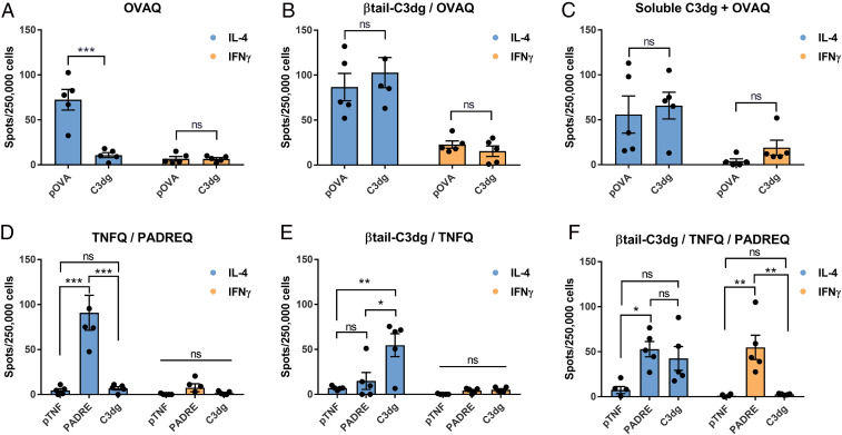
Complement protein C3dg, a key linkage between innate and adaptive immunity, is capable of stimulating both humoral and cell-mediated immune responses, leading to considerable interest in its use as a molecular adjuvant. However, the potential of C3dg as an adjuvant is limited without ways of controllably assembling multiple copies of it into vaccine platforms. Here, we report a strategy to assemble C3dg into supramolecular nanofibers with excellent compositional control, using β-tail fusion tags. These assemblies were investigated as therapeutic active immunotherapies, which may offer advantages over existing biologics, particularly toward chronic inflammatory diseases. Supramolecular assemblies based on the Q11 peptide system containing β-tail-tagged C3dg, B cell epitopes from TNF, and the universal T cell epitope PADRE raised strong antibody responses against both TNF and C3dg, and prophylactic immunization with these materials significantly improved protection in a lethal TNF-mediated inflammation model. Additionally, in a murine model of psoriasis induced by imiquimod, the C3dg-adjuvanted nanofiber vaccine performed as well as anti-TNF monoclonal antibodies. Nanofibers containing only β-tail-C3dg and lacking the TNF B cell epitope also showed improvements in both models, suggesting that supramolecular C3dg, by itself, played an important therapeutic role. We observed that immunization with β-tail-C3dg caused the expansion of an autoreactive C3dg-specific T cell population, which may act to dampen the immune response, preventing excessive inflammation. These findings indicate that molecular assemblies displaying C3dg warrant further development as active immunotherapies.

Keywords: active immunotherapy; immune engineering; immunoengineering; self-assembly; vaccine.


Conflict of interest statement

Competing interest statement: J.H.C. is an inventor on a US patent describing the β-tail technology.

Figures

Fig. 1.
Fig. 1.
Engineered β-tail–C3dg integrates into self-assembled peptide nanofibers. (A) Schematic representation of the β-tail’s transitional behavior into a β-sheet conformation (24). (B) Schematic of nanofiber components. (C) Schematic of coassembled nanofibers. (D) SDS-PAGE of expressed, purified β-tail–C3dg protein. (E) Negative-stained TEM images of nanofibers containing 20 μM β-tail–C3dg (prepared at 2 mM total Q11 peptide concentration and diluted to 0.2 mM for imaging). (F) β-tail–C3dg was integrated into Q11 nanofibers over the range of 12.5 to 50 μM in a β-tail–dependent manner, as measured by loss of protein from the supernatant. (G) Quantification of β-tail–C3dg and C3dg assembly into nanofibers, expressed as the percentage of total protein incorporated. n = 3, ± SD for F and G. (Scale bar, 200 nm.) Statistical significance (G) was tested by multiple t tests with Holm–Sidak correction, **P < 0.01, ***P < 0.001.
Fig. 2.
Fig. 2.
β-tail-C3dg activates B cells in vitro in a dose-dependent manner and enhances humoral responses in vivo. (A) Nanofibers containing increasing amounts of β-tail–C3dg enhanced B cell activation in a dose-dependent manner. Signal was normalized by subtracting the initial mean florescent intensity measurement from all subsequent data points. Arrow indicates time of nanofiber application. (B) Total calcium signaling integrated over 90 s poststimulation for the same groups as in A. (C) β-tail–C3dg assembled into Q11 nanofibers elicited faster and stronger calcium signaling than soluble C3dg. (D) Total calcium signaling integrated over 90 s poststimulation for the same groups as in C. OVA peptide-specific (E) and C3dg protein-specific (F) IgG responses in the sera of mice immunized with formulations containing OVAQ, soluble C3dg, and supramolecular β-tail–C3dg. TNF peptide-specific (G) and C3dg protein-specific (H) IgG responses in the sera of mice immunized with various formulations containing TNFQ, PADREQ, soluble C3dg, and β-tail–C3dg. (AD) Combination of two independent experiments in which sample sizes were n = 2 mice. In E, mice were immunized at week 0 and boosted at week 3 and 6. In F, mice were immunized at week 0 and boosted at week 4. n = 5 for each group in EH. Mean ± SD is shown. Statistical significance was tested by one-way ANOVA with Tukey’s multiple comparison test (B), Student’s two-tailed, unpaired t test (D), or two-way ANOVA with Tukey’s multiple comparison test (EH). ns = not significant, *P < 0.05, **P < 0.01, ***P < 0.001, ****P < 0.0001.
Fig. 3.
Fig. 3.
Immunization with peptide nanofibers protected in a model of acute TNF-mediated inflammation, with the optimized formulation preventing the development of shock-like symptoms. (A) The difference between the highest and lowest measured temperature per mouse. (BI) Body temperature and overall survival of individual mice in each group. Letters at the lower right-hand corner of each graph indicate other groups that are statistically different (P < 0.05). (I and J) The lowest recorded temperature of each mouse graphed against its anti-TNF (J) and anti-C3dg (K) titers from serum collected 1 wk before the LPS Challenge. Combination of two independent experiments in which n = 5 each (soluble C3dg + TNFQ group was only included in one experiment). Statistical comparisons of temperature changes (A) and temperature/titer correlations (I and J) were made using one-way ANOVA with Tukey’s multiple comparison test. Statistical comparisons of survival between all groups in CH were made using log-rank test; ns = not significant, *P < 0.05, ****P < 0.0001.
Fig. 4.
Fig. 4.
β-tail–C3dg in nanofibers induces the production of TH2-biased, C3dg-specific helper T cells. Cytokine-secreting cells from mice immunized with OVAQ formulations (AC) and TNFQ formulations (DF) were quantified ex vivo using ELISpot. Lymphocytes from spleens were suspended and restimulated with peptide epitope or C3dg. n = 5 for all groups; data points represent individual mice. In AC, statistical significance was tested by multiple two-tailed t tests with Holm–Sidak correction. In DF, statistical significance was tested by one-way ANOVA with Tukey’s multiple comparison test. ns: not significant, *P < 0.05, **P < 0.01, ***P < 0.001.
Fig. 5.
Fig. 5.
Immunization with peptide nanofibers reduces the amount of epidermal thickening in a model of imiquimod-induced psoriasis. Representative histology images and epidermal thickening of skin collected from C57BL/6 (AB) and TNF-KO (CD) mice are shown. (B) Mice treated with anti-TNF antibody 3 and 0 h prior to the first application of imiquimod exhibited similar levels of reduction in epidermal thickening when compared to mice immunized with β-tail–C3dg/TNF/PADRE nanofibers. (D) TNF-KO mice immunized with the β-tail–C3dg/TNF/PADRE formulation also showed reduced epidermal thickening. Data points represent individual mice. (B) Combination of two independent experiments in which n = 5 for each group (not all groups were included in the repeated experiment). (D) Single experiment with n = 5 for each group. Statistical significance was tested using one-way ANOVA with Tukey’s multiple comparison test. ns: not significant, *P < 0.05, **P < 0.01, ****P < 0.0001. (Scale bar, 200 μm.)
Fig. 6.
Fig. 6.
CD4+ T cells play a critical role in the prevention of shock-like symptoms, whereas α-C3dg antibodies do not provide protection in an LPS challenge. Serum from (A) unimmunized mice or (B) β-tail–C3dg immunized mice was administered via tail vein injection on day −1 before the LPS challenge. (C) C3dg protein-specific IgG responses in the sera of mice (D and E) collected immediately prior to the start of the LPS challenge. (DF) Mice immunized with β-tail–C3dg assembled into nanofibers or unimmunized mice were treated either with a CD4-depleting antibody or isotype control at days −3 and −1 before the start of the LPS challenge. (D and E) Mice were immunized at week 0 and boosted at week 4. n = 5 for each group. (C) Statistical significance tested by Student’s two-tailed, unpaired t test. Statistical comparisons of survival between groups DF were made using log-rank test. Letters at the lower right-hand corner of each graph indicate other groups that are statistically different (P < 0.05). ****P < 0.0001.

References

    1. Fearon D. T., The complement system and adaptive immunity. Semin. Immunol. 10, 355–361 (1998). - PubMed
    1. Dempsey P. W., Allison M. E., Akkaraju S., Goodnow C. C., Fearon D. T., C3d of complement as a molecular adjuvant: Bridging innate and acquired immunity. Science 271, 348–350 (1996). - PubMed
    1. De Groot A. S., et al. ., C3d adjuvant effects are mediated through the activation of C3d-specific autoreactive T cells. Immunol. Cell Biol. 93, 189–197 (2015). - PMC - PubMed
    1. Green T. D., Montefiori D. C., Ross T. M., Enhancement of antibodies to the human immunodeficiency virus type 1 envelope by using the molecular adjuvant C3d. J. Virol. 77, 2046–2055 (2003). - PMC - PubMed
    1. Ross T. M., Xu Y., Green T. D., Montefiori D. C., Robinson H. L., Enhanced avidity maturation of antibody to human immunodeficiency virus envelope: DNA vaccination with gp120-C3d fusion proteins. AIDS Res. Hum. Retroviruses 17, 829–835 (2001). - PMC - PubMed

Publication types

LinkOut - more resources