. 2021 Apr 13;118(15):e2026804118.
doi: 10.1073/pnas.2026804118.

Capture and metabolomic analysis of the human endometrial epithelial organoid secretome

Affiliations

Capture and metabolomic analysis of the human endometrial epithelial organoid secretome

Constantine A Simintiras et al. Proc Natl Acad Sci U S A. .

Abstract

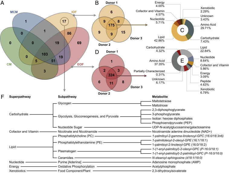
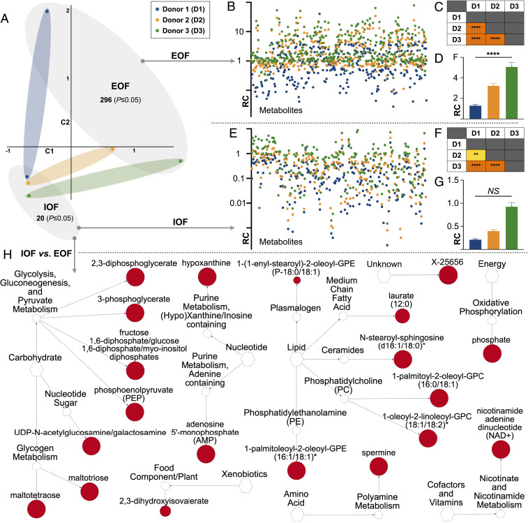
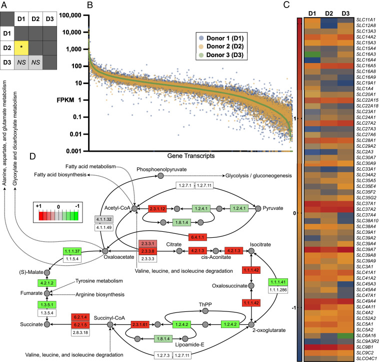
Suboptimal uterine fluid (UF) composition can lead to pregnancy loss and likely contributes to offspring susceptibility to chronic adult-onset disorders. However, our understanding of the biochemical composition and mechanisms underpinning UF formation and regulation remain elusive, particularly in humans. To address this challenge, we developed a high-throughput method for intraorganoid fluid (IOF) isolation from human endometrial epithelial organoids. The IOF is biochemically distinct to the extraorganoid fluid (EOF) and cell culture medium as evidenced by the exclusive presence of 17 metabolites in IOF. Similarly, 69 metabolites were unique to EOF, showing asymmetrical apical and basolateral secretion by the in vitro endometrial epithelium, in a manner resembling that observed in vivo. Contrasting the quantitative metabolomic profiles of IOF and EOF revealed donor-specific biochemical signatures of organoids. Subsequent RNA sequencing of these organoids from which IOF and EOF were derived established the capacity to readily perform organoid multiomics in tandem, and suggests that transcriptomic regulation underpins the observed secretory asymmetry. In summary, these data provided by modeling uterine luminal and basolateral fluid formation in vitro offer scope to better understand UF composition and regulation with potential impacts on female fertility and offspring well-being.

Keywords: endometrium; human; organoid; reproduction; uterine fluid.


Conflict of interest statement

The authors declare no competing interest.

Figures

Fig. 1.
Fig. 1.
Study design and EEO monitoring. (A) Schematic depiction of study-dependent variables. (B) Time-lapse live imaging over 60 h of representative EEO formation from each donor. (Scale bars, 100 μm.) (C) Schematic depiction of the EEO preparation regimen including a 48-h 17β-estradiol (E2) exposure. Mean (±SEM) EEO (D) count and (E) areas on days 0 and 6 (n = 12); P ≤ 0.0001 (****). Representative EEO on days 0 (F) and 6 (G) at ×40 magnification. (Scale bars, 500 µm.)
Fig. 2.
Fig. 2.
EEO IOF isolation validation. (A) Schematic depiction of the preparation for extracting IOF by MMN and transferring it to PBS. (B) Sequential images of IOF retrieval by MMN. (Scale bar, 250 μm.) (C) Mean normalized metabolomic profiles of IOF obtained by MMN vs. HTC from EEO vs. CM. Green depicts undetected metabolites. IOF profiles obtained by MMN and HTC were statistically identical (P = 0.735), whereas the CM metabolome differed from MMN and HTC counterparts (P ≤ 0.0001). (DF) Metabolomic profiles of IOF obtained by MMN vs. HTC by donor vs. CM profiling. Legends and statistical comparisons between groups are provided above each plot.
Fig. 3.
Fig. 3.
Qualitative organoid fluid metabolomics. (A) Venn diagram depicting the number of metabolites identified in organoid CM, MCM, IOF, and EOF across donors. (B) IOF metabolite presence by donor, in addition to (C) the mean superpathway breakdown of the IOF metabolites common to all donors. (D) EOF metabolite presence by donor, in addition to (E) the mean superpathway breakdown of the EOF metabolites common to all donors. (F) The 17 IOF-exclusive metabolites, arranged by pathway, wherein italicized metabolites are predicted.
Fig. 4.
Fig. 4.
Quantitative intraorganoid fluid metabolomics. (A) Principal component analysis of IOF and EOF quantitative metabolic profiles from each donor in addition to the number of metabolites elevated (P ≤ 0.05) in each. (B) EOF metabolomic signatures by donor (relative concentration [RC]). (C) Corresponding EOF donor vs. donor statistical comparisons [P ≤ 0.0001 (****)] are also provided. (D) Mean total metabolite levels in EOF by donor. (E) IOF metabolomic signatures by donor. (F) Corresponding IOF donor vs. donor statistical comparisons [P ≤ 0.01 (**)] are also provided. (G) Mean total metabolite levels in IOF by donor (not significant [NS]). (H) Network view by pathway of the metabolites elevated (P ≤ 0.05) in IOF vs. EOF wherein node diameter is proportional to fold-change magnitude.
Fig. 5.
Fig. 5.
Tandem organoid transcriptomics. (A) Statistical comparison [P ≤ 0.05 (*); not significant (NS)] of transcriptomic profiles of endometrial epithelial organoids obtained from all three donors. (B) Logarithmically transformed FPKM values per donor per gene (transcriptomic signatures) presented in descending order. (C) Relative transcript abundance of SLC protein genes, specifically, those exhibiting a ≥2.0-fold change between two or more donors. Green depicts undetected transcripts. (D) Pathview and Kyoto Ecyclopedia of Genes and Genomes (KEGG) database adapted extrapolation and rendering of transcriptomic profiles of donors 1 vs. 2 within the context of the citric acid cycle, wherein green denotes enzyme transcript-relative down-regulation and red depicts enzyme transcript-relative up-regulation.

Similar articles

Cited by

References

    1. Salamonsen L. A., Evans J., Nguyen H. P. T., Edgell T. A., The microenvironment of human implantation: Determinant of reproductive success. Am. J. Reprod. Immunol. 75, 218–225 (2016). - PubMed
    1. Spencer T. E., Biological roles of uterine glands in pregnancy. Semin. Reprod. Med. 32, 346–357 (2014). - PMC - PubMed
    1. Burton G. J., Watson A. L., Hempstock J., Skepper J. N., Jauniaux E., Uterine glands provide histiotrophic nutrition for the human fetus during the first trimester of pregnancy. J. Clin. Endocrinol. Metab. 87, 2954–2959 (2002). - PubMed
    1. Kelleher A. M., DeMayo F. J., Spencer T. E., Uterine glands: Developmental biology and functional roles in pregnancy. Endocr. Rev. 40, 1424–1445 (2019). - PMC - PubMed
    1. Spencer T. E., Kelleher A. M., Bartol F. F., Development and function of uterine glands in domestic animals. Annu. Rev. Anim. Biosci. 7, 125–147 (2019). - PubMed

Publication types