Case Reports
. 2021 Apr 13;118(15):e2102804118.
doi: 10.1073/pnas.2102804118.

Inherited deficiency of stress granule ZNFX1 in patients with monocytosis and mycobacterial disease

Affiliations
Case Reports

Inherited deficiency of stress granule ZNFX1 in patients with monocytosis and mycobacterial disease

Tom Le Voyer et al. Proc Natl Acad Sci U S A. .

Abstract

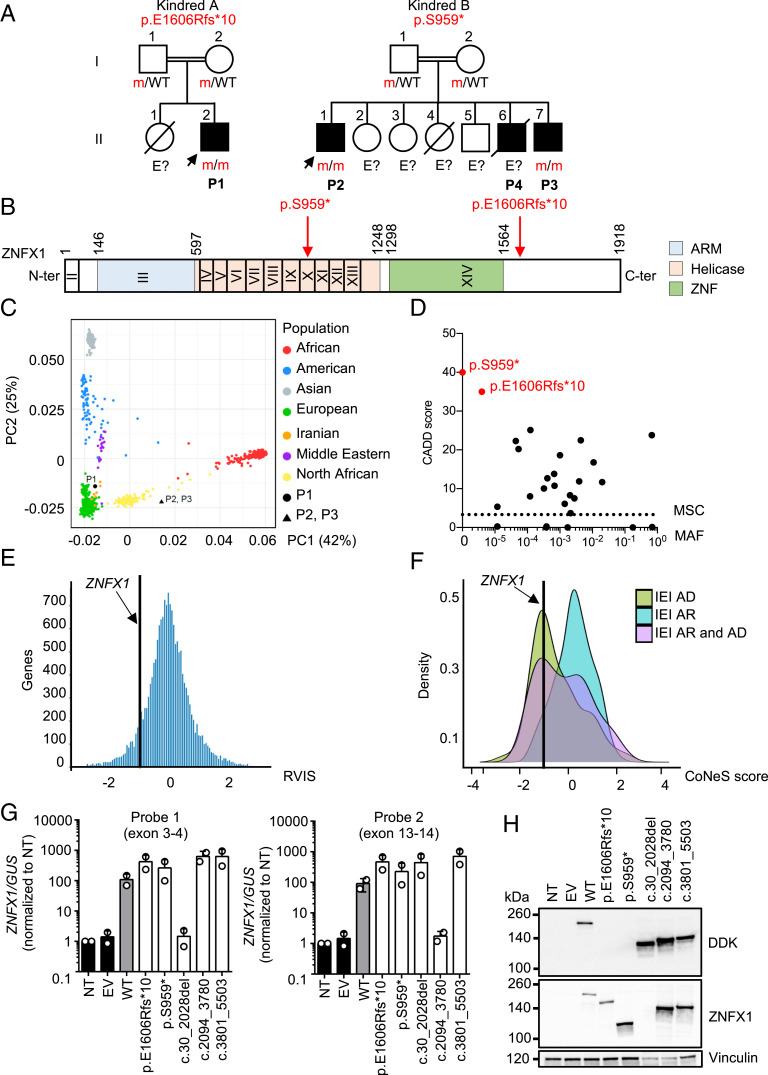
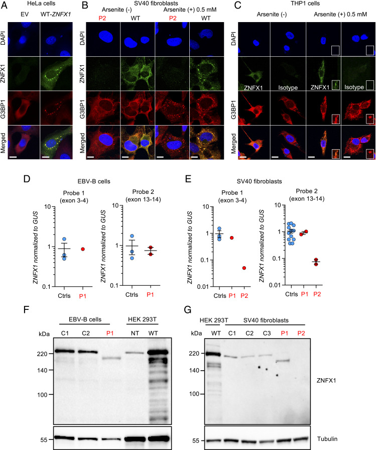
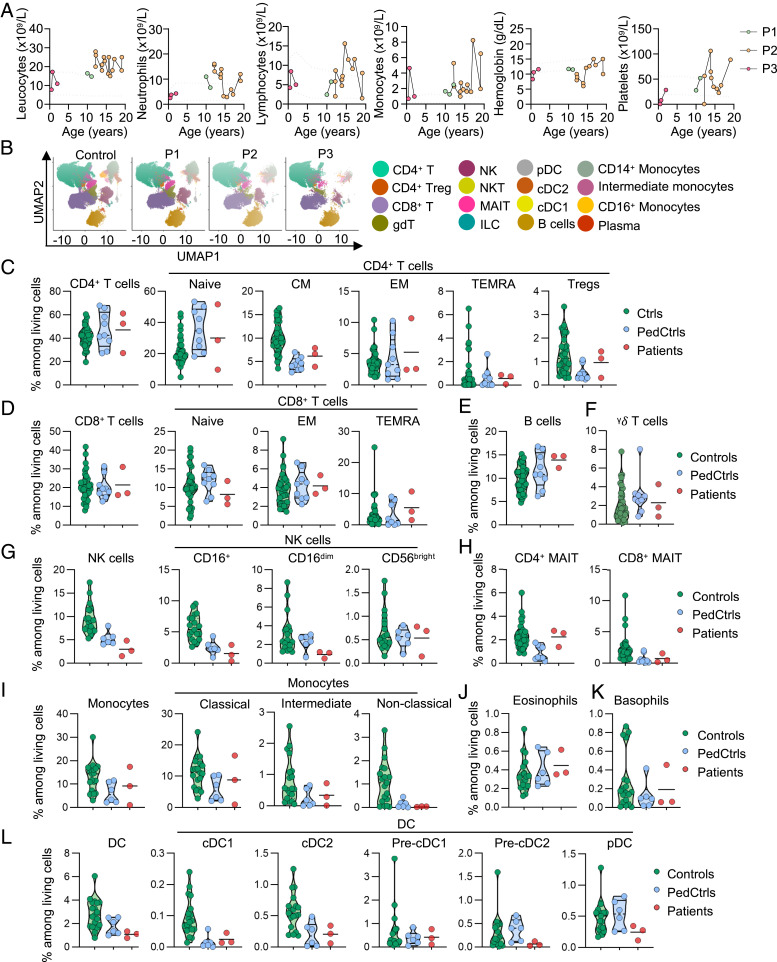
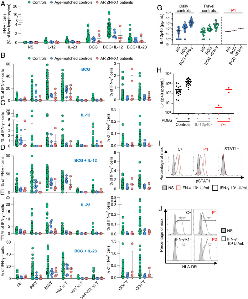
Human inborn errors of IFN-γ underlie mycobacterial disease, due to insufficient IFN-γ production by lymphoid cells, impaired myeloid cell responses to this cytokine, or both. We report four patients from two unrelated kindreds with intermittent monocytosis and mycobacterial disease, including bacillus Calmette-Guérin-osis and disseminated tuberculosis, and without any known inborn error of IFN-γ. The patients are homozygous for ZNFX1 variants (p.S959* and p.E1606Rfs*10) predicted to be loss of function (pLOF). There are no subjects homozygous for pLOF variants in public databases. ZNFX1 is a conserved and broadly expressed helicase, but its biology remains largely unknown. It is thought to act as a viral double-stranded RNA sensor in mice, but these patients do not suffer from severe viral illnesses. We analyze its subcellular localization upon overexpression in A549 and HeLa cell lines and upon stimulation of THP1 and fibroblastic cell lines. We find that this cytoplasmic protein can be recruited to or even induce stress granules. The endogenous ZNFX1 protein in cell lines of the patient homozygous for the p.E1606Rfs*10 variant is truncated, whereas ZNFX1 expression is abolished in cell lines from the patients with the p.S959* variant. Lymphocyte subsets are present at normal frequencies in these patients and produce IFN-γ normally. The hematopoietic and nonhematopoietic cells of the patients tested respond normally to IFN-γ. Our results indicate that human ZNFX1 is associated with stress granules and essential for both monocyte homeostasis and protective immunity to mycobacteria.

Keywords: ZNFX1; inborn error of immunity; inflammation; monocytosis; mycobacteria.


Conflict of interest statement

The authors declare no competing interest.

Figures

Fig. 1.
Fig. 1.
AR ZNFX1 deficiency in four patients from two unrelated kindreds. (A) Pedigrees of the two unrelated kindreds. Generations are indicated by Roman numerals (I and II), and each individual is indicated by an Arabic numeral (1 to 7). Affected patients are represented by closed black symbols, and index cases are indicated by an arrow. “E?” indicates individuals with an unknown genotype. Symbols crossed with a black diagonal line correspond to deceased individuals. (B) Schematic representation of the ZNFX1 gene. Coding exons are indicated by Roman numerals (II to XIV). The ZNFX1 protein is represented, with three predicted domains: the ARM, helicase, and ZNF domains. The reported mutations are indicated in red with an arrow. (C) PCA showing the origins of P1, P2, and P3 plotted on main ethnic origins extracted from the 1000 Genomes database and our own WES database. (D) MAF and CADD score of the homozygous ZNFX1 variants found in P1 to P3 (red symbols) and of all homozygous variants in gnomAD v.2.1.1 and BRAVO/TOPmed (black symbols). The dotted line corresponds to the MSC, with its 99% CI. The MSC for ZNFX1 is 3.3. (E) According to its RVIS, ZNFX1 is highly intolerant of genetic variation. (F) CoNeS for ZNFX1 and its distribution for genes causing IEI, according to disease mode of inheritance (AD). (G) RT-qPCR on cDNA from HEK293T cells nontransfected (NT) or transfected with an empty plasmid (EV), WT-ZNXF1, mutated-ZNXF1 (E1606Rfs*10 and S959*), or constructs with in-frame deletions of each ZNFX1 domain, for the ARM (c.30_2028del), helicase (c.2094_3780del), and ZNF (c.3801_5503del) domains, with a probe spanning the junction between exons 3 and 4 (probe 1, Left) or exons 13 and 14 (probe 2, Right). GUSB was used for normalization. Values are expressed as means ± SEM. (H) Western blot of total protein extracts from HEK293T cells either left NT or transfected with EV, WT, or mutated ZNXF1, or constructs with in-frame deletions of the ARM (c.30_2028del), helicase (c.2094_3780del), or ZNF (c.3801_5503del) domain, all inserted into pCMV6 with a C-terminal Flag-tag. ZNXF1 was detected with an mAb directed against the N terminus of ZNXF1 or an antibody directed against the C-terminal DDK (Flag) tag. An antibody against vinculin was used as a loading control. The results shown are representative of two independent experiments.
Fig. 2.
Fig. 2.
Expression of ZNFX1 in cell lines. (A) Confocal microscopy of HeLa cells transiently transfected with an EV or the WT-ZNFX1, stained with the anti-G3BP1 antibody, DAPI, or an antibody directed against the C terminus of ZNFX1. (Scale bar, 10 µm.) (B) Colocalization of ZNFX1 and G3BP1 in SV40 fibroblasts stimulated for 24 h with 25 µg/mL of poly(I:C) from a healthy control and a ZNFX1-deficient patient (P2), treated or not with 0.5 mM arsenite for 30 min. (Scale bar, 10 µm.) (C) Colocalization of ZNFX1 and G3BP1 in adherent THP1 cells treated or not with 0.5 mM arsenite for 30 min. An IgG isotype is used as a negative control. The results shown are representative of at least two independent experiments. (Scale bar, 10 µm.) (D) RT-qPCR for ZNFX1 with a probe spanning the junction between exons 3 and 4 (Left) or spanning the junction between exons 13 and 14 (Right) in EBV-B cells from healthy controls (Ctrls, n = 3) and a patient (P1). GUSB was used for normalization. Values are expressed as means ± SEM. (E) RT-qPCR for ZNFX1 with a probe spanning the junction between exons 3 and 4 (Left) and a probe spanning the junction between exons 13 and 14 (Right) in SV40 fibroblasts from healthy controls (n = 3) and patients (P1 and P2). GUSB was used for normalization. Values are expressed as means ± SEM. (F) Western blot of total protein extracts from EBV-B cells from healthy controls (C1 and C2) and a patient (P1). ZNFX1 was detected with an mAb directed against the N terminus. An antibody against tubulin was used as a loading control. The results shown are representative of two independent experiments. (G) Western blot of total protein extracts from SV40 fibroblasts from healthy controls (C1, C2, and C3) and patients (P1 and P2). ZNFX1 was detected with an mAb directed against the N terminus. An antibody against tubulin was used as a loading control. The results shown are representative of two independent experiments.
Fig. 3.
Fig. 3.
Hematological and immunological profile of patients with inherited ZNFX1 deficiency. (A) Absolute numbers of peripheral total leukocytes, neutrophils, lymphocytes, monocytes and platelets, and hemoglobin from P1, P2, and P3, determined from complete blood counts. (B) UMAP analysis of CyTOF showing the distribution of the main peripheral leukocyte populations identified in a healthy control, P1, P2, and P3. (C) Frequency of living CD4+ T cells and their subsets in adult controls (green circles), age-matched controls (PedCtrls, blue circles), and P1, P2, and P3 (red circles), as determined by CyTOF. Frequency of living (D) CD8+ T cells and their subsets, (E) B cells, (F) γδ T cells, (G) NK cells and their subsets, and (H) MAIT cells, and frequency of (I) monocytes and their subsets, (J) eosinophils, (K) basophils, and (L) dendritic cells (DCs) and their subsets, as determined by CyTOF. Results are expressed as a proportion of living single PBMCs.
Fig. 4.
Fig. 4.
Conserved IFN-γ immunity in ZNFX1-deficient patients. PBMCs from controls (green dots), age-matched controls (blue dots), and patients with AR ZNFX1 deficiency (red dots) were left unstimulated or were stimulated with IL-12, or IL-23, with or without bacillus Calmette–Guérin activation. (A) IFN-γ levels in the supernatant of PBMCs with and without stimulation with IL-12, IL-23, bacillus Calmette–Guérin, bacillus Calmette–Guérin+IL-12, and bacillus Calmette–Guérin+IL-23, as assessed by intracellular flow cytometry. Proportion of IFN-γ−producing lymphocytes of various subsets involved in innate (NK, iNKT, and MAIT cells), adaptive (CD4+ and CD8+ T cells), and both adaptive and innate (γδ T cells including Vδ1+, Vδ2+, and Vδ1Vδ2 subsets) immunity after stimulation with (B) bacillus Calmette–Guérin, (C) IL-12, (D) bacillus Calmette–Guérin+ IL-12, (E) IL-23, or (F) bacillus Calmette–Guérin+IL-23. (G) Secretion of IL-12p40 by whole blood from daily controls (n = 69), travel controls (n = 14), P1, with and without stimulation with bacillus Calmette–Guérin alone, or bacillus Calmette–Guérin and IFN-γ. ELISA was used to determine the levels of this cytokine. (H) Secretion of IL-12p40 by EBV-B cells from controls, patients with AR complete IL-12p40 deficiency (IL-12p40−/−), or P1, after stimulation with 10−7 M PDBu for 24 h. ELISA was used to determine the levels of these cytokines. (I) Phosphorylation of STAT1 after 20 min of stimulation with IFN-γ (104 IU/mL) or IFN-α (104 IU/mL) in EBV-B cells from a healthy control (C+), P1, and a patient with AR complete STAT1 deficiency (STAT1−/−), as determined by flow cytometry. (J) Induction of HLA-DR expression in SV40 fibroblasts from controls (C+), P1, P2, and a patient with AR complete IFN-γR1 deficiency (IFN-γR1−/−) after 48 h of stimulation with IFN-γ (104 IU/mL), as determined by flow cytometry. The results of I and J are representative of two independent experiments.

References

    1. Bustamante J., Mendelian susceptibility to mycobacterial disease: Recent discoveries. Hum. Genet. 139, 993–1000 (2020). - PMC - PubMed
    1. Bustamante J., Boisson-Dupuis S., Abel L., Casanova J. L., Mendelian susceptibility to mycobacterial disease: Genetic, immunological, and clinical features of inborn errors of IFN-γ immunity. Semin. Immunol. 26, 454–470 (2014). - PMC - PubMed
    1. Yang R., et al., Human T-bet governs innate and innate-like adaptive IFN-gamma immunity against mycobacteria. Cell 183, 1826–1847.e31 (2020). - PMC - PubMed
    1. Kerner G., et al., Inherited human IFN-γ deficiency underlies mycobacterial disease. J. Clin. Invest. 130, 3158–3171 (2020). - PMC - PubMed
    1. Nathan C. F., Murray H. W., Wiebe M. E., Rubin B. Y., Identification of interferon-gamma as the lymphokine that activates human macrophage oxidative metabolism and antimicrobial activity. J. Exp. Med. 158, 670–689 (1983). - PMC - PubMed

Publication types

MeSH terms