. 2021 Apr 1;62(4):26.
doi: 10.1167/iovs.62.4.26.

Posture-Dependent Collapse of the Optic Nerve Subarachnoid Space: A Combined MRI and Modeling Study

Affiliations

Posture-Dependent Collapse of the Optic Nerve Subarachnoid Space: A Combined MRI and Modeling Study

Petter Holmlund et al. Invest Ophthalmol Vis Sci. .

Abstract

Purpose: We hypothesize that a collapse of the optic nerve subarachnoid space (ONSAS) in the upright posture may protect the eyes from large translamina cribrosa pressure differences (TLCPD) believed to play a role in various optic nerve diseases (e.g., glaucoma). In this study, we combined magnetic resonance imaging (MRI) and mathematical modeling to investigate this potential ONSAS collapse and its effects on the TLCPD.

Methods: First, we performed MRI on six healthy volunteers in 6° head-down tilt (HDT) and 13° head-up tilt (HUT) to assess changes in ONSAS volume (measured from the eye to the optic canal) with changes in posture. The volume change reflects optic nerve sheath (ONS) distensibility. Second, we used the MRI data and mathematical modeling to simulate ONSAS pressure and the potential ONSAS collapse in a 90° upright posture.

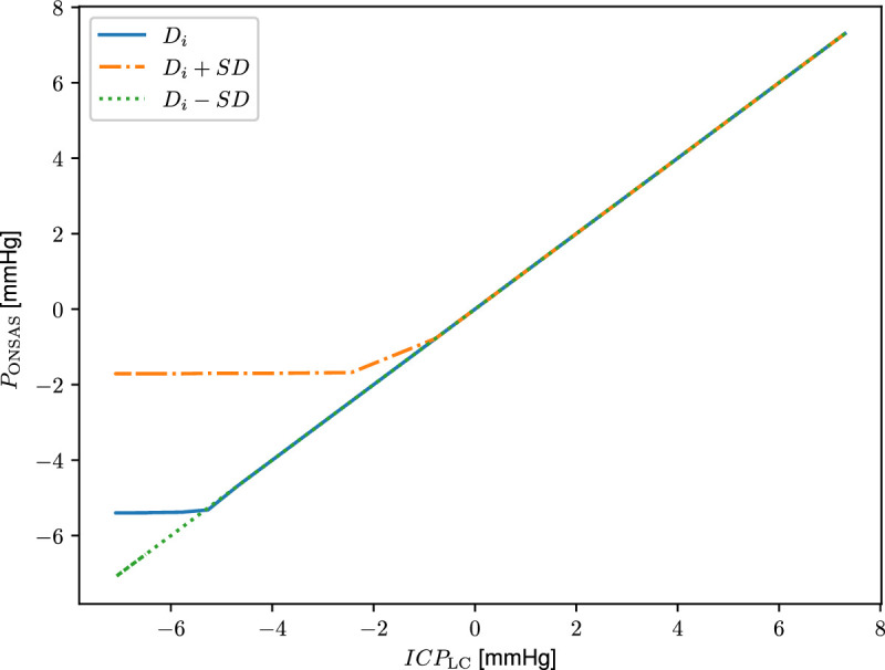
Results: The MRI showed a 33% decrease in ONSAS volume from the HDT to HUT (P < 0.001). In the upright posture, the simulations predicted an ONSAS collapse 25 mm behind lamina cribrosa, disrupting the pressure communication between the ONSAS and the intracranial subarachnoid space. The collapse reduced the simulated postural increase in TLCPD by roughly 1 mm Hg, although this reduction was highly sensitive to ONS distensibility, varying between 0 and 4.8 mm Hg when varying the distensibility by ± 1 SD.

Conclusions: The ONSAS volume along the optic nerve is posture dependent. The simulations supported the hypothesized ONSAS collapse in the upright posture and showed that even small changes in ONS stiffness/distensibility may affect the TLCPD.


Conflict of interest statement

Disclosure: P. Holmlund, None; K.-H. Støverud, None; A. Wåhlin, None; U. Wiklund, None; J. Malm, None; G. Jóhannesson, None; A. Eklund, None

Figures

Figure 1.
Figure 1.
MRI image of the ONSAS segmentation. (A) Volume. (B) Diameter (average of four directions). The ONSAS radius rONS and optic nerve radius rON are also highlighted.
Figure 2.
Figure 2.
The compartment model. The three compartments consist of the cranial SAS (c), spinal SAS (s), and ONSAS, each with a pressure, volume, and compliance. Changes in volume from supine equilibrium in each compartment serve as the dependent variables of the underlying ordinary differential equations (ODEs) of the model. The model includes cranial CSF production and CSF absorption to the venous system through arachnoid granulations (AG) and absorption across the ONS. Flow rates (indicated by arrows) are determined by the pressure difference and resistances between compartments. For all absorption sites, only outflow from the CSF compartments was allowed. The compliances depend on both internal CSF pressures and surrounding venous (and intraorbital) pressures, meaning that venous pressure plays a major role both in absorption and compliance. The ONSAS resistance (RONSAS) is based on the acquired MRI measurements of the ONSAS/ONS radius (rONS) and is allowed to vary. Postural changes are implemented through hydrostatic effects in the CSF and venous systems, including the collapse of the internal jugular veins and the potential collapse of the ONSAS. The reference level for compartment c is the auditory meatus, the venous hydrostatic indifference point is used for compartment s, and the level of the LC for the ONSAS compartment. The pressures were hydrostatically adjusted to the same level when calculating the flow rates to account for the height differences between the reference points. The main model outputs include ICP at the level of the LC (ICPLC), the ONSAS pressure (PONSAS) and rONS when going from supine to upright.
Figure 3.
Figure 3.
Average ONSAS volume per slice in the MRI data for all optic nerves. In all slices the volume decreased when changing from HDT to HUT.
Figure 4.
Figure 4.
MRI of the ONSAS in a test subject in HDT and HUT 3, 10, and 15 mm behind LC. The ONSAS is annulus-shaped and visible in all images.
Figure 5.
Figure 5.
MRI of the ONSAS in a second subject in HDT and HUT 3, 10, and 15 mm behind LC. In HUT, the ONSAS is visible in the bulbar region only.
Figure 6.
Figure 6.
Plot of pressure (AC) and minimum ONSAS radius (DF) versus time when changing body position from supine to upright at time = 5 minutes. Each column corresponds to different degrees of ONS distensibility to illustrate the sensitive relationship between distensibility and predicted ONSAS collapse: (A & D) the distensibility directly acquired from the measurements (Di), (B & E) represents increased distensibility (Di + 1 · SD), and (C & F) represents reduced distensibility (Di − 1 · SD). In the supine position, the ICP at the level of the eye (ICPLC) equals the ONSAS pressure (PONSAS). In the upright posture, occlusion of the ONSAS causes a pressure difference in (A) and (B), whereas in (C) there is no occlusion, and the ICP equals the ONSAS pressure. Note that the pressure difference between the cranial and optic nerve SAS increases with increased distensibility.
Figure 7.
Figure 7.
Plotting the ONSAS pressure as a function of the ICP for three different distensibility values. The curve shows a linear dependence when the ONSAS is open and that the ONSAS pressure is nearly constant after the collapse. For Di the collapse happens at an ICP of –5.4 mm Hg, for Di + 1 · SD the collapse happens at –1.7 mm Hg, and for Di − 1 · SD there is no collapse and there is a linear relationship between ONSAS pressure and ICP all the way.
Figure 8.
Figure 8.
Computed radii for 0 deg supine (red) and 90 deg upright (blue) postures. The computed collapse occurred in the last segment (furthest away from the LC). The optic nerve shape is based on the MRI measurements.

Comment in

Similar articles

Cited by

References

    1. Berdahl JP, Fautsch MP, Stinnett SS, Allingham RR.. Intracranial pressure in primary open angle glaucoma, normal tension glaucoma, and ocular hypertension: a case-control study. Invest Ophthalmol Vis Sci. 2008; 49: 5412–5418. - PMC - PubMed
    1. Ren R, Jonas JB, Tian G, et al. .. Cerebrospinal fluid pressure in glaucoma: a prospective study. Ophthalmology. 2010; 117: 259–266. - PubMed
    1. Lindén C, Qvarlander S, Jóhannesson G, et al. .. Normal-tension glaucoma has normal intracranial pressure: a prospective study of intracranial pressure and intraocular pressure in different body positions. Ophthalmology. 2018; 125: 361–368. - PubMed
    1. Guy AH, Wiggs JL, Turalba A, Pasquale LR.. Translating the low translaminar cribrosa pressure gradient hypothesis into the clinical care of glaucoma. Semin Ophthalmol. 2016; 31: 131–139. - PubMed
    1. Johannesson G, Eklund A, Linden C.. Intracranial and intraocular pressure at the lamina cribrosa: gradient effects. Curr Neurol Neurosci Rep. 2018; 18: 25. - PMC - PubMed

Publication types