. 2021 Apr 20;12(1):54.
doi: 10.1186/s13244-021-00992-w.

Identifying lumbosacral plexus nerve root abnormalities in patients with sciatica using 3T readout-segmented echo-planar diffusion weighted MR neurography

Affiliations

Identifying lumbosacral plexus nerve root abnormalities in patients with sciatica using 3T readout-segmented echo-planar diffusion weighted MR neurography

Osamah M Abdulaal et al. Insights Imaging. .

Abstract

Objectives: To investigate the accuracy of Diffusion Weighted Imaging (DWI) using the Readout Segmentation of Long Variable Echo-trains (RESOLVE) sequence in detecting lumbosacral nerve abnormalities.

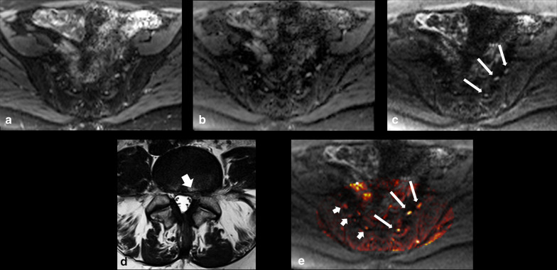
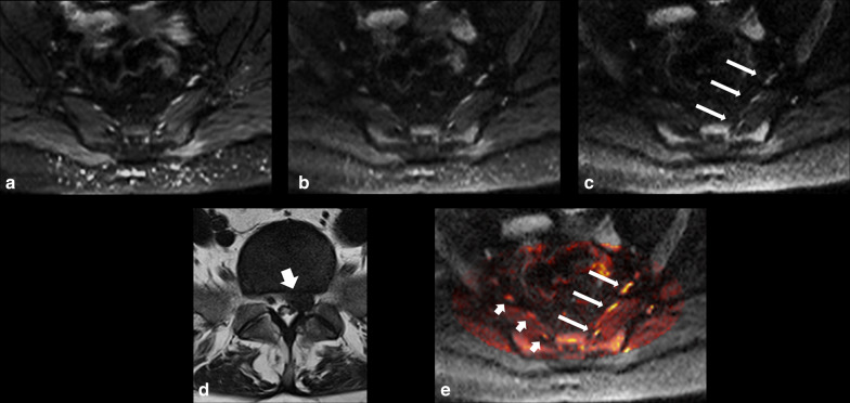
Methods: Following institutional ethics committee approval, patients with sciatica-type lower limb radicular symptoms (n = 110) were recruited and prospectively scanned using 3T MRI. Additional participants (n = 17) who underwent neurophysiological testing (EMG/NCV), were also prospectively studied. In addition to routine lumbar spine MRI, a DWI-RESOLVE sequence of the lumbosacral plexus was performed. Two radiologists, blinded to the side of patient symptoms, independently evaluated the MR images. The size and signal intensity changes of the nerves were evaluated using ordinal 4-point Likert-scales. Signal-to-noise ratio (SNR), apparent diffusion coefficient (ADC) and size were measured for affected and normal nerves. Inter-observer agreement was determined with kappa statistics; κ.

Results: In patients who did not undergo EMG/NCV testing (n = 110), the DWI-RESOLVE sequence detected lumbosacral nerve abnormalities that correlated with symptoms in 36.3% (40/110). This is a similar percentage to patients who underwent EMG/NCV testing, which was positive and correlated with symptoms in 41.2% (7/17). Inter-observer agreement for evaluation of lumbosacral nerve abnormalities was excellent and ranged from 0.87 to 0.94. SNR and nerve size measurements demonstrated statistically significant differences for the L5 and S1 nerves (p value < 0.05) for patients who did not undergo EMG/NCV testing.

Conclusion: The DWI-RESOLVE sequence is a promising new method that may permit accurate detection and localization of lumbar nerve abnormalities in patients with sciatica.

Keywords: Evidence-based practice; Magnetic resonance imaging; Sciatica; Spine.


Conflict of interest statement

The authors declare that they have no competing interests.

Figures

Fig. 1
Fig. 1
DWI-RESOLVE images on two different patients demonstrating typical nerve region of interest (ROI) placements (a) and dimensional calipers (b) for quantitative measurements
Fig. 2.
Fig. 2.
38-year-old female who presented with left leg pain and numbness. DWI-RESOLVE images (a) b = 50, (b) b = 500, (c) b = 800. The arrows on the b800 image correlate to the left L5, S1 and S2 nerves from anterior to posterior. The nerves were evaluated by both readers on axial DWI-RESOLVE images as positive for abnormality (grade II abnormal size and signal intensity). The left L5 nerve appears less involved compared to the left S1 and S2 nerve levels. Axial T2W TSE (d) of the lumbar spine at L5/S1 highlights a large left-sided disc herniation (arrow), which explains symptoms and correlates to nerve abnormalities. The color-mapped image e demonstrates the asymmetry of the lumbosacral plexus to better effect. Long arrows indicate abnormal left nerves and short arrows indicate normal contralateral side
Fig. 3.
Fig. 3.
45-year-old female suffering from left leg pain, severe weakness and numbness. DWI-RESOLVE images (a) b = 50, (b) b = 500, (c) b = 800. The arrows on the b800 image correlate to the left L5, S1 and S2 nerves from anterior to posterior. The nerves were evaluated by both readers on axial DWI-RESOLVE images as positive for abnormality (grade III abnormal size and signal intensity). Axial T2W TSE d of the lumbar spine at L5/S1 highlights a large left-sided disc herniation (arrow), which explains symptoms and correlates to nerve abnormalities. The color-mapped image e demonstrates the asymmetry of the lumbosacral plexus to better effect in this patient. Long arrows indicate abnormal left nerves and short arrows indicate normal contralateral side
Fig. 4.
Fig. 4.
63-year-old man suffering from severe right leg pain and numbness. DWI-RESOLVE images a b = 50, b b = 500, c b = 800. The arrows on each of the b value images correlate to the right L5 nerve root. The nerve was evaluated by both readers on axial DWI-RESOLVE images as positive for abnormality (grade IV abnormal size and signal intensity). Sagittal T2W TSE (d) of the lumbar spine demonstrates no significant disc herniation or other potential cause for symptoms. The color-mapped image e demonstrates the asymmetry of the right L5 nerve. Long arrows indicate abnormal right L5 nerve and short arrow indicates normal contralateral left L5 nerve
Fig. 5
Fig. 5
Demonstration of DWI-RESOLVE signal inhomogeneity. The b50 image (a) of a patient with normal neurophysiology testing demonstrates relatively prominent loss of signal on the left side of the body compared to the right. The left sciatic nerve (short arrow) is obscured due to this loss of signal. The normal right sciatic nerve (long arrow) appears asymmetric due to this spurious artifact. The b800 image (b) on a different patient demonstrates the hyperintense signal (arrow) that can occur at the edge of the field of view on some patients. This type of signal inhomogeneity does not typically obscure or distort the lumbosacral plexus
Fig. 6
Fig. 6
The relationship between ADC values for normal and abnormal lumbosacral nerves. Findings presented are the 7 nerve abnormalities for 7/17 patients who had lumbosacral nerve abnormalities on DWI-RESOLVE which matched both the clinical indications and the EMG/NCV findings. These 7 nerve abnormalities included L5 (n = 2) and S1 (n = 5). The ADC values at b-500 and b-800 for the abnormal nerves were generally higher than those for the normal nerves. However, in two (n = 2) patients, the calculated ADC values for abnormal nerves was lower than for the normal contralateral nerves

Similar articles

Cited by

References

    1. Deyo RA, Mirza SK. Clinical practice. Herniated Lumbar Intervertebral Disk. N Engl J Med. 2016;374:1763–1772. doi: 10.1056/NEJMcp1512658. - DOI - PubMed
    1. Brinjikji W, Luetmer PH, Comstock B, et al. Systematic literature review of imaging features of spinal degeneration in asymptomatic populations. AJNR Am J Neuroradiol. 2015;36:811–816. doi: 10.3174/ajnr.A4173. - DOI - PMC - PubMed
    1. Ropper AH, Zafonte RD. Sciatica. N Engl J Med. 2015;372:1240–1248. doi: 10.1056/NEJMra1410151. - DOI - PubMed
    1. Laporte C, Albert JD, Duvauferrier R, Bertaud V, Gouillou M, Guillin R. MRI investigation of radiating pain in the lower limbs: value of an additional sequence dedicated to the lumbosacral plexus and pelvic girdle. AJR Am J Roentgenol. 2014;203:1280–1285. doi: 10.2214/AJR.13.11884. - DOI - PubMed
    1. Reza Soltani Z, Sajadi S, Tavana B. A comparison of magnetic resonance imaging with electrodiagnostic findings in the evaluation of clinical radiculopathy: a cross-sectional study. Eur Spine J. 2014;23:916–921. doi: 10.1007/s00586-013-3164-z. - DOI - PMC - PubMed

LinkOut - more resources