Review
. 2021 Apr 20;12(1):53.
doi: 10.1186/s13244-021-00991-x.

Mimickers of breast malignancy: imaging findings, pathologic concordance and clinical management

Affiliations
Review

Mimickers of breast malignancy: imaging findings, pathologic concordance and clinical management

Mary S Guirguis et al. Insights Imaging. .

Abstract

Many benign breast entities have a clinical and imaging presentation that can mimic breast cancer. The purpose of this review is to illustrate the wide spectrum of imaging features that can be associated with benign breast diseases with an emphasis on the suspicious imaging findings associated with these benign conditions that can mimic cancer. As radiologic-pathologic correlation can be particularly challenging in these cases, the radiologist's familiarity with these benign entities and their imaging features is essential to ensure that a benign pathology result is accepted as concordant when appropriate and that a suitable management plan is formulated.

Keywords: Benign breast disease; Benign breast masses; Breast cancer; Inflammatory breast disease; Radiologic-pathologic concordance.


Conflict of interest statement

The authors declare that they have no competing interests.

Figures

Fig. 1
Fig. 1
Categories of benign breast diseases that can mimic breast cancer
Fig. 2
Fig. 2
Breast abscess. A 45-year-old woman presented with a palpable area in the right breast. Mediolateral oblique (a) mammogram shows a focal asymmetry in the upper outer breast (arrow) and associated trinagular palpable marker. Power Doppler ultrasound images (b, c) reveal two hypoechoic, oval masses with peripheral vascularity in the same region (asterisks). Axial post-contrast T1-weighted MRI (d), sagittal T2-weighted MRI (e), and postcontrast subtraction T1-weighted MRI (f) show two T2-hyperintense, rim-enhancing masses (arrows). Needle biopsy showed acute inflammatory cells consistent with abscess without evidence of malignancy
Fig. 3
Fig. 3
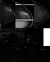
Granulomatous mastitis. A 34-year-old woman who is 2 years post-partum presented with a palpable left breast mass, diffuse breast swelling, tenderness, and erythema for several weeks. Bilateral mediolateral oblique mammogram (a) show diffuse skin thickening (solid arrow), global asymmetry, and trabecular thickening (dashed arrows) in the left breast, asymmetric from the right breast. Extended field of view grayscale ultrasound (b) shows an ill-defined hypoechoic, irregular mass (arrows). Color Doppler ultrasound (c) shows increased vascularity, edema (arrow head), and skin thickening (dashed arrow). T1weighted axial delayed post-contrast (d) and sagittal subtraction (e) post-contrast MRI shows diffuse skin thickening (solid arrow) and trabecular thickening with heterogenous enhancement involving the left superior breast (dashed arrows). There is associated axillary adenopathy (solid arrow in e). Axial PET/CT (f) shows diffuse fluorodeoxyglucose avidity involving the left breast (arrow). Findings and clinical presentation were suspicious for inflammatory breast cancer. The patient underwent three core needle breast biopsies of the mass in the left breast over the span of a month, and a skin punch biopsy. All biopsies showed dense stromal fibrosis, chronic inflammation, and features suggestive of granulomatous mastitis without atypia or malignancy
Fig. 4
Fig. 4
Diabetic mastopathy. A 56-year-old patient presented with bilateral palpable breast masses and an 8-year history of type 2 diabetes. Bilateral craniocaudal mammogram (a) shows bilateral non-calcified, obscured masses correlating with the palpable triangular markers (arrows). Grayscale right (b) and left (c) breast ultrasound shows irregular, hypoechoic masses with posterior acoustic shadowing (arrows). Power Doppler (d) ultrasound demonstrated internal vascularity (arrow) involving these masses. Core needle biopsy of both masses showed perilobular lymphocytic infiltration. Repeat core needle biopsy 8 months after the initial biopsy showed chronic lymphocytic lobulitis. No evidence of malignancy. Findings are consistent with diabetic mastopathy
Fig. 5
Fig. 5
Lymphocytic mastopathy. A 37-year-old woman presented with right breast palpable retroareolar mass, swelling, erythema, and tenderness. Craniocaudal and lateral mammography (a, b) shows a focal asymmetry (solid arrows) and skin thickening (dashed arrows) associated with the triangular palpable marker in the lateral breast. Grayscale (c) and color Doppler breast ultrasound (d) reveals multiple retroareolar, vascular masses (arrows). Core needle biopsy showed acute and chronic inflammation and granulation tissue. The patient’s symptoms resolved within a month. The patient presented 2 years later with a contralateral palpable breast mass and nipple inversion. Craniocaudal and lateral mammographic views (e, f) reveal a focal asymmetry in the central breast associated with the palpable marker (arrows). Grayscale (c) and color Doppler (d) breast ultrasound reveals a vascular heterogeneous mass (arrows). Core needle biopsy revealed benign breast tissue with lymphoplasmacytic infiltrate. The patient’s symptoms improved over the following 6 months. She did not have a history of diabetes or other known immunologic condition
Fig. 6
Fig. 6
Fat necrosis. A 52-year-old woman with remote history of mastopexy presented with a palpable breast mass. Mediolateral oblique mammogram (a) shows an irregular mass with spiculated margins, associated coarse rim calcifications (solid arrow), and focal skin thickening (dashed arrow). Longitudinal grayscale ultrasound (b) shows a non-parallel irregular hypoechoic mass (arrow). Fat-suppressed axial post-contrast T1-weighted breast MRI (c) shows an irregular mass with spiculated margins (arrow) with associated singal void artifact related to post-biopsy clip marker. Ultrasound-guided biopsy showed fat necrosis. Repeat biopsy under MRI guidance confirmed the diagnosis of fat necrosis
Fig. 7
Fig. 7
Stromal fibrosis. A 55-year-old woman presented for a focal asymmetry detected on screening mammography. Craniocaudal (a) and lateral (b) spot compression mammographic views show an oval non-calcified mass with angular margins (arrows). Grayscale transverse (c) and longitudinal (d) ultrasound show a mixed-echogenicity oval mass correlating with the mammographic finding (arrows). Ultrasound-guided biopsy yielded stromal fibrosis. Six-month follow-up mammography and ultrasound were recommended and demonstrated stability
Fig. 8
Fig. 8
Stromal fibrosis. A 48-year-old female presented with a palpable breast mass. Spot compression tangential (a), mediolateral (b), and craniocaudal (c) mammographic views did not show a corresponding abnormality. Grayscale (d) and power Doppler (f) ultrasound images reveal an irregular mass with posterior acoustic shadowing corresponding to the palpable area. Core needle biopsy of the mass showed stromal fibrosis without evidence of malignancy
Fig. 9
Fig. 9
Sclerosing adenosis. A 35-year-old woman presented with a palpable breast mass. Mediolateral, craniocaudal, and spot tangential mammographic views do not reveal an abnormality (ac, respectively). Grayscale and color Doppler ultrasound images (d, e) reveal a vascular, mixed cystic, and solid mass correlating with the area of palpable abnormality. Core needle biopsy showed sclerosing adenosis in a background of dense fibrosis without atypia or carcinoma. A 6-month follow-up ultrasound was rcommended and demonstrated stability
Fig. 10
Fig. 10
Hamartoma. A 14-year-old presented with unilateral breast enlargement. A mass was not palpable on examination. Color Doppler (a) and longitudinal panoramic ultrasound grayscale images (b) show a hypervascular, 17-cm isoechoic breast mass replacing the entire breast. T1-weighted non-fat-suppressed (c) and T1-weighted fat-suppressed (d) post-contrast MRI images show a well-circumscribed, heterogeneously enhancing breast mass suspicious for a sarcoma (arrow). T2-weighted sagittal images (e) show moderate T2 signal with marked heterogeneity (arrow). Pathology at the time of surgical excision showed hamartoma with areas of pseudoangiomatous stromal hyperplasia and fibroadenomatoid change
Fig. 11
Fig. 11
Pseudoangiomatous stromal hyperplasia. A 41-year-old woman presented with a palpable breast mass. Mediolateral oblique mammogram (a) shows a high-density well-circumscribed mass (arrow). Longitudinal grayscale ultrasound (b) shows a large, oval circumscribed mass. Core needle biopsy showed pseudoangiomatous stromal hyperplasia (PASH). A different patient, a 40-year-old woman, was found to have a new breast mass (arrow) on mammography, craniocaudal spot view (c). Transverse grayscale (d) ultrasound reveals a corresponding oval isoechoic circumscribed mass. Core needle biopsy showed PASH
Fig. 12
Fig. 12
Suspicious presentation of pseudoangiomatous stromal hyperplasia. A 56-year-old woman with a new focal asymmetry in the outer breast that was identified on her (a) screening mammogram and persisted on (b) additional diagnostic spot compression views (arrows). The area was palpable by the patient and the clinician. The focal asymmetry had developed since her prior available mammogram performed 4 years earlier (c). Breast ultrasound (d) shows a corresponding irregular hypoechoic mass with associated posterior acoustic shadowing (arrow). Breast MRI sagittal post-contrast T1-weighted delayed images (e) and axial post-contrast T1-weighted images (f) show irregular enhancing masses (arrows). Corresponding T2 hyperintense cystic components with slit-like spaces (arrow) are identified (g), which favors PASH when present. Ultrasound-guided and MRI-guided biopsies were performed in this case and showed PASH. The area remained mammographically stable for over 4 years
Fig. 13
Fig. 13
Tubular adenoma. A 36-year-old woman presented with a palpable breast mass. Spot cradiocaudal and lateral mammogram (a, b) shows a high-density irregular mass (solid arrow), adjacent to an incidental oil cyst (dotted arrow). Grayscale (c) and power Doppler ultrasound (d) images show an irregular hypoechoic mass with increased vascularity (arrows). Core needle biopsy showed tubular adenoma. The mass remained stable sonographically for 30 months and mammographically for 9 years
Fig. 14
Fig. 14
Desmoid fibromatosis. A 47-year-old woman presented with a palpable mass. Lateral spot compression mammogram (a) shows an irregular, high-density mass with spiculated margins adjacent to the chest wall (arrow). Grayscale ultrasound (b) shows a corresponding irregular, hypoechoic anti-parallel mass with indistinct margins. T1-weighted post-contrast fat-saturated MRI of the chest (c) shows invasion of the pectoralis major muscle by this irregular enahancing mass (dotted arrow). Core needle biopsy showed desmoid fibromatosis. The patient underwent surgical excision
Fig. 15
Fig. 15
Granular cell tumor. A 65-year-old woman presented for further evaluation of a focal asymmetry detected on screening mammopgrahy. Craniocaudal and lateral spot compression views (a, b) show a small irregular mass with spiculated margins (arrows). Color Doppler (c) and grayscale (d) ultrasound shows a corresponding irregular mass with internal vascularity (arrows). Core needle biopsy yielded granular cell tumor which was treated with surgical excision

Similar articles

Cited by

References

    1. Sabaté JM, Clotet M, Gómez A, De Las HP, Torrubia S, Salinas T. Radiologic evaluation of uncommon inflammatory and reactive breast disorders. Radiographics. 2005;25(2):411–424. - PubMed
    1. Trop I, Dugas A, David J, et al. Breast abscesses: evidence-based algorithms for diagnosis, management, and follow-up. Radiographics. 2011;31(6):1683–1699. - PubMed
    1. Schäfer P, Fürrer C, Mermillod B. An association of cigarette smoking with recurrent subareolar breast abscess. Int J Epidemiol. 1988;17(4):810–813. - PubMed
    1. Rizzo M, Gabram S, Staley C, et al. Management of breast abscesses in nonlactating women. Am Surg. 2010;76(3):292–295. - PubMed
    1. Berna-Serna JD, Madrigal M. Percutaneous management of breast abscesses. An experience of 39 cases. Ultrasound Med Biol. 2004;30(1):1–6. - PubMed

LinkOut - more resources