Review
. 2021 Apr 20;13(1):24.
doi: 10.1186/s13089-021-00222-w.

High-frequency ultrasound in clinical dermatology: a review

Affiliations
Review

High-frequency ultrasound in clinical dermatology: a review

Jack Levy et al. Ultrasound J. .

Abstract

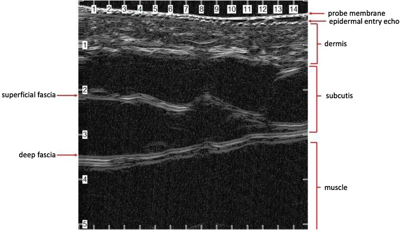
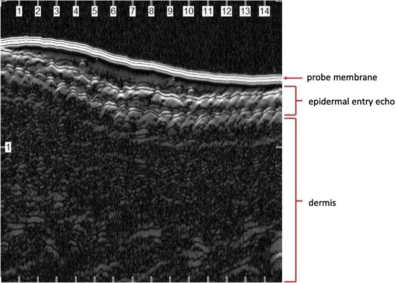
Background: Ultrasound was first introduced in clinical dermatology in 1979. Since that time, ultrasound technology has continued to develop along with its popularity and utility. Today, high-frequency ultrasound (HFUS), or ultrasound using a frequency of at least 10 megahertz (MHz), allows for high-resolution imaging of the skin from the stratum corneum to the deep fascia. This non-invasive and easy-to-interpret tool allows physicians to assess skin findings in real-time, enabling enhanced diagnostic, management, and surgical capabilities. In this review, we discuss how HFUS fits into the landscape of skin imaging. We provide a brief history of its introduction to dermatology, explain key principles of ultrasonography, and review its use in characterizing normal skin, common neoplasms of the skin, dermatologic diseases and cosmetic dermatology.

Conclusion: As frequency advancements in ultrasonography continue, the broad applications of this imaging modality will continue to grow. HFUS is a fast, safe and readily available tool that can aid in diagnosing, monitoring and treating dermatologic conditions by providing more objective assessment measures.

Keywords: Dermatology; Diagnostic imaging; High-frequency ultrasound; Ultrasonography.


Conflict of interest statement

The authors declare that they have no competing interest.

Figures

Fig. 1
Fig. 1
Normal skin from the anterior arm using a 50-MHz HFUS probe (Longport, Inc. EPISCAN). Note the fine hyperechoic epidermal entry echo. Deep to the entry echo is the hyperechoic dermis measuring about 1 millimeter (mm) with thicker horizontal bands in the deep dermis corresponding to the organized, horizontally arrayed collagen bundles in the reticular dermis. Deep to the dermis is the subcutis composed primarily of hypoechoic fat globules. Within this layer is the hyperechoic superficial fascia. Deep to the subcutis is the hyperechoic deep fascia overlying the biceps muscle
Fig. 2
Fig. 2
Glabrous skin from the palm using a 50-MHz HFUS probe (Longport, Inc. EPISCAN). Note the bilaminar hyperechoic entry echo due to the thickened stratum corneum in this anatomic location
Fig. 3
Fig. 3
Longitudinal view of a normal nail using a 50-MHz HFUS probe (Longport, Inc. EPISCAN). Note the bilaminar appearance of the nail plate consisting of the hyperechoic dorsal and ventral nail plates separated by a hypoechoic band. The top-most hyperechoic horizontal line represents the probe membrane
Fig. 4
Fig. 4
Nodular basal cell carcinoma measuring 5 mm by 7 mm on the left lower cheek of a 66-year-old man. Note the pearly borders and central ulceration in the gross photograph (a), the polymorphous vessels and ulceration in the dermatoscopic photo (b), and the 5 mm hypoechoic lesion in the superficial dermis across the short axis of this biopsy-supported nodular basal cell carcinoma using a 50-MHz UHFUS probe (Longport, Inc. EPISCAN)

Similar articles

Cited by

References

    1. Bhattacharya A, Young A, Wong A, Stalling S, Wei M, Hadley D. Precision diagnosis of melanoma and other skin lesions from digital images. AMIA Jt Summits Transl Sci Proc. 2017;2017:220–226. - PMC - PubMed
    1. Schneider SL, Kohli I, Hamzavi IH, Council ML, Rossi AM, Ozog DM. Emerging imaging technologies in dermatology: Part I: Basic principles. J Am Acad Dermatol. 2019;80(4):1114–1120. doi: 10.1016/j.jaad.2018.11.042. - DOI - PMC - PubMed
    1. Kane D, Grassi W, Sturrock R, Balint PV. A brief history of musculoskeletal ultrasound: 'From bats and ships to babies and hips'. Rheumatology. 2004;43(7):931–933. doi: 10.1093/rheumatology/keh004. - DOI - PubMed
    1. Parsons SK, Chan JA, Yu WW, Obadan N, Ratichek SJ, Lee J et al. Noninvasive Diagnostic Techniques for the Detection of Skin Cancers. AHRQ Comparative Effectiveness Technical Briefs. Rockville (MD)2011. - PubMed
    1. Barcaui Ede O, Carvalho AC, Lopes FP, Pineiro-Maceira J, Barcaui CB. High frequency ultrasound with color Doppler in dermatology. An Bras Dermatol. 2016;91(3):262–273. doi: 10.1590/abd1806-4841.20164446. - DOI - PMC - PubMed

LinkOut - more resources