Review
. 2021 Aug;37(8):1188-1202.
doi: 10.1007/s12264-021-00683-y. Epub 2021 Apr 20.

Emerging Role of PD-1 in the Central Nervous System and Brain Diseases

Affiliations
Review

Emerging Role of PD-1 in the Central Nervous System and Brain Diseases

Junli Zhao et al. Neurosci Bull. 2021 Aug.

Abstract

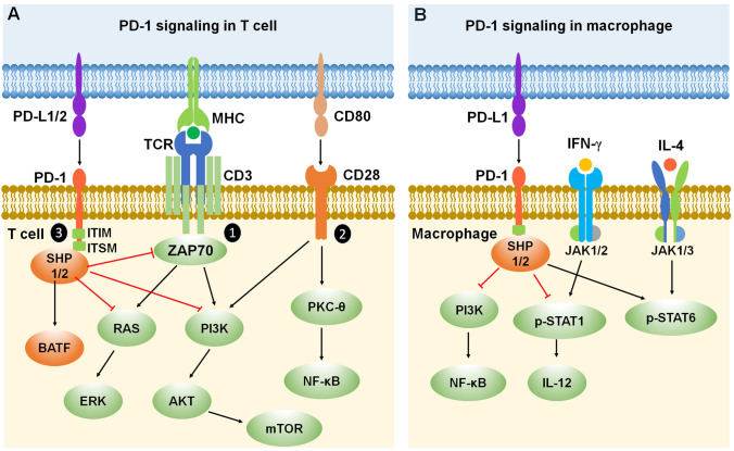
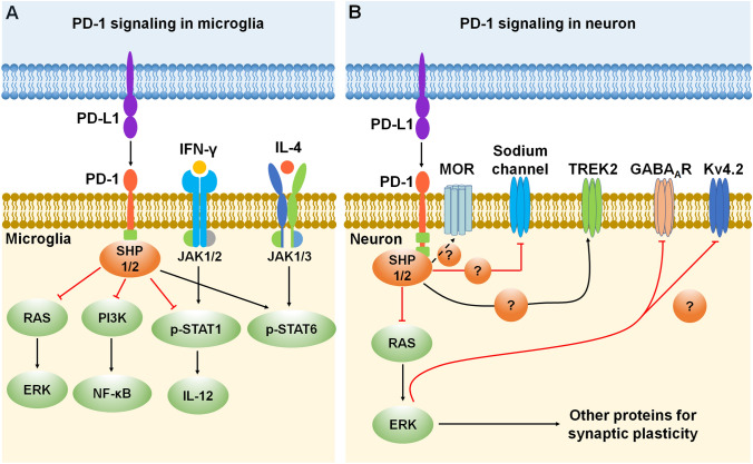
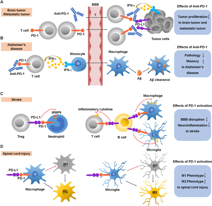
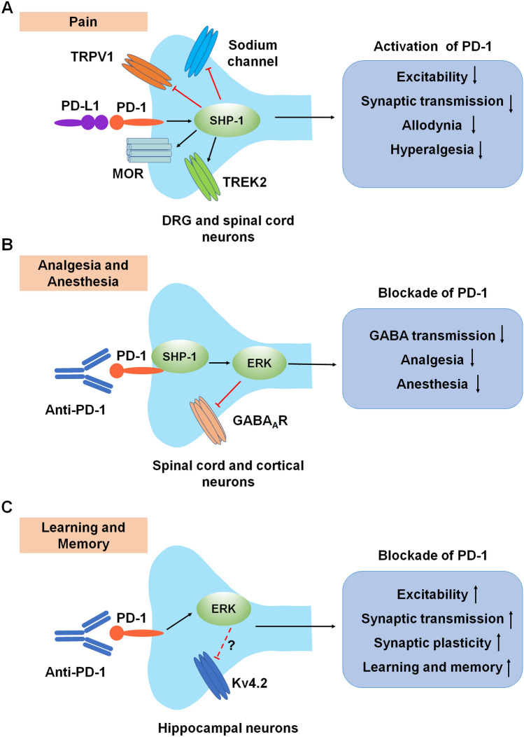
Programmed cell death protein 1 (PD-1) is an immune checkpoint modulator and a major target of immunotherapy as anti-PD-1 monoclonal antibodies have demonstrated remarkable efficacy in cancer treatment. Accumulating evidence suggests an important role of PD-1 in the central nervous system (CNS). PD-1 has been implicated in CNS disorders such as brain tumors, Alzheimer's disease, ischemic stroke, spinal cord injury, multiple sclerosis, cognitive function, and pain. PD-1 signaling suppresses the CNS immune response via resident microglia and infiltrating peripheral immune cells. Notably, PD-1 is also widely expressed in neurons and suppresses neuronal activity via downstream Src homology 2 domain-containing protein tyrosine phosphatase 1 and modulation of ion channel function. An improved understanding of PD-1 signaling in the cross-talk between glial cells, neurons, and peripheral immune cells in the CNS will shed light on immunomodulation, neuromodulation, and novel strategies for treating brain diseases.

Keywords: Central nervous system; Immune checkpoint; Immunotherapy; Neurotherapy; PD-1.


Conflict of interest statement

The authors claim that there are no conflicts of interest.

Figures

Fig. 1
Fig. 1
Timeline for major events leading to the development of PD-1 functions and PD-1-based immunotherapy.
Fig. 2
Fig. 2
PD-1 signaling in T cells and macrophages. A Mechanisms of PD-1 signaling in T cells. PD-1 inhibits T cell function by recruiting phosphatases SHP-1/SHP-2 to the ITIM/ITSM domain in the PD-1 tail and increasing the expression of transcription factor BATF. In addition, PD-1 inhibitory signaling antagonizes positive T cell signaling events triggered by (1) TCR interacting with MHC and (2) CD28 interacting with CD80. (3) PD-1 signaling inhibits ZAP70 and the RAS-ERK and PI3K-AKT-mTOR signaling pathways. B Mechanisms of PD-1 signaling in macrophages. PD-1 inhibits macrophage function by recruiting phosphatases SHP-1/SHP-2 to the ITIM/ITSM domain in the PD-1 tail, leading to inhibition of the PI3K-NF-κB signaling pathway. Moreover, PD-1 signaling suppresses IFN-γ-activated M1 macrophage polarization by reducing the phosphorylation of STAT1 and the secretion of IL-12, while promoting IL-4-activated M2 macrophage polarization by increasing STAT6 phosphorylation. Red lines ending in a bar represent inhibitory signaling, and black arrows indicate positive signaling. Abbreviations: PD-1, programmed cell death protein 1; PD-L1/2, PD-1 ligands, PD-L1 (B7-H1) and PD-L2 (B7-DC); ITIM, immunoreceptor tyrosine-based inhibitory motif; ITSM, immunoreceptor tyrosine-based switch motif; SHP, Src homology 2 domain-containing protein tyrosine phosphatase; BATF, basic leucine zipper ATF-like transcription factor; MHC, major histocompatibility complex; TCR, T cell receptor; CD, cluster of differentiation; ZAP70, zeta-chain-associated protein kinase 70; RAS, a small GTPase encoding RAS (retrovirus-associated DNA sequences); ERK, extracellular signal-regulated kinase; PI3K, type I phosphatidylinositol 3-kinase; AKT, serine/threonine-specific protein kinase; mTOR, mammalian target of rapamycin; PKC-θ, protein kinase C theta; NF-κB, nuclear factor kappa B; p-STAT1/6, phosphorylated signal transducer and activator of transcription 1/6; IL, interleukin; IFN-γ, interferon gamma; JAK, Janus kinase.
Fig. 3
Fig. 3
PD-1 signaling and expression in microglia and neurons. A Mechanisms of PD-1 signaling in microglia. PD-1 inhibits microglial function by recruiting phosphatases SHP-1/SHP-2 to the ITIM/ITSM domain in the PD-1 tail and then inhibiting the RAS-ERK and PI3K-NF-κB signaling pathways. Moreover, PD-1 signaling suppresses IFN-γ-activated M1 microglia polarization by reducing the phosphorylation of STAT1 and the secretion of IL-12, while promoting IL-4-activated M2 microglia polarization by increasing STAT6 phosphorylation. B Mechanisms of PD-1 signaling in neurons. Activation of the PD-1 pathway dampens neuronal excitation via activation of the phosphatase SHP-1/2 and resulting in the downstream modulation of sodium and potassium channels (TREK2 and Kv4.2), as well as GABAA receptors. Moreover, PD-1 signaling regulates mu-opioid receptor (MOR) function through activation of the phosphatase SHP-1. Red lines ending in a bar represent inhibitory signaling, and black arrows indicate positive signaling. Abbreviations: PD-1, programmed cell death protein 1; PD-L1, PD-1 ligand; SHP, Src homology 2 domain-containing protein tyrosine phosphatase; RAS, a small GTPase encoding RAS (retrovirus-associated DNA sequences); ERK, extracellular signal-regulated kinase; PI3K, type I phosphatidylinositol 3-kinase; NF-κB, nuclear factor kappa B; p-STAT 1/6, phosphorylated signal transducer and activator of transcription 1/6; IL, interleukin; IFN-γ, interferon gamma; JAK, Janus kinase; MOR, mu-opioid receptor; TREK2, TWIK-related K+ channel-2; GABAAR, gamma-aminobutyric acid A receptor; Kv4.2, potassium voltage-gated channel subfamily D member 2.
Fig. 4
Fig. 4
Immunomodulation by PD-1 in CNS diseases. A Anti-PD-1 antibody treatment induces IFN-γ–dependent activity and promotes T cell recruitment to the brain for anti-tumor immunotherapy. B Anti-PD-1 antibody treatment evokes a systemic IFN-γ–dependent immune response that enables the mobilization of monocyte-derived macrophages to the brain, thereby reducing pathology and improving memory in Alzheimer’s disease. C Activation of PD-1 signaling suppresses (1) the release of MMPs from neutrophils, protecting the BBB and (2) the release of inflammatory cytokines from T cell and microglia and macrophages, reducing neuroinflammation in stroke. D Activation of PD-1 signaling suppresses microglia and macrophage M1 polarization and promotes M2 polarization in spinal cord injury. Red lines ending in a bar represent inhibitory signaling, and black arrows indicate positive signaling. Abbreviations: PD-1, programmed cell death protein 1; PD-L1, PD-1 ligand; IFN-γ, interferon gamma; BBB, blood-brain barrier; Aβ, β-amyloid; MMP, matrix metallopeptidases.
Fig. 5
Fig. 5
Neuromodulation by PD-1 in the PNS and CNS. A Modulation of pain in primary sensory neurons and spinal dorsal horn neurons. Activation of PD-1 signaling in DRG neurons decreases neuronal excitability and synaptic transmission and inhibits physiological pain and pathological pain (allodynia and hyperalgesia) through modulation of ion channels. B Modulation of GABA-mediated analgesia and anesthesia in CNS neurons. C Modulation of learning and memory in hippocampal neurons. Anti-PD-1 antibody treatment increases hippocampal neuronal excitability, synaptic transmission, and synaptic plasticity, thereby enhancing learning and memory. Red lines ending in a bar represent inhibitory signaling, and black arrows indicate positive signaling. Abbreviations: PD-1, programmed cell death protein 1; PD-L1, PD-1 ligand; TRPV1, transient receptor potential subtype V1; MOR, mu-opioid receptor; TREK2, TWIK-related K+ channel-2; DRG, dorsal root ganglion; SHP-1, Src homology 2 domain-containing protein tyrosine phosphatase 1; ERK, extracellular signal-regulated kinase; GABAAR, gamma-aminobutyric acid A receptor; Kv4.2, potassium voltage-gated channel subfamily D member 2.

References

    1. Ishida Y, Agata Y, Shibahara K, Honjo T. Induced expression of PD-1, a novel member of the immunoglobulin gene superfamily, upon programmed cell death. EMBO J. 1992;11:3887–3895. doi: 10.1002/j.1460-2075.1992.tb05481.x. - DOI - PMC - PubMed
    1. Agata Y, Kawasaki A, Nishimura H, Ishida Y, Tsubata T, Yagita H, et al. Expression of the PD-1 antigen on the surface of stimulated mouse T and B lymphocytes. Int Immunol. 1996;8:765–772. doi: 10.1093/intimm/8.5.765. - DOI - PubMed
    1. Bardhan K, Anagnostou T, Boussiotis VA. The PD1:PD-L1/2 pathway from discovery to clinical implementation. Front Immunol. 2016;7:550. doi: 10.3389/fimmu.2016.00550. - DOI - PMC - PubMed
    1. Dong HD, Zhu GF, Tamada K, Chen LP. B7–H1, a third member of the B7 family, co-stimulates T-cell proliferation and interleukin-10 secretion. Nat Med. 1999;5:1365–1369. doi: 10.1038/70932. - DOI - PubMed
    1. Freeman GJ, Long AJ, Iwai Y, Latchman Y, Bourque K, Brown JA, et al. Engagement of the PD-1 immunoinhibitory receptor by a novel B7-family member leads to negative regulation of lymphocyte activation. Blood. 2000;96:810a–811a. - PMC - PubMed

Substances